Parathyroid Perfusion Optical Device
Clinical need:
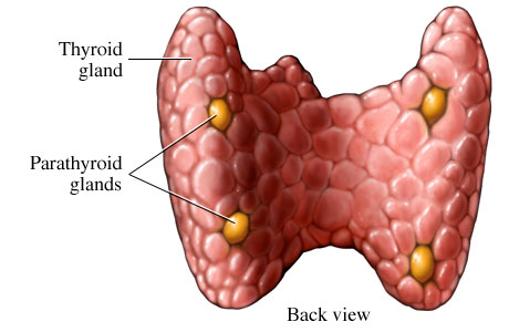
Endocrine surgeons at the Vanderbilt University Medical Center have highlighted an unmet clinical need for information regarding perfusion state in thyroid bed surgical settings. During removal of a thyroid gland, vascular tissues perfusing parathyroid glands may be damaged, causing eventual tissue death and impaired function of parathyroid. Understanding the perfusion state of the parathyroid glands is important to deciding whether to explant and then implant these tissues elsewhere in order to maintain reduced, but functional parathryoid hormonal output.
Team:
To meet this clinical need and provide an objective assessment of parathyroid perfusion, a team of senior undergraduate biomedical engineering students will design and build a device to quantify the perfusion state of thyroid bed tissues, particularly parathyroid glands. The team members are Gabriela Caires de Jesus, Tianhang Lu, Itamar Shapira, James Tatum, and Yu Zhou. The team is advised by Dr. Matthew Walker, Dr. Anita Mahadevan-Jansen, Dr. James T. Broome, MD, and PhD student Melanie McWade.
Proposed Solution:
A laser speckle imaging system is currently under design and preliminary implementation. Once complete, the system will be verified to generate proof of concept for further clinical use. The system will be presented at Vanderbilt University School of Engineering’s Senior Design Day, on April 21, 2014.
Actualized Solution:
The team is presenting a completed laser speckle imaging system on Monday, April 21, 2014. Proof of concept has been established using both a microfluidic phantom and animal model.


©2024 Vanderbilt University ·
Site Development: University Web Communications