Quantitative MRI in Muscle Disease
Project Description
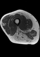 In muscle diseases such as the muscular dystrophies and inflammatory myopathies, there is a significant and urgent need for quantitative, objective biomarkers that can be used to understand disease natural history and perform clinical trials. Advanced, quantitative magnetic resonance imaging methods are sensitive to relevant aspects of disease state; but these methods are typically used only in research contexts, not in clinical radiology clinics. Instead, clinical radiology exams are based on qualitative descriptions of images obtained using conventional imaging methods. This precludes the quantitative comparison of MRI findings to other medically relevant data. As a result, the development of biomarkers that can advance the understanding and treatment of human muscle disease is impaired. However, we have recently obtained promising preliminary results suggesting that image texture analysis methods can be applied to conventionally obtained radiology images and used to predict the degree of muscle wasting and fat infiltration in muscle diseases.
Our future goals for this project are:
- To develop improved statistical models for predicting the degree of muscle wasting and fat infiltration in muscle diseases from image texture measurements;
- To understand the relationship between image texture measurements and other medically and functionally relevant data; and
- To investigate the diagnostic and prognostic potential of image texture measurements.
Collaborators
We’re working with Krista Vandenborne, Glenn Walter, and the Imaging DMD team at the University of Florida to expand our database and improve our models.
Funding
This project was initiated under an R01 award from the National Institute of Arthritis and Musculoskeletal and Skin Diseases and further developed using an SEC Faculty Travel Grant.
Representative Publications
Strijkers GJ, Araujo ECA, Azzabou N, Bendahan D, Blamire A, Burakiewicz J, Carlier PG, Damon B, Deligianni X, Froeling M, Heerschap A, Hollingsworth KG, Hooijmans MT, Karampinos D, Loudos G, Madelin G, Marty B, Nagel AM, Nederveen AJ, Nelissen JL, Santini F, Scheidegger O, Schick F, Sinclair C, Sinkus R, de Sousa PL, Straub V, Walter G, Kan HE. (2019). Exploration of new contrasts, targets, and MR imaging and spectroscopy techniques for neuromuscular disease – A workshop report of working group 3 of the Biomedicine and Molecular Biosciences COST Action BM1304 MYO-MRI. Journal of Neuromuscular Diseases. 6:1-30.
Damon BM, Li K, Dortch RD, Welch EB, Park JH, Buck AKW, Towse T, Does MD, Gochberg DF, Bryant N. (2016). Quantitative magnetic resonance imaging of skeletal muscle disease. Journal of Visualized Experiments. 118:e52352, doi:10.3791/52352.
Bryant ND, Li K, Does MD, Barnes S, Gochberg DF, Yankeelov TE, Park JH*, Damon BM*. (2014). Multi-parametric MRI characterization of inflammation in murine skeletal muscle. NMR in Biomedicine. 27: 716-725. *Co-senior authors.
Li K, Dortch RD, Welch EB, Bryant ND, Buck AKW, Towse TF, Gochberg DF, Does MD, Damon BM*, Park JH*. (2014). Multi-parametric MRI characterization of human thigh muscles at 3.0T – relaxation, magnetization transfer, fat/water, and diffusion tensor imaging. NMR in Biomedicine. 27: 1070-1084. *Co-senior authors.