Progress Report 4 1/26/18
Since our last progress report, we have made significant progress on the realization of our direct aspiration thrombectomy model. We have ordered the larger-diameter silicone tubing as planned in our previous report to allow for the passing of catheters through the tubing. Additionally, differential pressure gauges have been ordered to transduce intracranial pressure (ICP), which is modeled by a plastic bag inflated by a syringe, and pressure across a simulated clot in the cranial arteries we wish to model through thin-walled tubing.
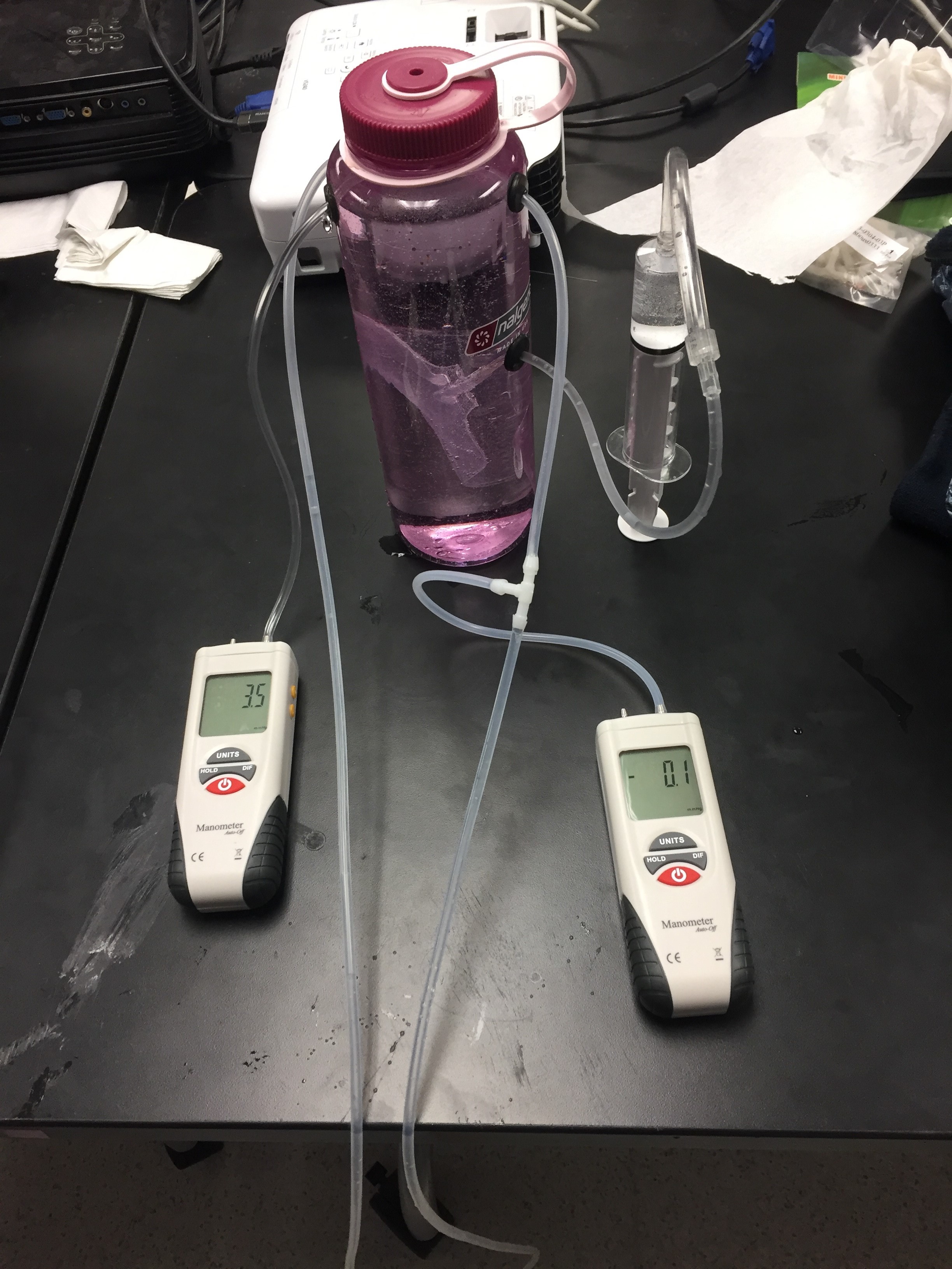
We built our second model using the new materials, feedback from Dr. Walker and Dr. Froehler, as well as our own criticisms of the first prototype. The significant changes made include moving the model Middle Cerebral Artery (MCA) closer to the lid of the water bottle for easier access, as well as adding a pressure gauge to measure the pressure inside the water bottle (the intracranial pressure). We also changed tubing to a 3mm ID x 5mm OD silicone walled tubing which more accurately represents the MCA. We recently ordered even thinner silicone tubing (3mm ID x 4mm OD) at the suggestion of Dr. Froehler. We hope that the thin walled tubing will allow pressure changes in the ICP to transduce across the wall of the tube and into the artery. We used a ½” drill bit to drill the hole for the plastic bag to enter into the water bottle through a ⅝” grommet with a 9/32” sized hole drilled in it. We used a 15/32” drill bit to drill two holes on either side of the bottle for the silicone tube (modeling the MCA) to pass through ½” grommets with 13/64” sized holes drilled through the middle.

We also began more thorough research into physical blood vessel properties and the anatomical background of common ischemic strokes in the brain. These included relationships between arterial pressure and intracranial pressure, and other physiological factors. In addition, we investigated common vessel parameters, including length, diameter, and material properties.
On Wednesday January 17, we presented our progress in the form of an oral presentation to Dr. Walker. He provided us with feedback and reminded us to maintain consistency with our project. Our mentor, Dr. Froehler, also provided us with feedback on how to improve from our current model in two meetings at the beginning of the semester, which can be seen in how we approach our future directions.
Our next goals for our prototype are to completely seal our current prototype so that we do not leak water over time and to ensure that our pressure measurements are accurate. We intend to better emulate the cranial arteries with silicone tubing by decreasing the thickness of the tubing while maintaining the current inner diameter. Additionally, we hope to simulate the actual composition of a clot, to provide a better model to measure differential pressure. We plan to use a small piece of pliable material, ideally foam from an earplug or similar, to accomplish this. We would then place a strain gage within this clot that could measure the pulling force applied by the aspiration catheter. In terms of future prototypes we plan on fabricating a see-through plastic container that is also 1.5L, portable, cheap, and rigid. Due to the flat walls this container could potentially be less likely to leak. Most importantly this switch to have a wider top of the container would allow for greater medical provider compatibility so that the user could more easily reach in to the model. We will also implement a motorized pump to simulate the flow of blood (through water) in the tubing, as depicted in the model below.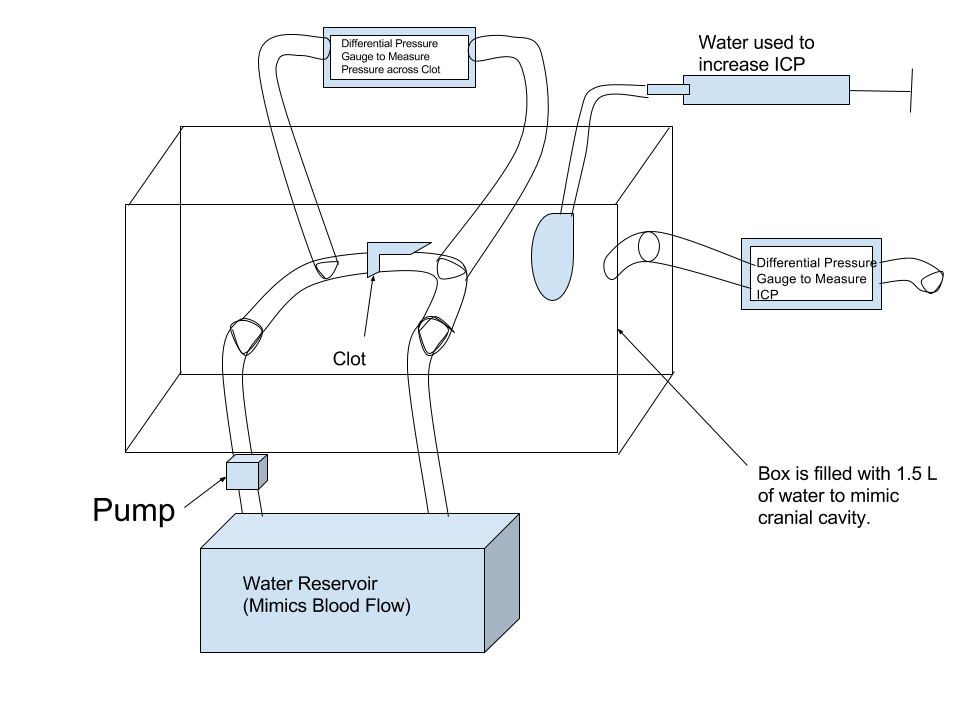
©2026 Vanderbilt University ·
Site Development: University Web Communications