Integrating histology and MRI in the first digital brain atlas of the common squirrel monkey, Saimiri sciureus
Peizhen Sun, Prasanna Parvathaneni, Yurui Gao, Kurt G. Schilling, Vaibhav A. Janve, Adam W. Anderson, Bennett A. Landman. “Integrating histology and MRI in the first digital brain atlas of the common squirrel monkey, Saimiri sciureus.” In Proceedings of the SPIE Medical Imaging Conference. Orlando, Florida, February 2015. †
Full text: https://www.ncbi.nlm.nih.gov/pmc/articles/PMC4405811/
Abstract
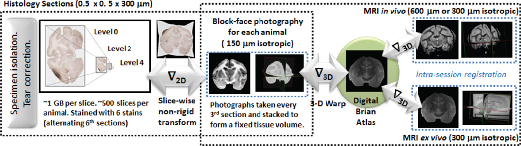
This effort is a continuation of development of a digital brain atlas of the common squirrel monkey, Saimiri sciureus, a New World monkey with functional and microstructural organization of central nervous system similar to that of humans. Here, we present the integration of histology with multi-modal magnetic resonance imaging (MRI) atlas constructed from the brain of an adult female squirrel monkey. The central concept of this work is to use block face photography to establish an intermediate common space in coordinate system which preserves the high resolution in-plane resolution of histology while enabling 3-D correspondence with MRI. In vivo MRI acquisitions include high resolution T2 structural imaging (300 µm isotropic) and low resolution diffusion tensor imaging (600 um isotropic). Ex vivo MRI acquisitions include high resolution T2 structural imaging and high resolution diffusion tensor imaging (both 300 µm isotropic). Cortical regions were manually annotated on the co-registered volumes based on published histological sections in-plane. We describe mapping of histology and MRI based data of the common squirrel monkey and construction of a viewing tool that enable online viewing of these datasets. The previously descried atlas MRI is used for its deformation to provide accurate conformation to the MRI, thus adding information at the histological level to the MRI volume. This paper presents the mapping of single 2D image slice in block face as a proof of concept and this can be extended to map the atlas space in 3D coordinate system as part of the future work and can be loaded to an XNAT system for further use.