Multi-Atlas Segmentation for Abdominal Organs with Gaussian Mixture Models
Ryan P. Burke, Zhoubing Xu, Christopher P. Lee, Rebeccah Baucom, Benjamin K. Poulose, Richard G. Abramson, Bennett A. Landman. “Multi-Atlas Segmentation for Abdominal Organs with Gaussian Mixture Models.” In Proceedings of the SPIE Medical Imaging Conference. Orlando, Florida, February 2015.
Full text: https://www.ncbi.nlm.nih.gov/pmc/articles/PMC4405670/
Abstract
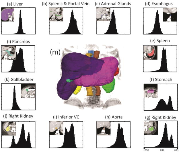
Abdominal organ segmentation with clinically acquired computed tomography (CT) is drawing increasing interest in the medical imaging community. Gaussian mixture models (GMM) have been extensively used through medical segmentation, most notably in the brain for cerebrospinal fluid/gray matter/white matter differentiation. Because abdominal CT exhibit strong localized intensity characteristics, GMM have recently been incorporated in multi-stage abdominal segmentation algorithms. In the context of variable abdominal anatomy and rich algorithms, it is difficult to assess the marginal contribution of GMM. Herein, we characterize the efficacy of an a posteriori framework that integrates GMM of organ-wise intensity likelihood with spatial priors from multiple target-specific registered labels. In our study, we first manually labeled 100 CT images. Then, we assigned 40 images to use as training data for constructing target-specific spatial priors and intensity likelihoods. The remaining 60 images were evaluated as test targets for segmenting 12 abdominal organs. The overlap between the true and the automatic segmentations was measured by Dice similarity coefficient (DSC). A median improvement of 145% was achieved by integrating the GMM intensity likelihood against the specific spatial prior. The proposed framework opens the opportunities for abdominal organ segmentation by efficiently using both the spatial and appearance information from the atlases, and creates a benchmark for large-scale automatic abdominal segmentation.