Revealing Latent Value of Clinically Acquired CTs of Traumatic Brain Injury Through Multi-Atlas Segmentation in a Retrospective Study of 1,003 with External Cross-Validation
Andrew J. Plassard, Patrick D. Kelly, Andrew J. Asman, Hakmook Kang, Mayur B. Patel, Bennett A. Landman. “Revealing Latent Value of Clinically Acquired CTs of Traumatic Brain Injury Through Multi-Atlas Segmentation in a Retrospective Study of 1,003 with External Cross-Validation” In Proceedings of the SPIE Medical Imaging Conference. Orlando, Florida, February 2015.
Full text: https://www.ncbi.nlm.nih.gov/pmc/articles/PMC4405676/
Abstract
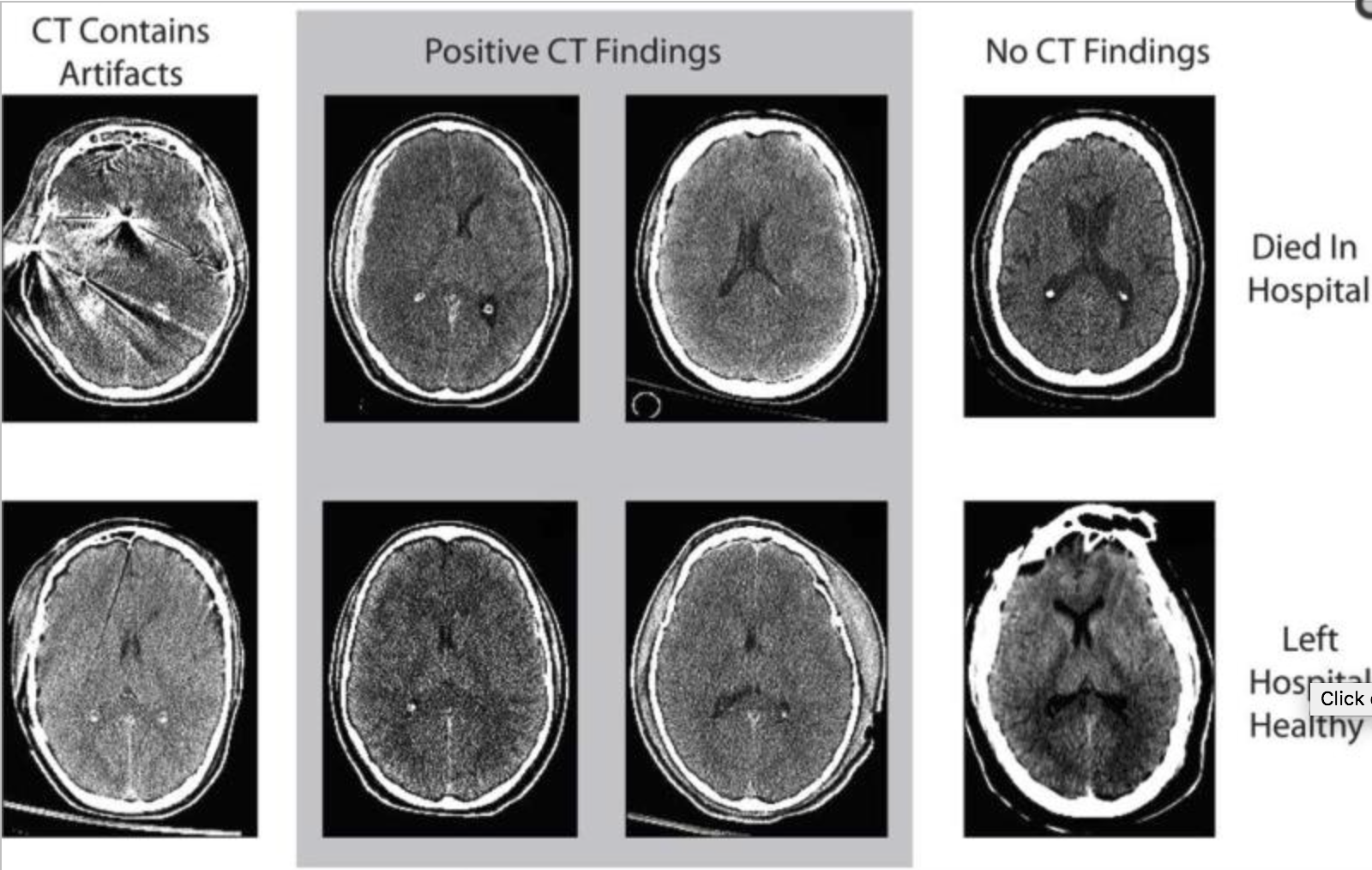
Medical imaging plays a key role in guiding treatment of traumatic brain injury (TBI) and for diagnosing intracranial hemorrhage; most commonly rapid computed tomography (CT) imaging is performed. Outcomes for patients with TBI are variable and difficult to predict upon hospital admission. Quantitative outcome scales (e.g., the Marshall classification) have been proposed to grade TBI severity on CT, but such measures have had relatively low value in staging patients by prognosis. Herein, we examine a cohort of 1,003 subjects admitted for TBI and imaged clinically to identify potential prognostic metrics using a “big data” paradigm. For all patients, a brain scan was segmented with multi-atlas labeling, and intensity/volume/texture features were computed in a localized manner. In a 10-fold cross-validation approach, the explanatory value of the image-derived features is assessed for length of hospital stay (days), discharge disposition (five point scale from death to return home), and the Rancho Los Amigos functional outcome score (Rancho Score). Image-derived features increased the predictive R2 to 0.38 (from 0.18) for length of stay, to 0.51 (from 0.4) for discharge disposition, and to 0.31 (from 0.16) for Rancho Score (over models consisting only of non-imaging admission metrics, but including positive/negative radiological CT findings). This study demonstrates that high volume retrospective analysis of clinical imaging data can reveal imaging signatures with prognostic value. These targets are suited for follow-up validation and represent targets for future feature selection efforts. Moreover, the increase in prognostic value would improve staging for intervention assessment and provide more reliable guidance for patients.
