Multi-Modal and Targeted Imaging Improves Automated Mid-Brain Segmentation
Andrew J. Plassard, Pierre F. D’Haese, Srivatsan Pallavaram, Allen T. Newton, Daniel O. Claassen, Benoit M. Dawant, Bennett A. Landman. “Multi-Modal and Targeted Imaging Improves Automated Mid-Brain Segmentation” In Proceedings of the SPIE Medical Imaging Conference. Orlando, Florida, February 2017. Oral presentation.
Abstract
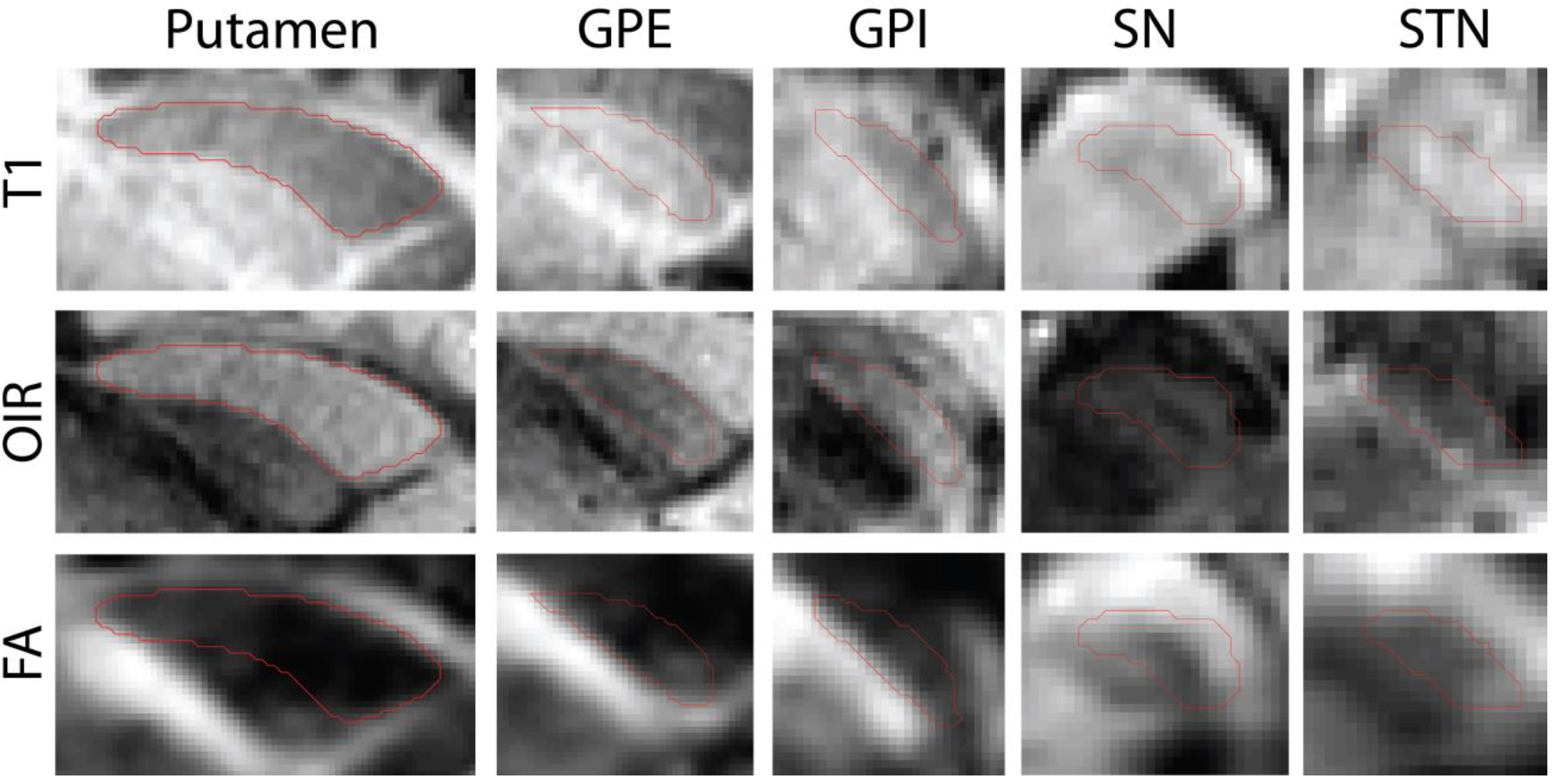
The basal ganglia and limbic system comprise a relevant network for Parkinson’s Disease. In order to manually trace these structures, specialized sequences at 7T are used. These sequences are not feasible for clinical patients. Targeted imaging sequences at have been presented to enhance contrast in a select group of these structures. In this work, we show that a series of atlases generated at 7T can be used to accurately segment mid-brain structures at 3T using a combination of imaging sequences. On average, using T1 and optimized inversion recovery together produced significantly improved segmentation results than any individual modality.