Our Design
Near-Infrared Spectroscopy (NIRS)

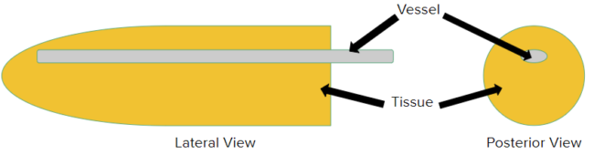
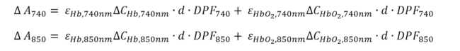
Using the Modified Beer-Lambert’s Law (Figure 2), our dual wavelength NIRS device converts raw voltage signal values to lactate concentrations ratiometrically. Utilizing this concept, our design focused IR light from the LED into the capillary bed of the finger, where it would be absorbed by lactic acid within the bloodstream. Then, any remaining non-absorbed light would be directed back into the photoconductor, where its intensity would be measured and compared to the input. This difference in values would be related to, and could allow for the calculation of, circulating lactic acid concentration.
Circuitry
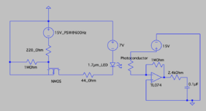
Design of the circuit involved developing optimal IR-light sensing modality, following traditional optical sensing methods. Main circuitry included the Thorlabs 1µm – 3µm photoconductor and two different Marktech IR-LEDs, one of 1650 nm and another at 1720 nm. We designed an LED-photoconductor optical system composed of 2 components: a pulsed current source (PCS) to drive the LED and a current to voltage (C2V) converter to read the photoconductor signal.
Phantom
Several designs were considered as phantoms to represent the pertinent real-life properties of the finger. At the outset, several design iterations included utilizing thin, IR-transparent catheters surrounded by various polymer mixtures that could approximately summarize the traits of the various layers of tissue from nail to vessel. Due to restraints of phantom dimensions and IR penetration depth, the design shifted to a more precise PDMS (polydimethylsiloxane) microfluidics phantom. To fulfill properties of blood, initial plans included increasing the complexity of lactate solutions with every successful absorbance trial. These solutions were, in order of increasing similarity to blood, were water, acetone, deuterated chloroform, PBS (phosphate buffered saline), FBS (fetal bovine serum), and ultimately defibrinated sheep blood.