Cyanotic Congenital Heart Disease
Cyanotic Congenital Heart Defect (CCHD)
- CCHD is a broad category of heart defects that encompasses conditions in which heart defects lead to a decrease in the amount of oxygen that is able to circulate throughout the body. A common indication of this condition is a blue tint of the skin caused by the amount of deoxygenated blood in the system. The five major CCHDs are triscupid atresia, tetralogy of fallot, transposition of the great arteries, total anomalous pulmonary venous return, and truncus arteriosus. The link below will take you to the Cleveland clinic website where you can find more information broadly about CCHD. For more information on specific CCHDs, please visit CDC’s website and search the specific CCHD. They provide statistics, descriptions, treatment, etc.
- https://my.clevelandclinic.org/health/diseases/22441-cyanotic-heart-disease#:~:text=Cyanotic%20congenital%20heart%20disease%3A%20Cyanotic,a%20bluish%20tint%2C%20called%20cyanosis.
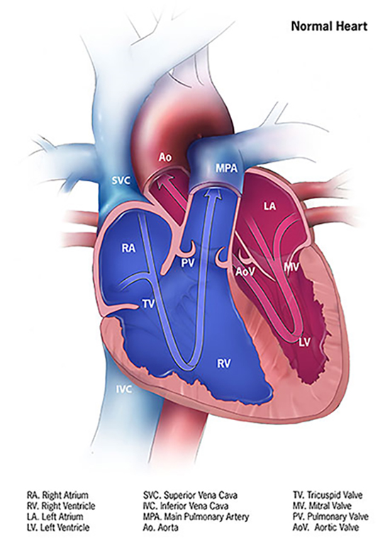
Normal Heart Flow
Tricuspid Atresia

Triscupid atresia is a congenital heart defect where the triscupid valve does not form connecting the right atrium and right ventricle. Often times this is presented with an obligate atrial septal defect and/or ventricular septal defect that connects the two atria or ventricles respectively. This leads to a mixing of oxygenated and deoxygenated blood. Because of the lack of blood flow to the right ventricle from the right atrium, the right ventricle is often underdeveloped.
Tertralogy of Fallot
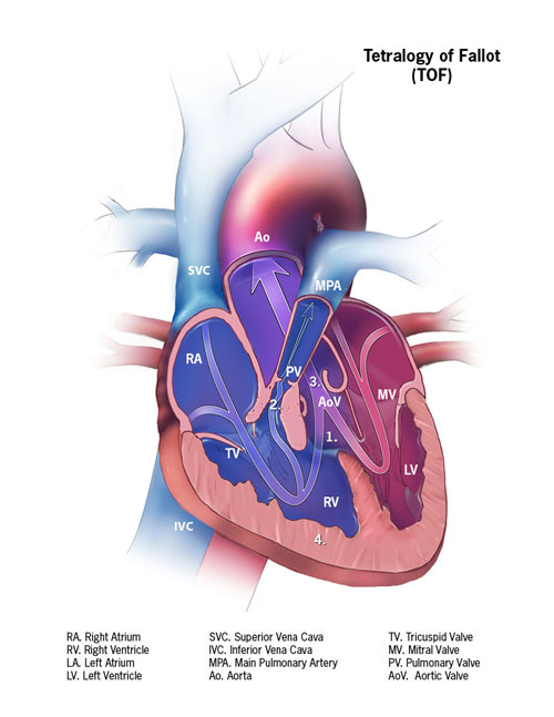
Tetralogy of fallot is characterized by having four heart/vessel defects. The first of these is the presence of a ventricular septal defect (VSD) which means a hole in the wall separating the right and left ventricles. This allows for mixing of oxygenated blood from the left ventricle and deoxygenated blood from the right ventricle. In addition to the VSD, the right ventricle itself thickens which is known as ventricular hypertrophy. Regarding the blood vessels, the pulmonary artery and valve both are narrower impeding and slowing blood flow to the lungs to be oxygenated. Lastly, the aortic valve is situated on top of the VSD instead of being located only over the left ventricle. It is also enlarged which allows it to encompass both ventricles leading to the return of mixed blood throughout the body.
Transposition of the Great Arteries

Transposition of the great arteries in which the aorta and pulmonary artery switch position and connect to the wrong ventricles. The aorta will connect to the right ventricle leading to systemic circulation of deoxygenated blood, and the pulmonary artery will connect to the left ventricle leading to a return of oxygenated blood back to the lungs. The presence of a patent ductus arteriosus, a connection between the aorta and pulmonary artery, allows for the mixing of blood and subsequent delivery of partially oxygenated blood to the body. Furthermore, the aorta is positioned in front of the pulmonary artery instead of being behind it in a normal heart.
Total Anomalous Pulmonary Venous Return
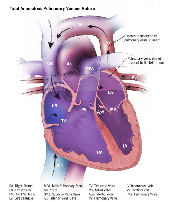
Total anomalous pulmonary venous return (TAPVR) is a condition in which recently oxygenated blood is delivered to the right atrium instead of the left atrium. Without an additional defect like an atrial septal defect, this condition can be lethal due to the lack of systemic circulation with oxygenated blood. This condition can be less severe and be partial instead of total if not all of the pulmonary veins have altered connections. There are three different forms of TAPVR that are different based on how the pulmonary veins abnormally connect to the heart. The pulmonary veins can connect to the inferior vena cava, superior vena cava, or right atrium directly.
Truncus Arteriosus

This condition occurs as a result of the failure of the aorta and main pulmonary artery to separate. As a result, the heart is left with one major blood vessel leading out of the heart. In addition to the one major blood vessel, the heart usually will have a VSD. This leads to larger volumes of blood being delivered to the lungs, and the heart works harder to contract and circulate this amount of blood.