The Kidney
The Vertebrate Kidney:
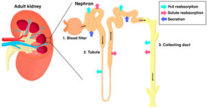
In vertebrates the kidney acts as the blood filtration system by removing wastes and excess fluids from the body. It consists of a very complex labyrinth of different types of tubes. Vascular tubes bring blood to the capillary bed of the kidney/nephron – aka the glomeruli which filters the blood and returns it back to the general circulation. The epithelial tubes (proximal tubule, loop of henle, and collecting duct) receive the blood filtrate and then are responsible for the waste extraction, salt recovery, and water balancing (Drummond 2003).
Kidney Formation in Vertebrates
Human Kidney Organogenesis:
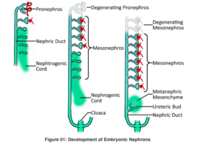
In humans, the process of kidney development starts as early as the 3rd week of embryogenesis/gestation with the emergence of the pronephros (the most anterior and primitive kidney) this is soon followed by the mesonephros (the adult kidney of fish and amphibians), in the 4th week of gestation and lastly just directly caudal, the metanephros forms in the 5th week of gestation – this become the permanent and functional kidney on humans (Reidy & Rosenblum, 2009).
Zebrafish Kidney Organogenesis:
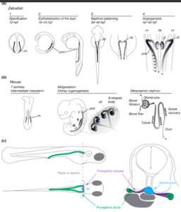
In zebrafish, the process of kidney development starts as early as the 5 somite stage (~12 hpf) however it is not until the 28 somite stage (24+ hpf) in the embryo where a complete pronephros comprised of two bilaterally paired parallel nephrons has developed. Overall the full functioning pronephros forms from events between 24- 48 hpf in the embryo and hatches with only a pronephros. The mesonephros forms during the larval stage (10-14 dpf).
More specifically, the zebrafish kidney forms in a stepwise fashion, the pronephric ducts are the first too form and are complete by around 24 hours post-fertilization (hpf), followed by the tubules between 30 hpf and 40 hpf, and a fully functional blood filtering glomerulus by 48 hpf (Drummond 2003).
Cellular Structure of the Zebrafish Kidney:
One of the most important regions of the zebrafish kidney is the renal corpuscle – one of the defining features of the corpuscle – the glomerulus. The glomerulus consist of fenestrated endothelium which have small pores in the capillary walls and the Bowman’s capsule which contains a parietal and visceral layer of epithelia. In zebrafish, the glomerular filtrate passes into another important structure known as the proximal tubule which consists of proximal tubular epithelial cells (PTECs) which reabsorb the essential substances such as glucose and amino acids back into the blood. Zebrafish also contain a distal tubule which is responsible for reabsorption of H2O and concentration of urine, and finally the collecting duct – which consists of principle (sodium absorption) and intercalated cells (Drummond et al., 2023).
Differences Between Human and Zebrafish Kidney:
The key difference between the cellular structure of the zebrafish and human kidney is that humans have podocytes in the glomeruli which help regulate filtration because they form filtration slits that allow small molecules to pass through without losing larger ones. In terms of larger structures zebrafish lack a distinct loop of henle – instead they just have a proximal and distal tubule that aid in water maintenance, nutrient reabsorption and concentration of urine (Drummond et al., 2023).