VU BreakThru

Home » News » MASI Lab: Connecting clinical neuroscience with advanced technological imaging analysis
MASI Lab: Connecting clinical neuroscience with advanced technological imaging analysis
Posted by anderc8 on Friday, February 17, 2017 in News, TIPs 2015.
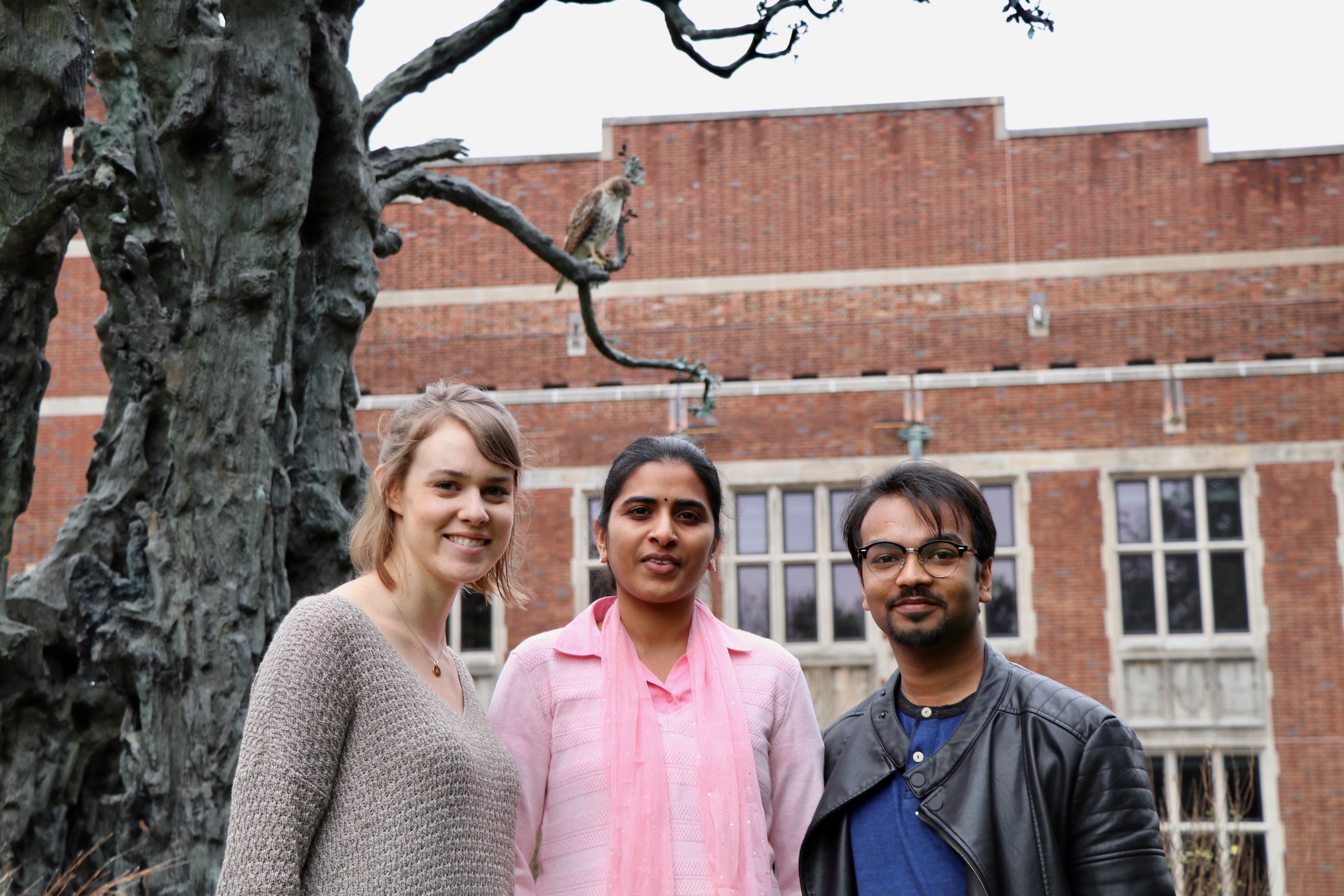
Three members of the MASI lab: Allison Hainline, Prasanna Parvathaneni, and Vishwesh Nath.
Written by:
Vishwesh Nath, Vanderbilt University graduate research assistant (Masters)
Prasanna Parvathaneni,Vanderbilt University graduate research assistant (Ph.D)
Allison Hainline,Vanderbilt University graduate research assistant (Ph.D)
At the Medical Imaging and Statistical Interpretation (MASI) lab the first thing you notice is the active environment encouraging, friendly interaction. The principal investigator of this project, Bennett Landman, Ph.D., is right across the hall and his open door policy encourages students to drop in for quick questions and science talks. He is a leading scientist in the field of Diffusion Imaging and his academic background consists of an intersection between Bio-Medical (BME), Electrical Engineering (EE), Computer Science (CS) and Radiology. The expertise of the PI in multiple domains combined with the diverse team of graduate students with various backgrounds translates into effective research. We have representation from CS (Vish), EE (Prasanna), BME (Kurt), Biostatistics (Allison) and BME (Cam), just to name a few. We have the opportunity to collaborate and learn from those with different strengths and perspectives while honing our own skills. These strengths are particularly useful when someone new arrives in the lab with limited expertise in medical imaging. For example, “I recently joined the lab and soon found that image analysis has a steep learning curve. Imagine trying to understand MR physics, acquiring data from scanners and designing metrics at the same time. However, my fellow MASI lab members made the transition much more smooth. I’m still learning new things every day, but knowing that I have people to turn to for help makes it much less daunting!” – Allison
In addition to encouraging interdisciplinary research within the lab, we also have close relationships with several other Vanderbilt departments including the Vanderbilt University Medical Center Additionally, these direct relationships allow our lab to use the cutting-edge technology that enables us to work on real world problems and advance the analysis techniques to have a higher impact. One notable result of these collaborations is the automated processing pipelines (raw data processed to quantified statistics ready for analysis) our lab has created being implemented across a variety of research and clinical projects.
The ongoing research at MASI lab gives us the ability to ask deep research questions and make them applicable to the neurological conditions we care about like Schizophrenia, multiple sclerosis, Autism, Alzheimer’s, depression, deep brain stimulation, etc. “Having seen friends and family suffer with various neurological disorders and the therapeutic healing offered in alternate medicine instigated my interest to work in this field. Moving to academic research after being in the software industry for years is a challenge in itself, in addition to not having any exposure to medical imaging or neuroscience. However, getting into MASI lab has tremendously helped in making this transition as smooth as possible. The skills acquired from this lab have equipped me to work with confidence on a brain tumor project at UCSF and has given me an opportunity to make good friends. The focus of our project to understand the structure and function of the brain using advanced diffusion MRI methods keeps the motivation going!” – Prasanna
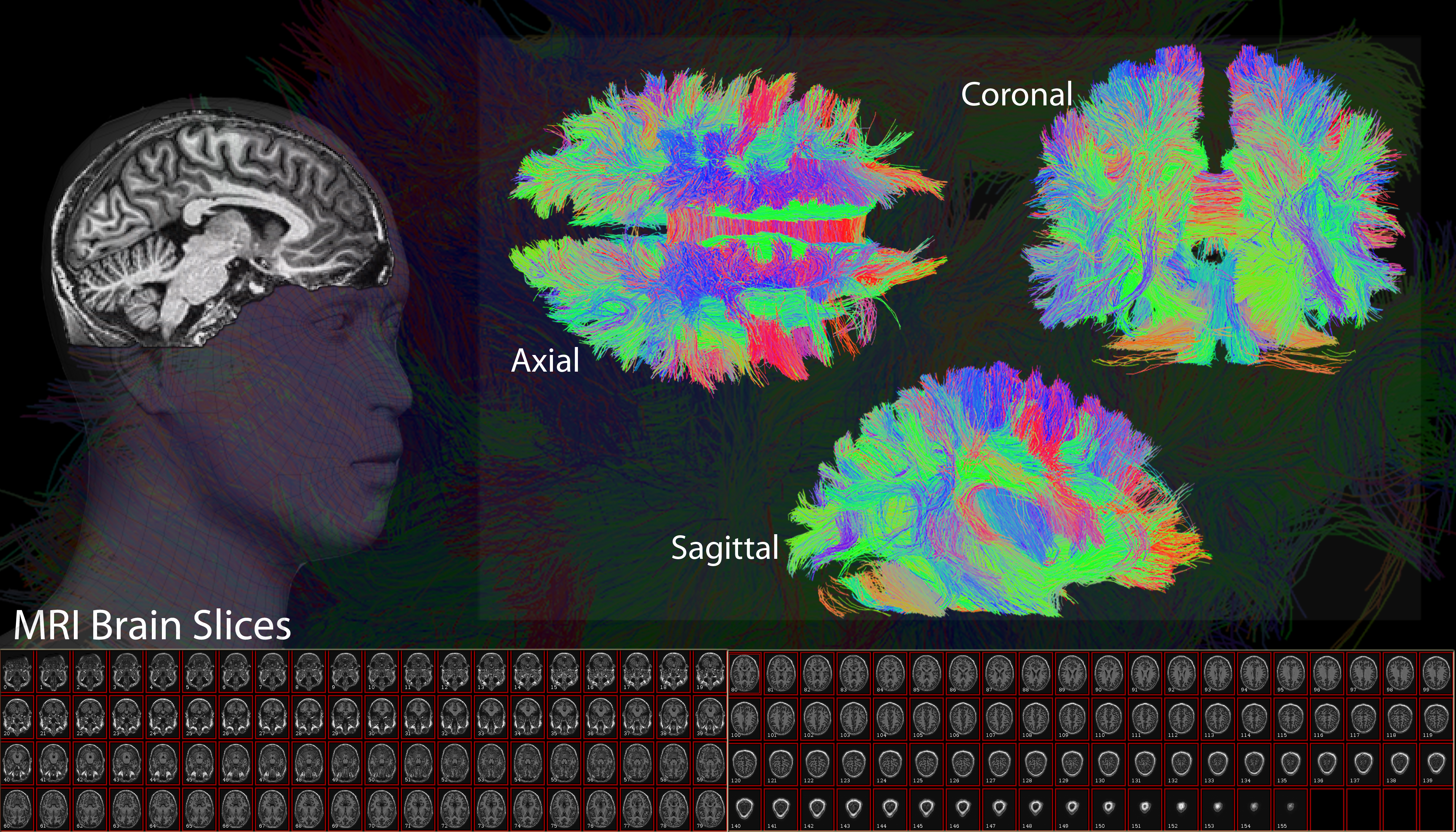
White matter fiber tracts of the human brain reconstructed from MRI data.
One of the most interesting projects from the MASI lab is focused on advancing the methods applicable to Diffusion Weighted Magnetic Resonance Imaging (DW-MRI) neuroimaging data. Diffusion MRI could accurately detect early onset of pathophysiologic changes compared to regular MRI, revealing damage to the underlying microstructure that might have otherwise gone undetected in regular MRI. Numerous studies across the globe are adapting advanced diffusion MRI techniques to analyze structure and function of the brain in healthy and disease conditions. However, identifying the appropriate technique that is optimal or advantageous given practical considerations remains an active area of concern. The specific aim of our project is to address this important gap in the brain imaging community.
A future goal for this project is to quantify the pros and cons of various DW-MRI methods and create a system in which scans from any location can be easily compared. Currently, it is extremely difficult to compare results across different scanners and scan locations due to various implementation methods and scanner idiosyncrasies. We are working to quantify and correct these differences through a comprehensive quality assurance protocol. The ability to directly compare scans from all across the world would be highly influential on the kinds of research that can be performed and the clinical problems that could be solved.
We have the unique opportunity to experiment with different acquisition sequences for MRI scanners. For example “when I joined the lab, I was new to acquiring scanner data and was suddenly swirled into acquiring one of the largest single subject MR data of the human brain. It would not have been possible without the supportive environment of the lab. But being able to acquire 6,000 scans at my will was a gargantuan task. Moreover, working on it has been an amazing experience. There is so much yet to be quantified from that dataset!” – Vish
The ability to acquire and use such a high-quality dataset has driven much of the research that is conducted in our lab. It has also led us to organize the TraCED challenge, which allows for the comparison of different tractography methods. The challenge is intended to complement simulation efforts and pre-clinical validation studies, which suffer from limitations in capturing physiological and imaging considerations of in vivo human studies.
Interested in learning more?
VISE includes 10 technical laboratories spanning three engineering departments (Biomedical Engineering, Mechanical Engineering and Electrical Engineering & Computer Science) and the Otolaryngology department as well as clinical departments that include Surgery, Neurological Surgery, Radiology, Otolaryngology, Hearing and Speech, Oncology, Gastroenterology, Surgical and Radiological Oncology, Ophthalmology, Urology and Thoracic Surgery.
Learn more about VISE by visiting www.vanderbilt.edu/vise and follow us on our social media channels:
Facebook: https://www.facebook.com/visevanderbilt
Instagram: https://www.instagram.com/visevanderbilt/
Twitter: https://twitter.com/ViseVanderbilt
YouTube: http://vanderbi.lt/viseyoutube
If you’re interested in learning more about what the MASI lab is doing, please visit https://my.vanderbilt.edu/masi/. On our website, you can see the rich blend of research being conducted by our friends in other MASI projects.
2 Comments on “MASI Lab: Connecting clinical neuroscience with advanced technological imaging analysis”
Raj on February 23rd, 2017 at 4:45 pm
Good work and impressive team!
Turner on March 5th, 2017 at 9:13 pm
Very Impressive, especially the scope of investigation.
Leave a Reply
Recent Posts
 Perceptions of AIs, Humans, and Others (Part 2) » 5.28.19
- Perceptions of AIs, Humans, and Others (Part 1) » 5.23.19
- Esteemed Writer Ted Chiang visits Vanderbilt (Part 2) » 4.17.19
- Esteemed Writer Ted Chiang visits Vanderbilt (Part 1) » 4.15.19
- Sublating Binaries: Smart Vehicles as Defensive Drivers » 4.1.19
- Sublating Binaries and an Intelligent Vehicles Case Study » 3.28.19
 HPV Symposium Set For March 1 » 1.28.19