VU BreakThru

Home » News » Collaboration Between the Departments of Otolaryngology, Biomedical Engineering and Medicine for Earlier Diagnosis of Throat Cancer
Collaboration Between the Departments of Otolaryngology, Biomedical Engineering and Medicine for Earlier Diagnosis of Throat Cancer
Posted by anderc8 on Wednesday, July 25, 2018 in News, TIPs 2017.
Written by G. Thomas, C. Burton Wood, MD, Justin R. Shinn, MD and Young J. Kim, MD
HPV-related cancers of the tonsils and the base of the tongue have increased rapidly over the last few decades. These cancers are often small and do not cause many symptoms in early stages, so many patients are not diagnosed until the tumor has already spread to lymph nodes in the neck. We are always trying to identify these tumors earlier to treat patients with less intensive therapies while still aiming to cure them of their cancer and minimize the side effects.
Raman spectroscopy has been used to diagnose difficult-to-locate cancers in other parts of the body, such as the cervix, gastrointestinal tract and the oral cavity, but has never been used to investigate cancers in the oropharynx (tonsils and base of tongue). This technique involves using a flexible fiber-optic probe illuminating the tissue with near infrared light and measuring the scattered light with the same probe. The incident light is scattered by the tissue in a distinct manner depending on the biochemical profile of the tissue. These changes in light intensity are recorded as a ‘Raman spectra’ by a detector that is controlled by a computer system, processed and then analyzed to determine whether the tissue is cancerous or non-cancerous. Using this technique, the Raman signatures are able to identify cancers without needing to take biopsies. There are no known side-effects using this technique.
We explored the feasibility of this technique as a collaborative project between Professor Anita Mahadevan-Jansen from the Vanderbilt Biophotonics Center (VBC) and Department of Biomedical Engineering, Dr. Young Kim (Department of Otolaryngology) and Dr. Krystle Kuhs (Division of Epidemiology, Department of Medicine) from the Vanderbilt University Medical Center as part of the TIPs grant on HPV-associated cancers. We initially performed ex vivo testing on biopsy samples with a Raman microscope (Renishaw) and subsequently used a portable clinical system with a 2.1 mm diameter optical probe, developed at the VBC that could easily fit within a laryngoscope. Our preliminary findings suggest that this technique has the potential to sensitively differentiate between cancer and non-cancerous oropharyngeal tissues. Additionally, we have enrolled three patients undergoing surgical biopsy for diagnosis of their oropharyngeal tumors for in vivo measurements using the clinical system shown in the figure above.. This new technique adds only 5-7 minutes to the total time of the procedure. The early results are promising and studies are currently underway in more patients to determine the effectiveness of Raman spectroscopy as a non-invasive real method for detecting oropharyngeal cancers. IN particular, we hope to be able to develop this technology for identifying which cancers are associated with the human papilloma virus (HPV) and which ones are not.
This study would not be possible without the TIPs award, as it provides the funding required to develop and build probes specifically designed to be used through our scopes during biopsy. We are excited about the future of this project and the collaborative effort it represents.
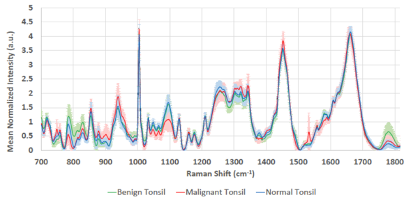
Representative spectra obtained using the benchmark Raman Spectroscopy (Renishaw) system.
Leave a Reply
Recent Posts
 Perceptions of AIs, Humans, and Others (Part 2) » 5.28.19
- Perceptions of AIs, Humans, and Others (Part 1) » 5.23.19
- Esteemed Writer Ted Chiang visits Vanderbilt (Part 2) » 4.17.19
- Esteemed Writer Ted Chiang visits Vanderbilt (Part 1) » 4.15.19
- Sublating Binaries: Smart Vehicles as Defensive Drivers » 4.1.19
- Sublating Binaries and an Intelligent Vehicles Case Study » 3.28.19
 HPV Symposium Set For March 1 » 1.28.19