Valor Tech

Progress Report #3
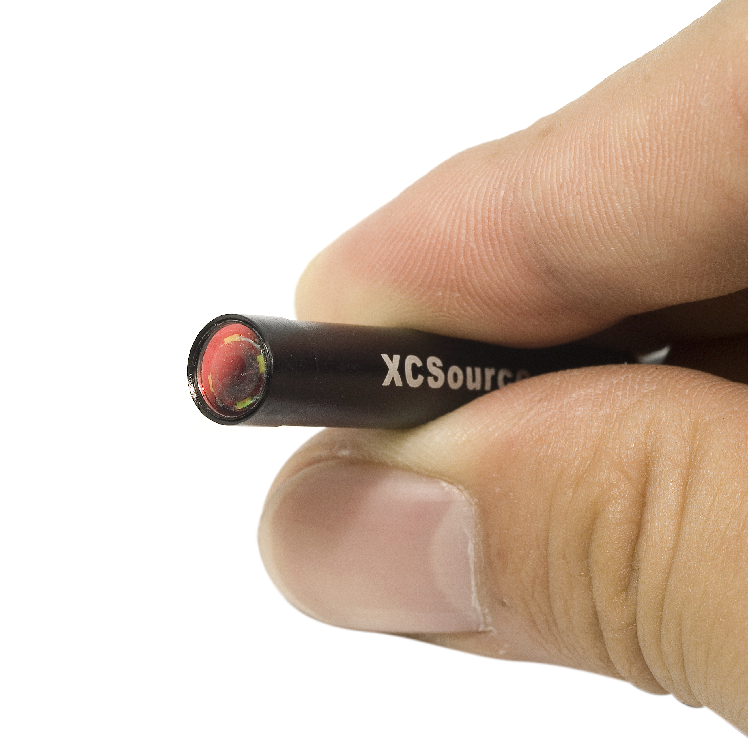 The XCSOURCE 7mm Waterproof 200x USB Microscope Inspection Borescope Endoscope Pipe Inspection Snake Video Camera is produced by Supereyes. The XSource camera has a resolution of 640*480*30 ftps, a ⅙ CMOS Image Sensor, a length of 2 meters, camera head outer diameter of 7 mm, 4 LEDS placed around the head of the camera, and built in functionality to modulate LED brightness. The XSource camera can be both interfaced with Mac and Windows operating systems. In Mac, PhotoBooth or Facetime can be used to process the video but video recording requires the camera to be used with Windows operating systems. The Supereyes video recording software is freely available online and is compatible with Windows operating systems. Interfacing the XSource with a MacBook Pro, we have visualized the interior of the artificial uterine model. Next steps include interfacing the camera with Windows in order to test the video recording capabilities as well as further exploring the utility of the XSource in the larger goal of simulating hysteroscopy.
The uterine model consists of an external hard plastic shell and a pink internal lining, simulating the size, shape, and general appearance of an actual uterus. Using the new camera system, we were able to explore the internal compartment of the uterine model and have, for the first time, gained a basic understanding of the limitations of hysteroscopic surgery. Within the model, we were able to see the different walls of the uterus and the insertion points where the fallopian tubes enter through the rear wall. By manipulating the camera externally, we were able to guide the scope to different areas in the uterus providing a greater appreciation for the limited movement available during the surgery. The test setup is similar to our expected final product as we hope to use a hysteroscopic scope to navigate a uterine model, completing goals under similar movement constraints.
Our next step will be opening the uterine model in order to explore its potential as an early clamshell prototype for our design. By using the current uterine model as a gold standard, we can begin to develop our own clamshell design. The uterine model is made of two plastic half-shells that are fixed together with adhesive. Using careful application of tools such as chisels, coping saws, and other fine blades, we should be able to open it with minimal damage to the interior. Once open, we will be able to design and construct the first series of our quantitative tests. In addition, we will gain more insight into the design parameters that will be required of our first model hysteroscope – which will be paired with the clamshell uterine model.
©2026 Vanderbilt University ·
Site Development: University Web Communications