Update on 16 April 2020:
If you would like to continue to submit results, or use/analyze data for your own studies, please send an email to whitematterchallenge@gmail.com to request data. You will not be able to access data through the QMENTA website.
Update on 8 April 2020:
We are happy to announce that we had over 110 submissions from 13 different research groups across our various sub-challenges! We have posted the challenge results and participants presentations to both the ISBI Whova app (http://2020.biomedicalimaging.org/virtual-event-instructions) and Vimeo channel (https://vimeo.com/channels/1562465) and would love to invite you to view our virtual program!
Overview:
We are excited to announce the multi-year ISBI 2019/2020 MRI White Matter Reconstruction Challenge, carried out on the QMENTA platform. The first year of this challenge is dedicated to designing the challenge, building the appropriate dataset(s), and presenting it for the first time at the 2019 ISBI conference (Thursday afternoon, April 11 at Venice, Italy). The core of the challenge will then take place in the second year, giving research groups and clinicians a full year to implement, and potentially revise, their submissions and algorithms.
The white matter reconstruction challenge is composed of 4-subchallenges, based on 4datasets reflecting the same (or similar) underlying biology. We aim to evaluate the ability to predict unseen signal (signal representation; sub-challenge #1), to estimate microstructural measures (signal modeling; sub-challenge #2 and sub-challenge #4), and evaluate sensitivity and specificity of potential biomarkers (biomarker evaluation; sub-challenge #3).
The challenges are based on the 4 datasets:
(1) The MASSIVE brain dataset (Froeling et al., Magn Reson Med. 2017): and in vivo human dataset based on diffusion weighted PGSE, including multiple shells (b-values), multiple cartesian q-space grids, and over 8000+ volumes
(2) A high resolution ex vivo mouse dataset (Ianus et al., Neuroimage. 2018): a high resolution, high SNR state-of-the art dataset with both double diffusion encoding (DDE), double oscillating diffusion encoding (DODE), with 5 different b-values, 6 different diffusion times and gradient frequencies.
(3) Realistic 3D simulated substrates of white matter tissue, designed using the methods described in (Hall, M. G., & Alexander, D. C.. IEEE transactions on medical imaging) which provides flexibility and control of a variety of microstructural features, including axonal caliber, density, and permeability.
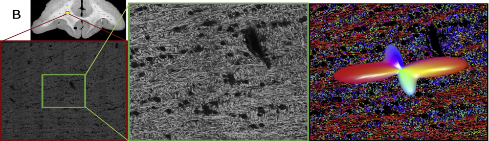
(4) Simulated dMRI signal in the realistic intra-axonal space, with tissue microstructure informed by electron microscopy (Lee et al., Brain Structure and Function (2019). doi.org/10.1007/s00429-019-01844-6).
We encourage you to visit the individual sub-challenge pages for detailed information, challenge guidelines, and data access.