
Research Snapshot: New microscopy technique unveils feature that can shape applications of a class of quantum materials
A team of researchers led by Oak Ridge National Laboratory microscopist Miaofang Chi and Vanderbilt theoretical physicist Sokrates Pantelides has used a new Scanning Transmission Electron Microscope technique to image the electron distribution in ionic compounds known as electrides— especially the electrons that float loosely within pockets and appear separate from the atomic network.
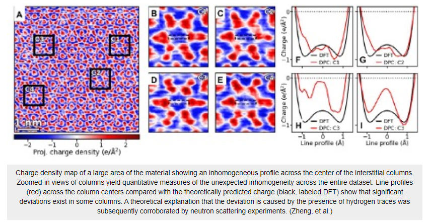
The new technique, Differential Phase Contrast in STEM, measures and maps electric fields and charge distributions inside a material. The study is the first time that DPC has been used in this way. By analyzing charge images of dozens of such channels, the team found that only some contain the negative charge predicted by theoretical calculations, while others have significantly less negative or even a small concentration of positive charge. Pantelides’ decades of experience with hydrogen led to the suggestion that traces of hydrogen, which are essentially impossible to eliminate, are responsible for the observed inhomogeneity, and subsequent detailed calculations confirmed the hypothesis. Neutron scattering experiments provided evidence in support of the hydrogen scenario.
Read the full article HERE