
Winners of the 2023 VINSE Summer Image Competition
VINSE is excited to unveil the twelve winning images from our 2023 summer image competition. Since 2017, VINSE celebrates the work of our researchers, seeking images of materials or devices that have been fabricated, characterized or imaged using VINSE equipment with an annual image competition. Congratulations to this year’s winners. Each will receive a $250 cash prize and will have their art displayed in the Engineering Science Building.

Magnolia leaf freeze fracture imaged at -140 C
Imaged in the VINSE facilities using the Helios Dual Beam Focused Ion Beam, Scanning Electron Microscope Original image credit: Al Coritz of Electron Microscopy Services and James McBride
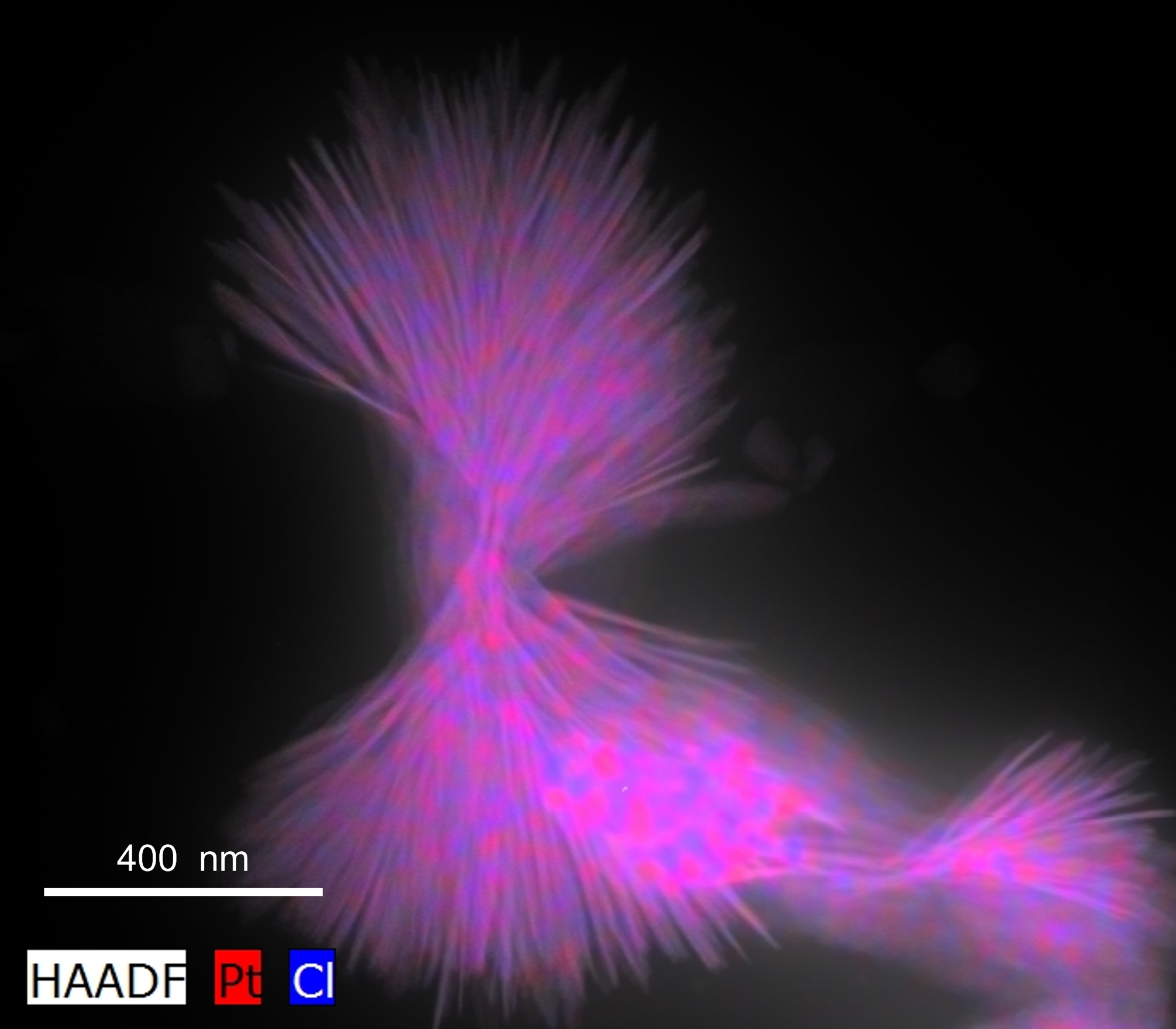
Crystallization of small molecule cisplatin
Imaged in the VINSE facilities using the FEI Tecnai G2 Osiris S/TEM

Polymethyl methacrylate (PMMA) residue on graphene after transferring to sulfonated poly-ether ether ketone (SPEEK) and soaking in acetone.
Imaged in the VINSE facilities using the Zeiss Merlin Scanning Electron Microscope

Edge of photomask used in photolithography for microfluidic device application
Fabricated and imaged in the VINSE facilities using the cleanroom and the Olympus microscope

4,4′-Bis(N-carbazolyl)-1,1′-biphenyl (CBP) nanostructures on a silicon chip
Imaged in the VINSE facilities using the Merlin Zeiss Scanning Electron Microscope

Plastic photolithography mask for microfluidics mixer devices where channel diameter and presence of teeth for increased mixing are changed between the four patterns.
Fabricated using the Bungard Filmstar Photoplotter in the VINSE Cleanroom

Sputtered vanadium dioxide patches grown on silicon on insulator (SOI) wafers
Fabricated in the VINSE facilities using the Raith e-Line Electron Beam Lithography System & Imaged in the VINSE facilities using the Zeiss Merlin Scanning Electron Microscope

30 nm slotted silicon nanoarrays supporting a radiation-less anapole state
Fabricated in the VINSE facilities the Raith e-Line Electron Beam Lithography System

ZnO nanowires grown hydrothermally on GaN. Image taken at the sample’s edge.
Imaged in the VINSE facilities using the Zeiss Merlin Scanning Electron Microscope

Immunocytochemistry/fluorescence microscopy of human embryonic stem cells (hESCs) cultured in 500um microdiscs and stained for germ layer markers. Yellow – SOX2 (pluripotency/ectodermal marker), pink – brachyury (mesendodermal/primitive streak marker).
Fabricated in the VINSE facilities using the Jelight M42 UV Ozone Cleaner

The image depicts the boundary between porous silicon formation and the un-etched silicon substrate, illustrating the edge effects of the electric field which drives the electrochemical etching process.
Imaged in the VINSE facilities using the Zeiss Merlin Scanning Electron Microscope

Photonic chip mechanically polished at Vanderbilt Earth and Environmental Science and etched at VINSE using buffered hydroflouric acid
Imaged in the VINSE facilities using the Merlin Zeiss Scanning Electron Microscope