Anisotropic Diffusion Phantom (Challenge #1)
Phantom Construction
The Anisotropic Diffusion Phantom is a biomimetic phantom containing complex geometries of anisotropic fibers that mimic the tissues of the brain. The phantom contains 14 flexible fiber bundles. Pathways are aligned in orthogonal planes, as well as in curved (both 90 degrees and helical curving), and kissing geometries (as well as a resolution bank) to mimic complex nerve fibers of the brain, with bundle dimensions of magnitudes comparable to major white matter pathways in the human brain.
MRI Imaging
MR scans were performed on two scanners, both Philips 3.0T systems. The 16 cm diameter phantom (matrix fluid filled) was imaged for both structural and diffusion contrasts. The structural scan utilized an 3D MPRAGE sequence to acquire a T1 contrast (TE/TR = 3.6/8ms, Matrix = 256 * 256, Resolution = 0.88*0.88mm, slice thickness = 1.0mm). A low-resolution diffusion contrast was acquired using a 2D EPI diffusion weighted sequence (TE/TR = 75ms/9.65s, Matrix = 72*72, resolution = 2.25*2.25mm, slice thickness = 2.5mm). 96 diffusion directions were acquired, uniformly sampled over a sphere, at b-values of 1,000 s/mm2 and 2,000 s/mm2. Non-diffusion weighted images were acquired between every 8 diffusion weighted images. Sampling was performed with phase encoding both anterior to posterior, and repeated posterior to anterior, in order to allow pre-processing for motion, eddy currents, and susceptibility distortions. This series of scans (2 b-values, 96 uniformly distributed directions, with two phase encoding direction each) was repeated 7 times on each scanner.
MRI data processing
Diffusion MRI pre-processing was performed in the coordinate system that the data were acquired in. Steps included correction for movement, susceptibility induced distortions, and eddy currents using FSLs topup and eddy algorithms [5]. The gradient tables were rotated based on the transformations obtained from the corrections.
Squirrel Monkey + Histological ground truth data (Challenge #2)
Tracer Injection
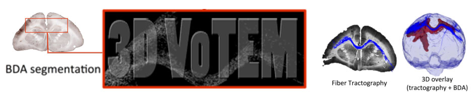
The histological ground truth data is acquired on a squirrel monkey brain. Here we utilize a commonly used neuroanatomical tracer for studying neuronal pathways, biotinylated dextrane amine (BDA). Because it is transported both anterograde and retrograde, BDA yields sensitive and detailed labeling of both axons and terminals, as well as neuronal cell bodies. This tracer relies on axonal transport systems; thus BDA injection is performed prior to ex vivo imaging. Under general anesthesia using aseptic techniques, BDA was injected into left hemisphere M1 cortex of two monkeys. Eight injections were made in each animal in order to cover a large M1 region representing the forearm as identified by intracortical microstimulation. After surgery, the monkey was allowed to recover from the procedure, giving the tracer sufficient time to be transported along axons to all regions connected to M1.
MRI Imaging
For ex vivo scanning, the brains were perfusion fixed with 4% paraformaldehyde preceded by rinse with physiological saline. The brain was removed from the skull and stored in buffered saline overnight. The next day, the brain was scanned on a 9.4 Tesla Varian scanner. Diffusion weighted imaging was performed using a pulsed gradient spin echo multi-shot spinwarp imaging sequence with full brain coverage (TR = 5.2s, TE = 26 ms, number of diffusion gradient directions = 31, b = 0, 1200s/mm2, voxel size = 300×300× 300 µm3, data matrix = 128×128×192, number of acquisitions = 10, SNR≈25, scanning time≈50 hr). The b value used in this experiment was lower than is optimal for diffusion studies in fixed tissue, due to hardware limitations. A low b value decreases the available diffusion contrast-to-noise ratio (CNR) in the image data, which has the same effect as higher image noise. To compensate for this shortcoming, we extended the scan time to 50 hours, which yielded a CNR comparable to in vivo human studies (equivalent to an in vivo study with mean diffusivity = 0.7×10⁻³ mm2/s and SNR≈20).
MRI data processing
Diffusion MRI pre-processing was performed in the coordinate system the data were acquired in. Steps included correction for movement, susceptibility induced distortions, and eddy currents using FSLs topup and eddy algorithms [5]. The gradient tables were rotated based on the transformations obtained from the corrections. All challenge data will be distributed and analyzed directly in the space in which diffusion data were acquired.
Histological Acquisition
Following ex vivo MRI scanning, the brain was frozen and cut serially on a microtome in the coronal plane at 50 um thickness. Prior to cutting every third section (i.e., at 150 mm intervals), the surface of the frozen tissue block was photographed using a Canon digital camera (image resolution = 50 um/pixel, image size = 3330×4000 pixels, number of images per brain ~ 280), mounted above the microtome. These block-face images have been shown to produce a more robust inter-modality registration results by providing a relatively undistorted intermediate reference space between the histological and MRI data [6]. Every 6th section (approximately the size of an MR voxel) is processed for BDA to trace pathways associated with the M1 cortex.
Whole-slide Brightfield microscopy was performed using a Leica SCN400 Slide Scanner at 20x magnification, resulting in a maximum in-plane resolution of 0.5um/pixel.
Registration
The multi-step registration utilized here is very similar to the registration procedure validated in an earlier study [7], which showed that the accuracy of the overall registration was approximately one MRI voxel (~0.3 mm). From the Leica image file, the TIFF image stored at 128 um/pixel (down-sample factor 256) was extracted and registered to the down-sampled photograph (256×256 pixels at a resolution of approximately 128 um/pixel) of the corresponding tissue block using a 2D affine transformation followed by a 2D non-rigid transformation, semi-automatically calculated via the Thin-Plate Spline algorithm [8]. Next, all down-sampled block face photographs were assembled into a 3D block volume and registered to the corresponding 3D MRI volume using a 3D affine transformation followed by a non-rigid transformation automatically calculated via the Adaptive Bases Algorithm [9]. The deformation fields produced by all registration steps were applied to processed histological data (see Section on Reference standard) in order to transfer the ground truth histological pathways into the diffusion space for comparisons with tractography.
Macaque NIH Data (Challenge #3)
Data Description
The provided dataset is the one used in the manuscript titled “Anatomical accuracy of brain connections derived from diffusion MRI tractography is inherently limited” by C. P. Thomas et al. published in PNAS (2014). The images were acquired from an ex-vivo fixed macaque brain at 0.25mm isotropic resolution. The diffusion weighted images (DWIs) contain 7 volumes with b=0 s/mm2 and 114 volumes with b=4900 s/mm2 (with small variations due to the effects of the imaging gradients). The provided DWIs were corrected for frequency drifts and eddy-currents distortions as described in the manuscript.
Ground Truth Pathways
Two ground truth pathways were derived from the anterograde tracer injections placed in (i) the precentral gyrus corresponding to the foot region of the primary motor cortex and (ii) rostroventral part of the occipital region corresponding to the ventral part of area V4 (V4v) and the adjacent ventral area V3 – as described and characterized in the book “Fiber pathways of the Brain” (Oxford University Press, 2009) by JD Schmahmann and D Pandya. The tracer-labeled regions of interest were transferred to the same space as the diffusion data. In addition, gray matter and white matter regions of interest were manually delineated on the high resolution data in order to assess agreement between tracer and tractography results.