Anisotropic Diffusion Phantom (challenge #1)
Participants will be given all diffusion-weighted data as training AND testing data. This can include any combination of b-values and number of directions, as well as seed points for fiber bundle tractography. However, the high resolution structural images, from which the ground truth fiber bundle volumes are defined (see Section: Reference Standard) will be hidden. We ask the participants to submit their ground truth fiber bundles for each of 16 pathways in this dataset. Both raw MRI acquisitions and pre-processed data will be made available. *note that you can use as little or as much of the 3080 diffusion weighted volumes as you would like, but please indicate in the text file which data was used (see Submission page)*
Histological Validation (challenge #2)
Participants will be given all data for a single subject for testing. This includes the full complement of diffusion data, the BDA injection region (to be used as a seed region if desired), and the ground truth histologically-defined BDA density throughout the brain. Testing will be performed on the second subject. Participants will be given the full complement of diffusion weighted images (from which any combination of b-value and number of directions can be utilized), as well as the BDA injection region. However, the ground truth BDA densities will be hidden from participants.
Macaque Validation (challenge #3)
Participants will be given three sets of data. First, the diffusion weighted images will be given in a large 4D NIFTI file encapsulating the images, along with a bvecs/bvals file pair describing the diffusion gradients and b-values in conventional row-major FSL format. Second, anatomical images including T1W and a magnetization transfer ratio image (MTR) which contains T1W contrast. Finally, seed ROIs, containing four binary NIFTI images, which were used as tractography seed points in Thomas et al., PNAS 2014. This includes two regions of interest, namely case21 and case28 from the anatomical work of Schmahmann and Pandya (Fiber Pathways of the Brain, Oxford University Press, 2009), with two different ROI sizes as described in the paper.
Anisotropic Diffusion Phantom – ground truth
The gold standard pathways for the fiber phantom are defined by the physical configuration of fibers throughout the container. The gold standard will be traced manually using the high resolution structural image, resulting in 14 binarized pathways throughout the container defined in structural space. Data will be registered to diffusion space, enabling direct comparison of tractography results.
In addition to binarized tractographic results, we will also request streamlines in Camino fiber format maps from participants (although not required) in order to facilitate comparisons of individual tracts and streamline densities (or probabilistic certainty measures) with true paths. The raw streamline format is 32 bit float. For each streamline, the format begins with the number of points N in the streamline, the index of the seed point, followed by the (x,y,z) coordinates (in mm) of each point: [<N>, <seed point index>, <x_1>, <y_1>, <z_1>,…,<x_numPoints>, <y_N>, <z_N>, <N>,…,<z_N>], where the is the point on the streamline where tracking began.
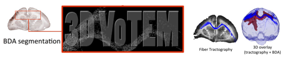
Histological Ground Truth
The reference data for the histological validation will be defined based on the high-resolution BDA stained slices, offering exquisite sensitivity and specificity for fibers connected to the injection region. After digitization, standard image processing techniques will segment BDA fibers in each slice (based on the dark brown staining intensity), resulting in BDA density maps defined in histological space (i.e., the total volume of BDA stain in each voxel, calculated from the number of segmented BDA pixels in each 300*300um sized region of the 2D histological slice). The multi-step registration procedure (describe above) will be used to transform BDA density maps into MRI space (with appropriate Jacobian corrections). These maps will be binarized, resulting in a volume of space occupied by fibers connected to M1 cortex. Both binarized maps and density maps represent the ground truth pathways, and facilitate direct comparisons with tractography results.
In addition to binarized tractographic results, we will also request streamlines in Trackvis (.trk) or Mrtrix (.tck) fiber format from participants (although not required) in order to facilitate comparisons of individual tracts and streamline densities. Streamlines can also be provided in FSL fiber format maps to allow comparing probabilistic certainty measures with BDA densities.
Macaque NIH Validation – submission format
For a direct comparison with the tractography results of different algorithms published by Thomas et al., the contestants are directed to use the provided seed ROIs. For deterministic tractography methods, the challengers must submit a tractography file for each of the four ROIs, either in TRACKVIS’ “.trk” format or MRTRIX’s “.tck” format (note that this is the same for all challenges). For probabilistic tractography methods, the submitted files must be in NIFTI format. These NIFTI images should have the same header information as the DWI file and their voxels should contain the “visitation counts” for the proposed probabilistic tractography method. Additionally, for the probabilistic tractography methods, please ensure that your track density image is thresholded at the visitation count that you consider optimal to determine whether a region is connected to another.