Olympus FV-1000 Inverted Confocal Microscope
Why Use This Microscope?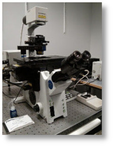
This microscope is well suited for viewing polarized cells and tissue samples on sealed, inverted slides or in 35mm Matek dishes with a #1.5 coverglass. Imaging through a non-glass surface is discouraged. All slides MUST be sealed, clean and dry prior to placing on the stage.
This microscope is well suited for viewing polarized cells and tissue samples on sealed, inverted slides or in 35mm Matek dishes with a #1.5 coverglass. Imaging through a non-glass surface is discouraged. All slides MUST be sealed, clean and dry prior to placing on the stage.
Imaging Modes:
- Laser scanning fluorescence and DIC (Nomarski)
- 3-D “Z-series”
- Time-series
- Simultaneous bleach/scan for PA-GFP and FRAP
Fluorescence Filters: (excitation/emission)
- NearUV/Blue (405nm for UV dyes e.g, DAPI)
- Blue/Green (457,488,514nm for blue, green, yellow dyes,
e.g., CFP,YFP,GFP, Cy-2, FITC-like dyes) - Green/Red (543nm for Cy-3, Texas Red, or rhodamine-like dyes)
- Red/Far-Red (633 nm for far-red dyes like Cy-5)
Objective Lenses:
- 10x / 0.30 Plan Neofluar
- 20x / 0.80 SPlan-UApo
- 40x / 1.30 Plan-Neofluar OIL
- 60x / 1.45 Plan-Apochromat OIL
- 100x / 1.40 SPlan-UApo OIL
Charges: $49.50 per hour
Note: To use the microscope you must have an active iLab account. Reservations are made through our on-line iLab calendar. Contact Sean to arrange training.
Location
- MRBIV 10434