
Electron Microscopy is one of the most important tools used for characterizing nanomaterials. Specifically, Transmission Electron Microscopy (TEM) and Scanning Transmission Electron Microscopy (STEM) can be used to determine the crystal structure and size of the nanocrystals synthesized in our lab. Information of size and shape homogeneity can be readily obtained.
At Vanderbilt University, we have a newly installed Tecnai Osiris TEM/STEM. This modern analytical electron microscope is equipped the Super-X EDS system, which allows for mapping of the entire periodic table (down to and including boron) with sub-nm spatial resolution. Combined with the high brightness X-FEG field emission gun, high resolution mapping of nanomaterials is achieved in minutes, rather than hours. Further, the microscope is equipped with a tomography holder for 3D imaging as well as an Aduro 300 heating stage for in-situ imaging up to 1300 °C. The Rosenthal group also has a long standing relationship with Oak Ridge National Laboratory and has utilized the aberration-corrected Nion UltraSTEMs for imaging nanocrystals with sub-angstrom resolution.
We continue to utilize state-of-the-art electron microscopy techniques to further elucidate structure-property relationships of colloidal nanocrystals. We utilized this imaging technique to reveal the true coverage of ZnS shell on colloidal quantum dots. In collaboration with Quantum Dot Corporation (Invitrogen/LifeTech), we helped develop core/shell nanocrystals with fluorescence quantum yields approaching unity. The materials that resulted in this work are now the commercially available Qdot® 655 and Qdot® 605 probes.
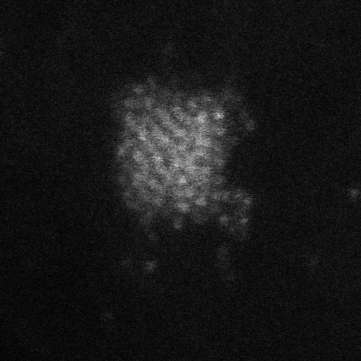
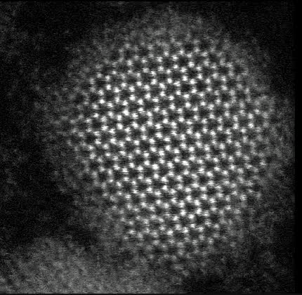
Recently, in collaboration with Stephen Pennycook and ORNL, we obtained Z-STEM videos of various size CdSe nanocrystals. Intriguingly, the atoms on the surface of the nanocrystals are dynamic with a crystalline core for larger nanocrystals, whereas all atoms are fluxional for sub-2 nm nanoclusters. This has led us to consider the possibility and implications of dynamic nanoparticle surfaces.
Selected Publications
Ng, A.; Poplawsky, J. D.; Li, C.; Pennycook, S. J.; Rosenthal, S. J., Direct Electronic Property Imaging of a Nanocrystal-Based Photovoltaic Device by Electron Beam-Induced Current via Scanning Electron Microscopy. J. Phys. Chem. Lett. 2014, 5 (5), 856-860.
McBride, J. R.; Pennycook, T. J.; Pennycook, S. J.; Rosenthal, S. J., The Possibility and Implications of Dynamic Nanoparticle Surfaces. ACS Nano 2013, 7 (10), 8358-8365.
Pennycook, T. J.; McBride, J. R.; Rosenthal, S. J.; Pennycook, S. J.; Pantelides, S. T., Dynamic Fluctuations in Ultrasmall Nanocrystals Induce White Light Emission. Nano Lett. 2012, 12 (6), 3038-3042
McBride, J. R.; Lupini, A. R.; Schreuder, M. A.; Smith, N. J.; Pennycook, S. J.; Rosenthal, S. J., Few-Layer Graphene as a Support Film for Transmission Electron Microscopy Imaging of Nanoparticles. ACS Appl. Mater. Interfaces 2009, 1 (12), 2886-2892.
McBride, J.; Treadway, J.; Feldman, L. C.; Pennycook, S. J.; Rosenthal, S. J., Structural Basis for Near Unity Quantum Yield Core/Shell Nanostructures. Nano Lett. 2006, 6 (7), 1496-1501.
McBride, J. R.; Kippeny, T. C.; Pennycook, S. J.; Rosenthal, S. J., Aberration-corrected Z-contrast scanning transmission electron microscopy of CdSe nanocrystals. Nano Lett. 2004, 4 (7), 1279-1283.