Parathyroid Perfusion Optical Device

Progress Report 4 (Second Semester)
Efforts in the weeks of 12 and 19 January have focused on implementation of a lab based laser speckle system and technological actualization of the design. Communications and Organizational Leader Itamar Shapira met with a biophotonics graduate student expert well acquainted with perfusion imaging, Kristin Poole. Kristin provided guidance using her experience with laser speckle imaging, particularly concerning image processing methodologies. Lastly, Kristin, displayed a commercially developed laser speckle imaging system in use at Vanderbilt University Medical Center for clinical trials.
The following are recommendations resulting from the meeting, taken from a memo distributed to the design team.
- Trying to use superficial human tissues is a poor perfusion model, even for initial work. We need to start using the microfluidic phantom as soon as possible. Doing so will allow for easy contrast between dark, flow regions and bright PDMS material. Later, an intraoperative animal model will be desirable.
- We need to improve our speckling by using a lens to spread out the beam. An optical filter will also allow our system to be used in ambient operating room lighting.
- Adopt a He-Ne laser. Such lasers have been successfully employed in the past for speckle imaging.
- Suspend open source code troublshooting and attempt self-implementation of code. Doing so will mean we understand our own code and have created added value with the code.
- Think very carefully about how our system is mounted, this will become a clinical concern.
Additional observations from meeting:
- Given that we are using relative perfusion units, we may need to image the parathyroid and then perform a comparison to a control which has no flow.
- Another possibility is examining the threshold of detection. We should use our microfluidic phantom at different flow rates. Hopefully no flow/low flow will not produce enough speckle contrast to be imaged, such that the imaging system will only pick up flowing fluid rather than stagnant fluid.
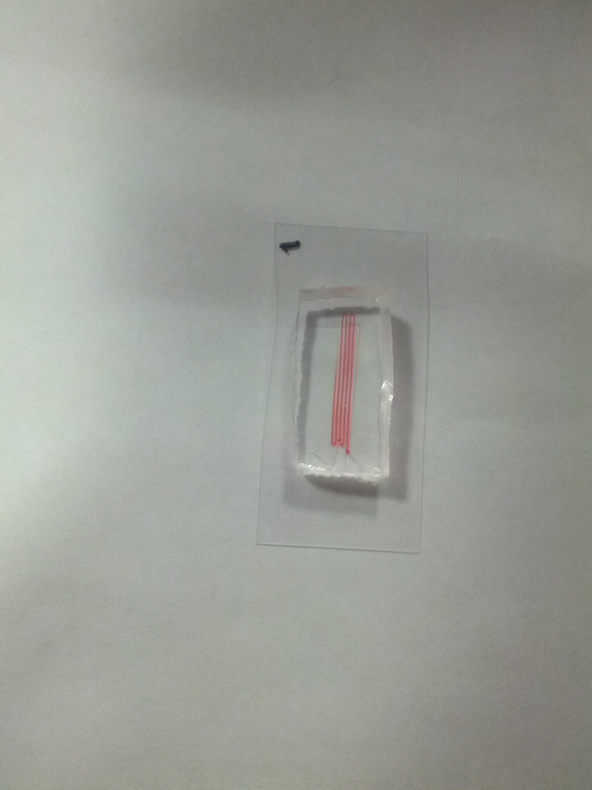
Michael Zhou has made progress on a microfluidic phantom model for device function diagnostics and for later proof of concept. Images are appended of the phantom being perfused with red dye. He is in deliberation with Dr. Kevin Seale about borrowing a microfluidic pump to quantify flow through the phantom.
James Tatum has made progress on the laboratory based system mounting and acquisition of appropriate lenses for speckling. He is currently considering a means of collumnating the light path to provide steady speckling at variable optical ranges.
Gabriella CDJ is spearheading the implementation of image processing code with Kevin Lu’s help to perform temporal and spatial laser speckle contrast analysis.
The focus remains on laboratory level implementation of technology such that clinical designs and proposals for funding may be undertaken.
©2026 Vanderbilt University ·
Site Development: University Web Communications