Home » News » The Ice Water Phantom Cometh
The Ice Water Phantom Cometh
Posted by anderc8 on Wednesday, August 23, 2017 in News, TIPs 2015.

Vishwesh Nath, PhD Student, MASI lab
Written by Vishwesh Nath, PhD Student, MASI lab, and Colin Hansen, PhD Student, MASI lab
A quick look at this device brings an unforgettable memory, and an initial reaction might leave one wondering: Is that dangerous? It is not. Known as diffusion phantoms, these spherical devices serve as a benchmark for assessing the performance of a magnetic resonance imaging (MRI) scanner. They were created to assess a scanner’s performance when the subject is a living being, though one can never have the exactness of what is present inside. While MRI has become a critical tool in the aid of diagnosing and understanding a variety of medical conditions, the technology is still constantly evolving.

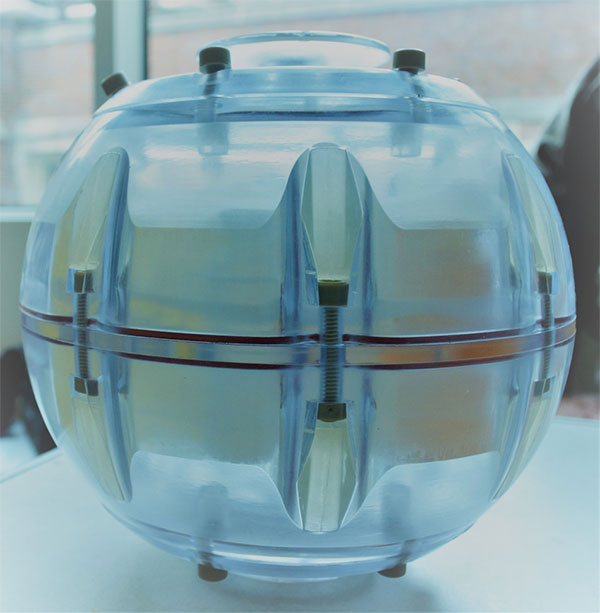
Side view of Ice Water Phantom. It is 194mm in diameter.
Diffusion MRI can lead us to beautiful nerve tracts that are interconnected via millions of axonal fiber bundles. The most important question that arises is: How do we calibrate a machine which is performing these complex measurements that allow us to image the in vivo (within living)? The analysis of the phantom data directly relates to the ongoing work at the MASI lab regarding clinical neuroscience. In brief, the MRI acquisition being performed of the human brain by a scanner is being calibrated by the use of phantoms.
“If the scanners are not calibrated effectively by the usage of phantoms, it will be even harder to analyze the MRI brain scans because we would be uncertain as to whether the acquired brain scans can be relied upon or not” – Vishwesh Nath
There can never be enough emphasis on the importance of minimizing error when clinical scans are being acquired on patients. It is critical that these scans do not lead to biased interpretation by radiologists and surgeons. Diffusion phantoms are used to assess how much error is being introduced in an imaging sequence. This can also be referred to as a ground truth, which can be acquired at different parameters of the scanner, while, at the same time, also providing an assessment of whether the scanner can acquire data with minimal error at the chosen parameters.

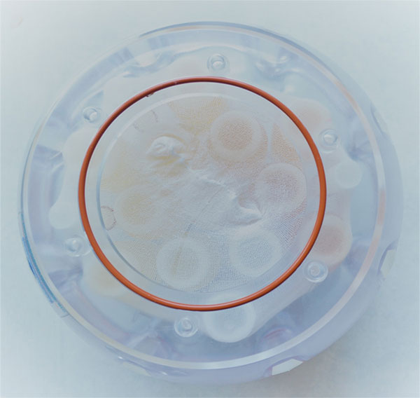
Top view of Ice Water Phantom with all vials visible.
This diffusion phantom uses ice water and requires a sufficient amount of ice so that water can be held at a temperature of 00 Fahrenheit for a period of 4-8 hours. The diffusion phantom has a complex configuration of different vials, with each vial serving as an indicator of different diffusion profiles which lead for a complex analysis of the assessment of scanner performance. Although it sounds easy, there are plenty of precautions to take when handling these expensive devices. For example, ice that is crushed too finely might lead to airgaps which may introduce artifacts into the data being acquired. While there are many moving parts when performing experiments of this intricate nature, there is also the motivation of a higher sense of purpose that drives us to keep our concentration when performing them.
“The importance of studying the phantom wasn’t clear to me at first, but I soon realized that under certain scan conditions the data collected from the diffusion scans of the phantom were quite informative. Now we are looking to collect even more scan data on the ice water phantom to develop an effective preprocessing pipeline to correct the data under these conditions. This potential contribution is exciting and will serve to drive me as we continue our work.” – Colin Hansen

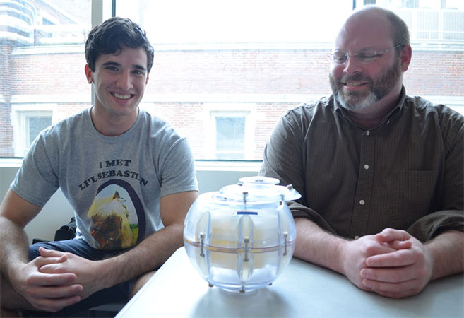
Colin Hansen (left) and Dr. Baxter Rogers.
With expertise specifically in the domain of medical physics, Dr. Baxter Rogers is the leading scientist regarding the phantom aspect of our project. Dr. Rogers has always been enthusiastic about this work and advises us when we need deep underlying explanations of the physics involved in this subject.
The diffusion phantom comes in various shapes and forms, and different types of phantoms are utilized depending on what the scanner is going to be used for. It could be used for optimizing the imaging sequence for the human brain or for optimizing the sequence of the abdomen and so on.
This project is a collaboration between Medical Imaging and Statistical Interpretation (MASI) Lab and Vanderbilt University Institute of Imaging Science (VUIIS). MASI is a VISE-affiliated lab. The project targets to bring the performance of a MRI scanner in light to clinicians and radiologists.
Learn more about VISE by visiting www.vanderbilt.edu/vise and be sure to follow us on our social media channels:
Facebook: https://www.facebook.com/visevanderbilt
Instagram: https://www.instagram.com/visevanderbilt/
Twitter: https://twitter.com/ViseVanderbilt
YouTube: http://vanderbi.lt/viseyoutube
