Home » Digital Humanities » Imaging Team Replicate Ancient Mesoamerican Artifacts for Gallery Exhibit
Imaging Team Replicate Ancient Mesoamerican Artifacts for Gallery Exhibit
Posted by vrcvanderbilt on Friday, June 7, 2019 in Digital Humanities, Events, Fine Arts Gallery, HART, News, Student/Alumni, Technology, VRC.
 When the Vanderbilt University Institute of Imaging Science (VUIIS) installed a new, state-of-the-art PET/CT scanner in early 2018, the team imagined a wide range of research opportunities — but none of them involved imaging artifacts for an art gallery exhibition.
This spring the VUIIS partnered with Vanderbilt’s Fine Arts Gallery to scan and create replicas of ancient Mesoamerican artifacts for a hands-on experience in the gallery’s current exhibition, Refuting “Noble Savages:” Reflections of Nature in Ancient Mesoamerican Artifacts.
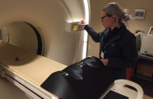
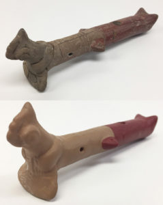
Using the scanner, the imaging team captured detailed pictures of each item’s surface and openings and sent the data to a 3D printer to print polymer replicas. Some of the replicas were then painted by undergraduate students in Markus Eberl’s spring semester course (Exhibiting Historical Art—Daily Life in Mesoamerica) to look like the original objects.
 Three replicas were created for the student-curated exhibition: an ancient flute and two ink stamps with varying designs.
“The Fine Arts Gallery has to secure its artifacts behind glass, so 3D replicas help us make these objects accessible to visitors,” said Eberl, associate professor of anthropology. “They can pick up the flute and play it. They can ink stamp replicas to reproduce the patterns engraved on the original stamps.”
“It’s a way for visitors to get closer not only to the objects, but also to the psychology and experiences of the people who originally used them,” added Emily Weiner, assistant curator of the Fine Arts Gallery.
According to Seth Smith, associate professor of radiology and radiological sciences and associate director of VUIIS, the collaboration demonstrates how imaging technology extends beyond clinical research, allowing the institute to be a resource for the greater Vanderbilt community.
“It was exciting for us to partner with the Fine Arts Gallery to do something completely out of the box. We were excited to develop a new relationship brought about because of the existence of a new imaging device within VUIIS. This is a bridge we don’t often get to see, and personally, I find it to be fascinating and encouraging,” said Smith.
 “It is exciting that our imaging technology can contribute not only to human health and the study of biological systems, but also to the humanities and our appreciation of ancient cultures,” said Todd Peterson, director of nuclear imaging. “While it may seem like a small detail, combining our tomographic imaging capabilities with 3D printing provides a true reproduction of these objects in a way that simply tracing the outer contours couldn’t.”
The partnership also allowed the team to learn more about the device’s capabilities. “This project was especially valuable as it provided the opportunity to test different aspects of our scanner’s imaging and reconstruction software which had not been previously explored,” said Anna Fisher, CNMT, nuclear medicine PET technologist II.
“We were able to acquire high-resolution CT images of each piece and successfully convert the data into 3D-printable formats all from the scanner itself. Discovering these unique capabilities has opened the door to new services we can provide here at VUIIS.”
The exhibition is on display through September 13 in the Vanderbilt Fine Arts Gallery in Cohen Memorial Hall on the western edge of the Peabody College campus. Gallery hours for the summer (now through August) are Tuesday-Friday, noon to 4 pm; Saturday, 1-5 pm; closed Sunday and Monday.—Kelsey Herbers courtesy of VUMC Reporter (May 9, 2019)
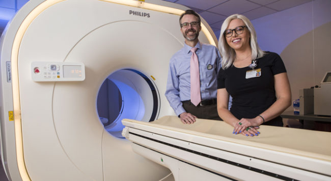
*Anna Fisher, certified nuclear medicine technologist, scans an ancient stamp using the PET/CT scanner. *Todd Peterson, director of nuclear imaging, and Fisher oversee the operation of the new research PET/CT scanner. (photos by Anne Rayner)

©2024 Vanderbilt University ·
Site Development: University Web Communications