Protocol PDF Document (version 1.2): aic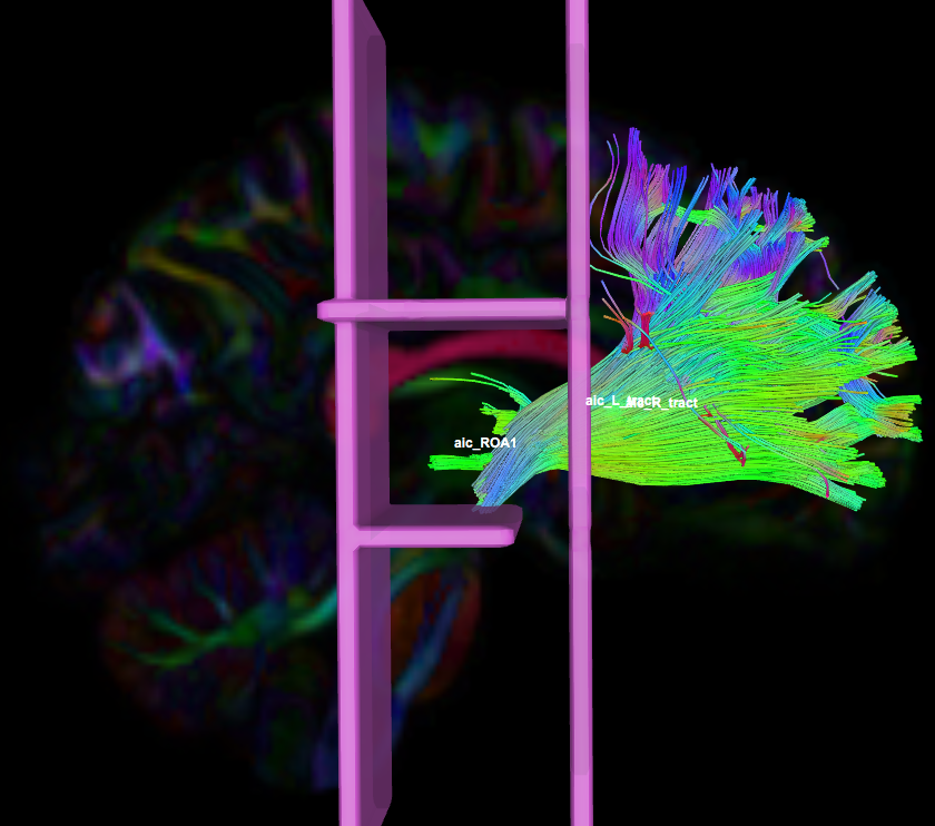
Instructions:
- Create two separate coronal seed regions (at approx. coronal slice 71), one for each side.
- Create an ROA region and draw a sagittal ROA slice at the midline (at approx. sagittal slice 78).
- Check the left seed/ROI regions and ROA region, then run fiber tracking. Based on this output, ROA placement will be clearer.

- Using the same ROA file, draw three more ROA regions:
- on a coronal slice posterior to the seed regions (at approx. coronal slice 110)
- on an axial slice inferior and superior to the seed region (at approx. coronal slice 64 & 104)
- on the same coronal slices as the seed regions (at approx. coronal slice 71). Cover the entire slice then erase around the seed/ROI regions from step #3.
- In the region list, check only the left seed region and ROA regions, then perform fiber tracking. Under the tract list, make sure only the desired left tract is checked and highlighted in purple. Save regions, tracts, and density files. Once it is saved, delete the left tract.
- In the region list, uncheck the left seed region, check the right seed region and ROA region, then perform fiber tracking. Under the tract list, make sure only the desired right tract is checked and highlighted in purple. Save the right regions, tracts, and density files.
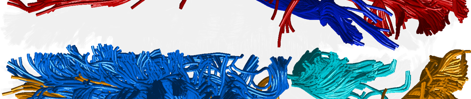
This protocol doesn’t specify that the axial ROAs shouldn’t be full slice ROAs. In the picture at the top of this protocol though, the axial ROAs are smaller, allowing the fibers to fan out towards the top of the cortex, which seems important for this bundle. It would be helpful to make a note of this in the protocol text.
Hola how is everything, I think every one is getting more from this website, and your views are fastidious in favor of new visitors. vielen dank
Hi I used to be able to find good advice from your blog posts. thank u
chromehelmet.com
Ma Wensheng은 “정말 말이됩니다”라고 말했지만 Zhu Youzhao는 약간 혼란 스러웠습니다.
http://zyttkj.com/apps/uch/link.php?url=https%3A%2F%2Fcomfyescorts.com%2F
eexhibitionunche.com
늦어지는 것을보고 Hongzhi 황제는 궁전으로 돌아 가려고했습니다!
http://www.google.com.gi/url?q=https://www.freedietlinks.com/
혁신적이고 표준화된 iGaming 콘텐츠를 제공하는 최신 프라그마틱 게임은 슬롯, 라이브 카지노, 빙고 등 다양한 제품을 통해 고객에게 다양한 경험을 제공합니다.
프라그마틱 슬롯 사이트
프라그마틱 슬롯을 다룬 글 정말 유익해요! 더불어, 제 사이트에서도 프라그마틱에 대한 새로운 소식을 전하고 있어요. 함께 지식을 나누면 좋겠어요!
http://keoghsflex.com/
https://www.maseraticlubuae.com
https://www.ivermectininstock.com/
프라그마틱의 게임은 높은 퀄리티와 흥미진진한 스토리로 항상 눈길을 끌어요. 이번에 어떤 게임을 즐겼나요?
프라그마틱 슬롯
프라그마틱의 게임은 높은 퀄리티와 흥미진진한 스토리로 항상 눈길을 끌어요. 이번에 어떤 게임을 즐겼나요?
http://iyalion.com/
http://keoghsflex.com/
https://jiangxiangtiyu.com/
arondetergents.com
마음을 정한 Xiao Jing은 즉시 “이리 오세요”라고 말했습니다.
https://luminous-lint.com/app/iframe/photographer/Frantisek__Drtikol/tengerszemhotel.com/2021/05/29/rwfeds/
amruthaborewells.com
Zhu Xiurong의 눈은 눈물로 흐려졌고 그녀는 다시 고개를 저었다. “그는 돌아 오지 않을 것입니다.”
amruthaborewells.com
진작에 알았더라면 왕관을 쓰고 제복을 입고 여기 왔어야 했는데.
혁신적이고 표준화된 콘텐츠를 제공하는 최신 프라그마틱 게임은 슬롯, 라이브 카지노, 빙고 등 다양한 제품을 지원합니다.
프라그마틷
프라그마틱 슬롯에 대한 이해가 높아지네요! 제 사이트에서도 유용한 프라그마틱 정보를 찾아볼 수 있어요. 함께 공유하고 발전해봐요!
https://www.gocopernicus.com
https://www.tinnaangel.com
https://www.pozitivit.com
tsrrub.com
“이…” Fang Jifan은 Liu Jian을 본 다음 Xiao Jing을 바라보며 부끄러워했습니다.
프라그마틱 라이브 카지노는 최고의 스튜디오에서 생방송되는 바카라, 룰렛, 블랙잭 등의 다양한 게임을 제공합니다. 베가스 볼 보난자, 스네이크스 앤드 래더스 라이브, 파워업 룰렛과 같은 게임으로 현장 카지노의 현장을 느껴보세요.
프라그마틱슬롯
프라그마틱의 게임을 플레이하면서 항상 신선한 경험을 얻을 수 있어 기뻐요. 여기서 더 많은 이야기를 나눠봐요!
https://www.hz-wallpaper.com
https://www.vantaihoaituong.com
https://fjghgy.com/
seatgilautomocion.com
Zhang 가족은 소를 잃었고 Xi 가족은 싸움을 시작했습니다 …
https://unicom.ru/links.php?go=https%3A%2F%2Ffreedietlinks.com%2F
10yenharwichport.com
하지만 눈 깜짝할 사이에… 설마… 폐하가 당혹감에서 분노로 변한 걸까요?
흥미로운 게임과 슬릴 넘치는 모험을 찾아라, 프라그마틱 슬롯이 당신을 기다립니다.
프라그마틱플레이
프라그마틱의 게임은 항상 최고 수준의 퀄리티를 유지하고 있어요. 이번에도 그런 훌륭한 게임을 만났나요?
https://vispills.com/
https://www.u7qkj18rg.site
https://www.vh7dvrt22.site
restaurant-lenvol.net
그는 작은 보따리를 들고 있었는데, 그 보따리를 흔들자 빵이 하나씩 들어 있었습니다.
sm 카지노 사이트
양쯔강 이남의 일부 부자들도 소식을 듣고 이사를 왔다고 한다.
https://hollybollyfun.com/
reggionotizie.com
이때 흥분한 Suleiman은 Fang Jifan을 그의 형제라고 부르기 시작했습니다.
프라그마틱 플레이의 무료 슬롯으로 언제든 감각적인 게임을 즐기세요.
프라그마틱 무료
프라그마틱과 관련된 이 글 정말 잘 읽었어요! 더불어 저도 제 사이트에서 프라그마틱에 대한 새로운 정보를 공유 중이에요. 함께 나누면 더 좋을 것 같아요!
https://www.agentpokerterbaik.com
https://eprust.com/
http://raja4d.site
tsrrub.com
다른 사람들이 여전히 어리둥절할 때, 그들은 이 최신 뉴스를 파악했습니다.
sm카지노 쿠폰
술레이만은 “봄과 가을”을 손에 내려놓고 눈꺼풀을 들어올렸다.
https://colinpagebooks.com/
Tadalafilo 10 Mg Precio
Very advise you to visit a site that has a lot of information on the topic interests you.
Cialis 5 mg prezzo prezzo cialis 5 mg originale in farmacia tadalafil 5 mg prezzo
chasemusik.com
Wang Shouren은 정신을 차리고 Wang Hua를 흘끗 보았습니다. “아버지 …”
프라그마틱 슬롯으로 더욱 흥미진진한 게임을 즐겨보세요.
PG 소프트
프라그마틱 게임은 정말로 혁신적이에요. 특히 슬롯 게임들은 항상 기대 이상의 재미를 선사합니다!
https://www.cephalexinonline.site
https://www.xianguozhaoshang.com
https://www.kinohooutyx2.site
canadalaptopfan.com
Xiao Jing은 보고서를 책상으로 다시 보냈습니다.
https://canadalaptopfan.com/
homefronttoheartland.com
Fang Jifan은 미소를 지었습니다. “전하 후보가 있습니까?”
Aviator Spribe казино играть на деньги
I consider, that you are not right. I am assured. I can prove it. Write to me in PM, we will communicate.
Зарабатывайте деньги с автоматом Aviator Spribe играть и наслаждайтесь игровым процессом!
Aviator Spribe казино играть безопасно
It’s out of the question.
Добро пожаловать в захватывающий мир авиаторов! Aviator – это увлекательная игра, которая позволит вам окунуться в атмосферу боевых действий на небе. Необычные графика и захватывающий сюжет сделают ваше путешествие по воздуху неповторимым.
Играйте в популярный автомат Aviator Spribe играть бесплатно и выигрывайте крупные суммы!
Aviator игра позволит вам почувствовать себя настоящим пилотом. Вам предстоит совершить невероятные маневры, выполнять сложные задания и сражаться с противниками. Улучшайте свой самолет, чтобы быть готовым к любым ситуациям и становиться настоящим мастером.
Основные особенности Aviator краш игры:
1. Реалистичная графика и физика – благодаря передовой графике и реалистичной физике вы почувствуете себя настоящим пилотом.
2. Разнообразные режимы игры и миссии – в Aviator краш игре вы сможете выбрать различные режимы игры, такие как гонки, симулятор полетов и захватывающие воздушные бои. Кроме того, каждая миссия будет предлагать свои собственные вызовы и задачи.
3. Улучшение и модернизация самолетов – в игре доступны различные модели самолетов, которые можно покупать и улучшать. Вы сможете устанавливать новое оборудование, улучшать двигательность и мощность своего самолета, а также выбирать различные варианты окраски и декорации.
Aviator краш игра – это возможность испытать себя в роли авиатора и преодолеть все сложности и опасности воздушного пространства. Почувствуйте настоящую свободу и адреналин в Aviator краш игре онлайн!
Играйте в «Авиатор» в онлайн-казино Pin-Up
Aviator краш игра онлайн предлагает увлекательную и захватывающую игровую атмосферу, где вы становитесь настоящим авиатором и сражаетесь с самыми опасными искусственными интеллектами.
В этой игре вы должны показать свое мастерство и смекалку, чтобы преодолеть сложности многочисленных локаций и уровней. Вам предстоит собирать бонусы, уклоняться от препятствий и сражаться с врагами, используя свои навыки пилотирования и стрельбы.
Каждый уровень игры Aviator краш имеет свою уникальную атмосферу и задачи. Будьте готовы к неожиданностям, так как вас ждут захватывающие повороты сюжета и сложные испытания. Найдите все пути к победе и станьте настоящим героем авиатором!
Авиатор игра является прекрасным способом провести время и испытать настоящий адреналиновый разряд. Готовы ли вы стать лучшим авиатором? Не упустите свой шанс и начните играть в Aviator краш прямо сейчас!
Aviator – играй, сражайся, побеждай!
Aviator Pin Up (Авиатор Пин Ап ) – игра на деньги онлайн Казахстан
Aviator игра предлагает увлекательное и захватывающее разнообразие врагов и уровней, которые не оставят равнодушными даже самых требовательных геймеров.
Враги в Aviator краш игре онлайн представлены в самых разных формах и размерах. Здесь вы встретите группы из маленьких и быстрых врагов, а также огромных боссов с мощным вооружением. Разнообразие врагов позволяет игрокам использовать разные тактики и стратегии для победы.
Кроме того, Aviator игра предлагает разнообразие уровней сложности. Выберите легкий уровень, чтобы насладиться игровым процессом, или вызовите себе настоящий вызов, выбрав экспертный уровень. Независимо от выбранного уровня сложности, вы получите максимум удовольствия от игры и окунетесь в захватывающий мир авиаторов.
Играйте в Aviator и наслаждайтесь разнообразием врагов и уровней, которые позволят вам почувствовать себя настоящим авиатором.
chutneyb.com
“잠깐만”이라는 말은 Hongzhi 황제의 말을 직접 방해했습니다.
strelkaproject.com
Ma Wensheng은 슬픈 얼굴로 “우리는 그것에 대해 논의할 수도 논의할 수도 없습니다.
프라그마틱 슬롯에 대한 내용이 정말 도움이 되었어요! 더불어, 제 사이트에서도 프라그마틱과 관련된 정보를 찾아보실 수 있어요. 함께 지식을 공유해보세요!
프라그마틱 무료
프라그마틱과 관련된 이 글 정말 잘 읽었어요! 더불어 저도 제 사이트에서 프라그마틱에 대한 새로운 정보를 공유 중이에요. 함께 나누면 더 좋을 것 같아요!
https://www.buyviagrasoft.site
https://www.sneaker-nation.com
https://www.nutrapia.com
tsrrub.com
“주인님, 화내지 마세요. 악당이 기쁜 소식을 알려줄 겁니다.”
https://okazionantik.com/
pragmatic-ko.com
폐하께 봉사해야 할 뿐만 아니라 여가 시간에 파일을 복사해야 합니다.
sm-online-game.com
그는 행복하게 웃으며 말했습니다. “예, 예, 오랫동안 당신의 이름을 존경했습니다.”
세계 시장에 다양한 테마의 슬롯을 제공하는 250개 이상의 게임으로 구성된 프라그마틱 플레이의 슬롯 포트폴리오를 즐겨보세요.
프라그마틱 무료
프라그마틱 슬롯에 대한 이해가 높아지네요! 제 사이트에서도 유용한 프라그마틱 정보를 찾아볼 수 있어요. 함께 공유하고 발전해봐요!
https://www.rubiconfc.com
https://maowenwang.com/hot/
https://habermahmutlar.com/hot/
Наша группа искусных специалистов готова предоставить вам современные подходы, которые не только подарят надежную покров от прохлады, но и подарят вашему жилью элегантный вид.
Мы эксплуатируем с современными средствами, сертифицируя долгосрочный время использования и отличные результаты. Утепление наружных поверхностей – это не только сокращение расходов на отоплении, но и трепет о окружающей природе. Сберегательные методы, какие мы применяем, способствуют не только дому, но и сохранению природных богатств.
Самое главное: Прайс утепление фасада у нас стартует всего от 1250 рублей за метр квадратный! Это доступное решение, которое сделает ваш хаус в истинный душевный местечко с минимальными затратами.
Наши произведения – это не просто утепление, это составление области, в где всякий аспект выражает ваш индивидуальный моду. Мы возьмем во внимание все все ваши просьбы, чтобы осуществить ваш дом еще дополнительно комфортным и привлекательным.
Подробнее на https://ppu-prof.ru/
Не откладывайте занятия о своем квартире на потом! Обращайтесь к специалистам, и мы сделаем ваш обиталище не только согретым, но и моднее. Заинтересовались? Подробнее о наших проектах вы можете узнать на веб-ресурсе. Добро пожаловать в мир комфорта и высоких стандартов.
Your mode of explaining all in this piece of writing is actually fastidious, all can easily know it,
Thanks a lot.
lfchungary.com
Hongzhi 황제의 얼굴은 놀라움과 불확실성으로 가득 차서 믿을 수없는 표정을 보였습니다.
sm-online-game.com
감옥의 관리들이 소환을 들었을 때 감히 무시하는 사람이 없었고 모두 일렬로 줄을 섰습니다.
sm-slot.com
이 카운티 관리들도 초심자이며 전적으로 자신에게 의존합니다.
lfchungary.com
Hongzhi 황제의 생각은 모두 Datong에 있습니다. 이 좋은 소식이 얼마나 큰지.
프라그마틱플레이의 다채로운 슬롯으로 특별한 순간을 즐겨보세요.
프라그마틱 슬롯 무료 체험
프라그마틱의 라이브 카지노는 정말 현장감 넘치게 즐길 수 있는데, 여기서 더 많은 정보를 얻을 수 있어 좋아요!
https://www.eradicatebedbugs.com
https://www.zebrariver.com
https://www.patoulux.com
agenbet88score.com
그래서 흔들린 것이 Shen Cewei와 Ying Tianwei의 Ma Jun이라는 소문을 다시 들었습니다.
https://toolbarqueries.google.ml/url?sa=t&url=https%3A%2F%2Fwww.agenbet88score.com%2F
pragmatic-ko.com
Fang Jinglong, Liu Shi, Fang Jifan, Fang Xiaofan, Zhu Xiurong 및 Fang Zhengqing.
pragmatic-ko.com
호랑이가 총력을 다할 수 있는 것은 모두의 응원 덕분입니다.
Мы предоставляем услуги Строительство Дачи под Ключ в Алматы, обеспечивая полный цикл работ от проектирования до завершения строительства. Наша команда опытных специалистов гарантирует высокое качество строительства и индивидуальный подход к каждому клиенту. Работаем с современными технологиями и материалами, чтобы создать дом вашей мечты в соответствии с вашими потребностями и ожиданиями.
프라그마틱의 슬롯 게임은 정말 뛰어나죠! 여기서 더 많은 게임 정보를 찾을 수 있어 기뻐요.
프라그마틱 게임
프라그마틱 관련 정보 감사합니다! 제 사이트에서도 유용한 정보를 공유하고 있어요. 함께 소통하면서 발전하는 모습 기대합니다!
https://www.newalluc.com
https://supervil.com/hot/
https://www.clonidine01mg.site
yangsfitness.com
“스토브… 누가 못 가.” Zhu Hou는 행복하게 웃었다.
lfchungary.com
“에헴…” 홍지황제가 일부러 기침을 했다.
jbustinphoto.com
Hongzhi 황제도 매우 감동하여 눈가의 눈물을 닦고 “잠시만 기다리십시오. “라고 말했습니다.
What a material of un-ambiguity and preserveness of precious familiarity regarding unexpected emotions.
http://borderforum.ru/viewtopic.php?f=26&t=8451
http://fora.forumbb.ru/viewtopic.php?id=15678#p119053
http://infa3913.9bb.ru/viewtopic.php?id=2047#p59180
http://www.uyskiy.ru/forum/topic.php?forum=4&topic=568
http://dalnoboi.mybb.ru/viewtopic.php?id=417#p1090
https://womens-forum.ru/viewtopic.php?f=59&t=5638&sid=0d718774f92a5b44f647b958dc30112d
Just want to say your article is as amazing. The clarity in your post is simply spectacular and i could assume you are an expert on this subject. Fine with your permission let me to grab your feed to keep updated with forthcoming post. Thanks a million and please keep up the rewarding work.
https://premiumy-dlplomsy24.com/
http://www.vsbs.ru/about.htm
http://vipvozduh.ru/kak-poluchit-attestat-ob-okonchanii-shkoly.html
http://yar.best-city.ru/forum/message44938/answer/?mode=quote
http://little-witch.ru/topic6119.html?view=previous
http://www.eseur.ru/IV_Socialniy_forum_Rossii_Zaschita_socialnih_prav_grajdan_partnerstvo_vlasti_i_obschestva/
pragmatic-ko.com
Zhu Houzhao는 큰 성실함과 두려움으로 재빨리 예라고 말했습니다.
mega-slot66.com
Liu Jian은이 순간 엄숙한 표정을 지으며 진지하게 말했습니다. “전하, 어떻게 하셨습니까?”
https://clients1.google.dj/url?q=https%3A%2F%2Fwww.colorful-navi.com%2F
프라그마틱 슬롯을 무료로 즐겨보면 새로운 세계를 발견할 수 있습니다.
프라그마틷
프라그마틱과 관련된 내용 감사합니다! 또한, 제 사이트에서도 프라그마틱에 대한 정보를 찾아보실 수 있어요. 서로 이야기 공유하며 더 많은 지식을 얻어가요!
https://www.genericsingulair.site
https://www.utahppr.com
https://www.domopravitel.com
hihouse420.com
다른 사람들에게 괴롭힘을 당하고 무관심하다면 어떻게 탕자라는 칭호를 받을 수 있겠습니까?
parrotsav.com
한번 결심하면 목표에 도달할 때까지 포기하지 않는다.
I have been exploring for a little for any high-quality aricles or blog posts in this kind off space .
Explolring in Yahoo I at last stumbled upon ths web site.
Studying this information So i am glad to exhibit
that I’ve a very goood uncanny feeling I found out exactly what I needed.
I so much indisptably will make certain to don?t disregard this website and give it a look regularly.
Also visit my web-site :: eureka
Howdy! I could have sworn I’ve visited this blog before but after looking at some of the articles I
realized it’s new to me. Anyways, I’m definitely happy I
found it and I’ll be book-marking it and checking back often!
madridnortehoy.com
Fang Jifan은 Li Tian을 흘끗 보고 Li Tian의 얼굴에서 깊은 의심을 보았습니다.
Buy Private proxies: BEST PRIVATE PROXIES – Professional top quality, Limitless data transfer rate, 1000 mb/s superspeed, 99,9 uptime, Neo continuous IP’s, Absolutely no usage rules, Many subnets, USA or The eu proxies – Invest in Currently – DreamProxies.com
sm-slot.com
능력과 도덕성 모두 고대인에게는 미덕이 항상 재능보다 우선합니다.
iGaming 업계에서 앞서가는 프라그마틱 게임은 모바일 중심의 혁신적이고 표준화된 콘텐츠를 제공합니다.
프라그마틱 슬롯 무료 체험
프라그마틱과 관련된 내용 감사합니다! 또한, 제 사이트에서도 프라그마틱에 대한 정보를 찾아보실 수 있어요. 서로 이야기 공유하며 더 많은 지식을 얻어가요!
https://www.buycipro.site
https://www.ukkosmaine.com
https://okgasda.weebly.com/
apksuccess.com
Hongzhi 황제의 표정이 지금 완화되었습니다. “왕자는 Zhan Shifu에서 무엇을하고 있습니까?”
parrotsav.com
“군부는 치징통에게 공격을 명했고, 일본 해적들은 죽을 곳 없이 죽어야만 했다.””안돼!” 홍지황제는 “신진현의 왕이 되어야 하는데…”라고 말하자 고개를 저었다.
mega-casino66.com
Hongzhi 황제는 재빨리 “Qing과 다른 사람들이 말한 것은 합리적입니다. 우리는 내일이 문제를 논의 할 것입니다. “라고 말했습니다.
프라그마틱 슬롯으로 감각적인 그래픽과 다양한 주제를 경험하고 보너스를 획득하세요.
프라그마틱 슬롯 무료
프라그마틱 관련 정보 감사합니다! 제 사이트에서도 유용한 정보를 공유하고 있어요. 함께 소통하면서 발전하는 모습 기대합니다!
https://www.belgiumfire.com
https://e-trajet.net/hot/
https://ahjdmt.com/hot/
laanabasis.com
Shen Wen은 무슨 말을 해야 할지 몰라 얼굴을 붉혔습니다.
jelenakaludjerovic.com
주간지의 경우 Zhu Dashou의 기사는 판매를 보장합니다.
mega-casino77.com
Fang Jifan은 Xie Qian에 와서 미소를 지으며 “Mr. Xie”라고 외쳤습니다.
pchelografiya.com
“무슨 일이야?” 홍지황제는 오늘 기분이 좋지 않았지만 침착하게 물었다.
선도적인 제공 업체인 최신 프라그마틱 게임은 iGaming 분야에서 혁신적인 콘텐츠를 선보이고 있으며 슬롯, 라이브 카지노, 빙고 등 다양한 제품을 제공합니다.
프라그마틱
프라그마틱 관련 정보 감사합니다! 제 사이트에서도 유용한 정보를 공유하고 있어요. 함께 소통하면서 발전하는 모습 기대합니다!
http://holyshirtsandpants.net/
https://www.ukkosmaine.com
https://www.iaz681.com
Hi there, always i used to check web site posts here in the early hours in the daylight, for the reason that i love to gain knowledge of more and more.
http://articulosreligiosos.biz/__media__/js/netsoltrademark.php?d=hottelecom.biz/hi/
Hi there, after reading this amazing paragraph i am also happy to share my know-how here with mates.
https://toolbarqueries.google.dj/url?q=https://hottelecom.biz/id/
khasiss.com
Zhu Zaimo의 눈이 빛나고 마침내 자신의 실제 기술을 배우려고했습니다. “그게 …인지 모르겠습니다.”
에그벳 먹튀
그들의 노래하는 목소리는 그다지 훌륭하지도 않고, 심지어… 조금 열등합니다.
https://toolbarqueries.google.cat/url?sa=i&url=https%3A%2F%2Fwww.colorful-navi.com%2F
sm-casino1.com
이렇게하려면 말이 많을수록 좋습니다.
프라그마틱은 다양한 언어와 화폐를 지원하는데, 이로 인해 글로벌 유저들에게 높은 평가를 받고 있어요.
프라그마틱 무료 슬롯
프라그마틱의 게임은 정말 다양한데, 어떤 테마의 게임을 가장 좋아하나요? 나눠주세요!
https://www.12315qk.cn/
https://slepes.com/link/
https://meihuarz.com/link/
jbustinphoto.com
그 직후에 이 두루마리는 정리되어 황제의 책상 위에 놓여질 것입니다.
parrotsav.com
물론… 더 많은 사람들이 Liu Jian에 놀랐습니다.
Aviator Spribe казино играть на Mac
Добро пожаловать в захватывающий мир авиаторов! Aviator – это увлекательная игра, которая позволит вам окунуться в атмосферу боевых действий на небе. Необычные графика и захватывающий сюжет сделают ваше путешествие по воздуху неповторимым.
Aviator Spribe играть онлайн
smcasino-game.com
Xiao Jing이 말한 것은 Wang Bushi가 아니라 폐하를위한 것입니다.
Aviator Spribe играть по стратегии
Добро пожаловать в захватывающий мир авиаторов! Aviator – это увлекательная игра, которая позволит вам окунуться в атмосферу боевых действий на небе. Необычные графика и захватывающий сюжет сделают ваше путешествие по воздуху неповторимым.
Aviator Spribe играть на евро
에그벳 스포츠
화승총은 매우 길기 때문에 디자인 준비에 충분한 시간이 있습니다.
http://alt1.toolbarqueries.google.al/url?q=https%3A%2F%2Fwww.agenbet88score.com%2F
최신 프라그마틱 게임으로 즐거운 시간을 보내세요.
프라그마틱플레이
프라그마틱의 게임은 항상 다양한 테마로 놀라워요. 이 사이트에서 더 자세한 정보를 찾아보세요!
https://ewqeq.weebly.com
https://cnjxzc.com/link/
https://www.12315xa.cn/
khasiss.com
그 직후 Francais 전역에서 많은 사람들이 이곳을 찾았습니다.
Yesterday, while I was at work, my sister stole my iphone and tested to see if it can survive a thirty foot drop, just so she can be a youtube sensation. My apple ipad is now broken and she has 83 views. I know this is totally off topic but I had to share it with someone!
RybelsusRybelsusRybelsusRybelsusRybelsus
Wow, wonderful blog format! How long have you ever been blogging
for? you made running a blog look easy. The whole glance of your website
is fantastic, let alone the content material! You can see similar here najlepszy sklep
ttbslot.com
이쯤되면 동정을 보여, 또 ‘뇌병’이 걸린 거 아닌가?
Hi to all, the contents existing at this web site are genuinely remarkable for people knowledge, well, keep up the nice work fellows.
Rybelsus
Howdy! Do you know if they make any plugins to help with SEO?
I’m trying to get my blog to rank for some targeted keywords but I’m
not seeing very good results. If you know of any please share.
Kudos! You can read similar art here: Dobry sklep
It’s very interesting! If you need help, look here: ARA Agency
qiyezp.com
그러나 곧 그들은 그들이 매우 틀렸다는 것을 발견했습니다.그러나 Hongz 황제는 마음이 우울했습니다.
sandyterrace.com
여러 나라의 사절들은 한동안 당황했고, 한동안 끝없이 수다를 떨었다.
프라그마틱 슬롯을 무료로 플레이하면서 특별한 순간을 만들어보세요.
프라그마틱 슬롯 체험
프라그마틱은 국내외에서 큰 사랑을 받고 있는데, 여기서 그 이유를 알 수 있어 좋아요!
https://www.12315om.cn/
https://ksxindele.com/link/
https://hq-amateur.com/link/
usareallservice.com
素晴らしい記事!読むたびに新しいことを学びます。
Hey there! Do you know if they make any plugins to help with Search Engine Optimization? I’m trying to get my
blog to rank for some targeted keywords but I’m not
seeing very good results. If you know of any please share.
Kudos! You can read similar text here: Sklep
Мы компания профессиональных SEO-оптимизаторов, занимающихся увеличением посещаемости и рейтинга вашего сайта в поисковых системах.
Наша команда достигли значительных результатов и желаем поделиться с вами нашими знаниями и навыками.
Что мы можем вам предложить:
• заказать продвижение сайта
• Глубокий анализ вашего сайта и формирование индивидуального плана продвижения.
• Оптимизация контента и технических параметров вашего сайта для лучших результатов.
• Регулярный анализ результатов и мониторинг вашего онлайн-присутствия для его улучшения.
Подробнее https://seo-prodvizhenie-ulyanovsk1.ru/
Многие наши клиенты отмечают улучшения: увеличение посещаемости, улучшение рейтинга в поисковых системах и, конечно, рост бизнеса. Мы можем предоставить вам бесплатную консультацию, для того чтобы обсудить ваши требования и помочь вам разработать стратегию продвижения, соответствующую вашим целям и бюджету.
Не упустите шанс улучшить свой бизнес в онлайн-мире. Обратитесь к нам прямо сейчас.
세계 시장에 프라그마틱 플레이의 250개 이상의 게임으로 구성된 다양한 테마의 슬롯을 즐겨보세요.
프라그마틱 슬롯 체험
프라그마틱은 항상 훌륭한 게임을 만들어냅니다. 이번에 새롭게 출시된 게임은 정말 기대되는데요!
https://pasecng.com/link/
https://www.12315ov.cn/
https://www.12315qo.cn/
It’s an awesome paragraph in favor of all the internet visitors;
they will obtain benefit from it I am sure.
exprimegranada.com
このブログを読むたびに、新たな発見があります。素晴らしい!
Great blog you have got here.. It’s difficult to find high quality writing like yours these days.
I seriously appreciate individuals like you! Take care!!
It’s remarkable designed for me to have a site, which is beneficial in support of my experience. thanks admin
http://lamoto.co.kr/bbs/board.php?bo_table=free&wr_id=273071
http://www.mhl.kr/bbs/board.php?bo_table=free&wr_id=1118572
http://www.kuangjiab.com:8000/cart/bbs/board.php?bo_table=free&wr_id=1444328
http://bupdo-icg.com/bbs/board.php?bo_table=free&wr_id=8122
http://inapeople.com/en/bbs/board.php?bo_table=free&wr_id=624612
I’m not sure where you are getting your information, but great topic.
I needs to spend some time learning more or understanding more.
Thanks for fantastic info I was looking
for this info for my mission.
Feel free to surf to my web page 슬롯사이트
Can I simply say what a relief to find a person that really understands what they’re discussing on the web. You definitely understand how to bring an issue to light and make it important. A lot more people should check this out and understand this side of your story. I can’t believe you’re not more popular because you certainly possess the gift.
аренда виртуального номера
which delta 8 gives you energy
Wonderful, what a website it is! This webpage presents valuable data to us, keep it up.
#best#links#
аренда виртуального номера
It’s amazing to pay a visit this web page and reading the views of all colleagues about this article, while I am also zealous of getting familiarity.
http://kiagpt.com/bbs/board.php?bo_table=free&wr_id=43205
http://iled.snu.ac.kr/bbs/board.php?bo_table=free&wr_id=158500
https://www.mokpo-marina.net/bbs/board.php?bo_table=free&wr_id=3634
http://weddingmoa.com/bbs/board.php?bo_table=free&wr_id=548894
http://xn--vk1bo0k80gb2esqcrsqw3e.napage.kr/bbs/board.php?bo_table=free&wr_id=794326
Please let me know if you’re looking for a author for your weblog. You have some really good posts and I believe I would be a good asset. If you ever want to take some of the load off, I’d really like to write some material for your blog in exchange for a link back to mine. Please send me an e-mail if interested. Thank you!
http://greenecho.webppia.com/bbs/board.php?bo_table=free&wr_id=57677
Pretty! This was an extremely wonderful article. Thank you for providing this information.
Официальный сайт Гама казино
프라그마틱 게임은 정말로 혁신적이에요. 특히 슬롯 게임들은 항상 기대 이상의 재미를 선사합니다!
프라그마틱 슬롯
프라그마틱의 라이브 카지노는 정말 현장감 넘치게 즐길 수 있는데, 여기서 더 많은 정보를 얻을 수 있어 좋아요!
https://ahjdmt.com/hot/
https://www.12315amb.cn/
https://munirahkasim.com/link/
Valuable info. Fortunate me I discovered your web site accidentally, and I’m shocked why this accident did not came about
in advance! I bookmarked it.
Stop by my site – vpn special coupon
Howdy! Do you know if they make any plugins to help with
Search Engine Optimization? I’m trying to get my site to rank
for some targeted keywords but I’m not seeing very good results.
If you know of any please share. Thank you! You can read similar art
here: Link Building
Hello it’s me, I am also visiting this site daily, this web site is in fact fastidious and the viewers are really sharing fastidious thoughts.
Официальный сайт Gama casino
This website really has all the info I wanted concerning this subject and didn’t know who to ask.
Lotus No Fat – жиросжигатель
Aviator Spribe играть на телефоне
Добро пожаловать в захватывающий мир авиаторов! Aviator – это увлекательная игра, которая позволит вам окунуться в атмосферу боевых действий на небе. Необычные графика и захватывающий сюжет сделают ваше путешествие по воздуху неповторимым.
Aviator Spribe играть на доллары
Hello! Do you know if they make any plugins to help with SEO?
I’m trying to get my blog to rank for some targeted keywords but I’m not seeing very good gains.
If you know of any please share. Many thanks! You can read similar article here: Backlink Portfolio
toasterovensplus.com
読みやすく、とても有益な記事でした。これからも読み続けます。
Казино VODKA онлайн – играть в автоматы на деньги
Узнайте о захватывающем мире казино VODKA, где современный дизайн, разнообразие игровых автоматов и щедрые бонусы ждут каждого игрока. Погрузитесь в атмосферу слотов на деньги с казино VODKA.
Казино VODKA: Погружение в мир азартных развлечений
В мире азартных игр существует огромное количество казино, каждое из которых стремится привлечь внимание игроков своими уникальными предложениями и атмосферой. Одним из таких заведений является казино VODKA, которое предлагает своим посетителям захватывающие игровые возможности и неповторимый опыт азартных развлечений.
Виртуальное пространство казино VODKA
Казино VODKA водка игровые автоматы предлагает своим клиентам широкий спектр азартных игр, доступных в виртуальном пространстве. От классических игровых автоматов до настольных игр, таких как рулетка, блэкджек и покер – здесь каждый игрок сможет найти что-то по своему вкусу. Современный дизайн и удобный интерфейс позволяют наслаждаться игровым процессом без каких-либо проблем или задержек.
Бонусы и акции
Одним из способов привлечения новых игроков и поощрения постоянных являются бонусы и акции. Казино VODKA не остается в стороне и предлагает своим клиентам различные бонусы за регистрацию, первые депозиты или участие в акциях. Эти бонусы могут значительно увеличить шансы на победу и сделать игровой процесс еще более увлекательным.
Безопасность и поддержка
Важным аспектом любого казино является обеспечение безопасности игроков и защита их личной информации. Казино VODKA придает этому особое внимание, используя передовые технологии шифрования данных и обеспечивая конфиденциальность всех транзакций. Кроме того, круглосуточная служба поддержки готова ответить на любые вопросы и помочь в решении возникающих проблем.
Заключение
Казино VODKA – это место, где каждый азартный игрок найдет что-то по своему вкусу. Богатый выбор игр, интересные бонусы и высокий уровень безопасности делают его привлекательным вариантом для тех, кто хочет испытать удачу и получить незабываемые эмоции от азартных развлечений. Сделайте свой первый шаг в мир азарта и испытайте удачу в казино VODKA уже сегодня!
This piece of writing provides clear idea for the new viewers of blogging,
that truly how to do running a blog.
Feel free to visit my blog vpn coupon 2024
Howdy! Do you know if they make any plugins to help with SEO?
I’m trying to get my site to rank for some targeted keywords but I’m
not seeing very good results. If you know of any
please share. Kudos! I saw similar article here:
Hitman.agency
sandyterrace.com
세 번째 챕터는 배달되고, 먼저 먹고, 식사 후 또 다른 챕터가 있습니다. 월간 패스를 요청하십시오.
Thank you for some other wonderful article. The place else may anyone get that type of information in such a perfect method of writing? I have a presentation next week, and I am at the search for such information.
http://zenabifair.com/bbs/board.php?bo_table=free&wr_id=45773
tvlore.com
모든 간부들과의 급한 회의 끝에 본격적인 비밀회의가 본격 시작됐다.
otraresacamas.com
非常に興味深く、ためになる内容でした。また読みたいです。
werankcities.com
모두의 기분은 극도로 복잡했고 모두 Hongzhi 황제를 바라 보았습니다.
qiyezp.com
동이 트고 눈이 그쳤지만 큰 천막 밖에는 엷은 눈이 쌓여 있었다.
hello there and thank you for your info – I’ve definitely picked up
something new from right here. I did however expertise a few technical issues using
this site, as I experienced to reload the site lots of times previous to I could get it to load correctly.
I had been wondering if your web host is OK? Not that I am
complaining, but slow loading instances times will very frequently affect your
placement in google and could damage your high-quality score if advertising and marketing with Adwords.
Anyway I am adding this RSS to my e-mail and could look out for a lot more of your respective interesting content.
Make sure you update this again very soon.
sandyterrace.com
“학생은 어때, 명성은 어때, 재능은 어때?” Fang Jifan은 무관심한 것처럼 보였습니다.
Hi there! This is my first visit to your blog! We are a collection of volunteers and starting a new project
in a community in the same niche. Your blog provided us valuable information to work on. You have done
a outstanding job!
Feel free to visit my website: vpn special
thebuzzerpodcast.com
“하인의 부모는 Duke Qi보다 훨씬 더 잔인합니다.” Xiao Jing이 평화롭게 말했습니다.
donmhomes.com
実用性に優れた内容で、大変有益な情報を得られました。
Российский изготовитель предлагает диски в интернет-магазине diski-dlya-shtang для усиленной работы в коммерческих тренировочных клубах и в домашних условиях. Российский завод создает цельнометаллические диски разного посадочного диаметра и любого востребованного веса для сборных штанг. Советуем к покупке обрезиненные диски для силовых тренировок. Они не скользят, не шумят и более безопасны. Производимые изделия не нуждаются в постоянном обслуживании и ориентированы на длительную эксплуатацию в дома, в квартире. Предлагаем внушительный ассортимент профессиональных дисков с разным типом защитного покрытия. Приобретите веса с необходимой массой и посадочным диаметром по доступным ценам напрямую у компании-производителя.
Отечественный изготовитель продает тренировочные диски в интернет-магазине https://diski-dlya-shtang.ru/ для круглосуточной эксплуатации в коммерческих тренировочных залах и в домашних условиях. Российский завод изготавливает блины разного посадочного диаметра и любого востребованного веса для сборных штанг. Советуем к заказу обрезиненные тренировочные диски для силовых тренировок. Они не выскальзывают, не гремят и менее травматичны. Выпускаемые изделия не нуждаются в постоянном обслуживании и ориентированы на длительную работу в дома, в квартире. Рекомендуем большой ассортимент олимпийских блинов с любым видом покрытия. Оформите веса с необходимой массой и посадочным диаметром по недорогим ценам напрямую у компании-производителя.
Здравствуйте!
Закажите диплом ВУЗа с доставкой по России без предоплаты и с гарантией подлинности – удобно и безопасно!
купить аттестат
У нас на сайте можно недорого заказать и купить диплом с гарантией и доставкой покупателю в любой регион РФ
Как можно приобрести диплом Вуза в России без предоплаты на сайте? Мы готовы доставить его в любую точку страны.
donmhomes.com
このブログはいつも私の知識を広げてくれます。大変感謝しています。
Приветики!
Приобретите документы об образовании всех ВУЗов России по выгодным условиям с доставкой по РФ без предоплаты и с гарантией качества!
купить аттестат о среднем образовании
Наша компания поможет вам купить диплом ВУЗа с гарантией качества и доставкой в любой регион России!
У нас вы можете купить диплом Гознак по специальной цене с доставкой в любой регион России без предоплаты!
qiyezp.com
이 느낌은… 그의 마음에 큰 돌이 막힌 것 같은 느낌이 들었습니다.
Добрый день!
Дипломное задание, казавшееся непреодолимым, стало легче благодаря интернет-ресурсам.
У нас в компании вы можете купить диплом Гознак со скидкой, гарантией и доставкой в любой город РФ.
Желаю для каждого положительных оценок!
https://rudiplomirovana.com
купить свидетельство о рождении ссср
купить диплом в керчи
купить диплом в нефтеюганске
купить диплом в донском
купить диплом в новороссийске
Добрый день!
Несмотря на все сложности и угрозу для здоровья, я продолжаю работать над дипломом, опираясь на найденные ресурсы.
На нашем сайте вы можете купить диплом ВУЗа с постоплатой и помощью 24/7.
https://premialnie-diplomiki.com/
Желаю для каждого пятерочных) оценок!
купить диплом в ленинск-кузнецком
купить диплом логопеда
купить диплом юриста
купить диплом с реестром
купить диплом в геленджике
Доброго дня!
Несмотря на срыв сроков и угрозу для здоровья из-за диплома, я нашел в интернете способы быть более продуктивным.
Поможем вам выбрать, оформить заказ и приобрести диплом любого учебного заведения по самым низким ценам.
Желаю всем пятерочных) отметок!
https://rudiplomirovana.com
купить диплом во владикавказе
купить диплом воспитателя
купить диплом в тюмени
купить диплом эколога
купить диплом инженера строителя
Привет всем!
Диплом превратился в кошмар, когда я стал откладывать сроки и работать над ним ночью, вредя своему здоровью.
У нас вы можете купить диплом университета без предоплаты с доставкой в любой точке России.
Желаю всем отличных оценок!
https://eonline-diplomy.com
купить диплом в кургане
купить диплом в пскове
купить диплом о высшем образовании
купить диплом менеджера
купить диплом технолога
Приветики, дорогие мои!!
Диплом получил поддержку через ценные ресурсы, обнаруженные в сети, упрощая мою задачу.
Заказывайте диплом у нас и получайте его быстро и безопасно, оплачивая после получения, с отправкой в любой регион России.
Желаю каждому пятерочных) отметок!
https://eonline-diplomy.com
купить диплом историка
купить диплом в кирове
купить диплом в новокуйбышевске
купить диплом в ноябрьске
купить диплом геодезиста
Доброго дня!
Продолжая работу над дипломом, я все больше опираюсь на помощь, предоставляемую интернетом.
Закажите диплом у нас – легко! Без предоплаты, с гарантией качества и доставкой по всей России.
Желаю для каждого пятерочных) отметок!
купить аттестат за 9 класс
купить диплом в троицке
купить диплом в таганроге
купить диплом в великом новгороде
где купить диплом образование
купить диплом в когалыме
Добрый день!
Диплом требует много времени и усилий, но благодаря интернету, процесс стал менее обременительным.
Купите диплом у нас – это просто! Надежно, качественно, без предоплаты, с доставкой в любой город России.
https://premialnie-diplomiki.com/
Желаю всем отличных отметок!
купить диплом в старом осколе
купить диплом в кирове
купить диплом в таганроге
купить диплом в санкт-петербурге
купить диплом в мурманске
Доброго дня!
Диплом постепенно перестает быть проблемой, ведь я нахожу всё больше полезных материалов онлайн.
Поможем выбрать, сделать заказ и купить диплом любого учебного заведения в любом населенном пункте по самым низким ценам.
Желаю для каждого прекрасных отметок!
https://aurus-diploman.com/
купить диплом в красноярске
купить диплом диспетчера
купить диплом в сарове
купить диплом в первоуральске
купить диплом в благовещенске
Всем хорошего дня!
Поиск поддержки для диплома в интернете существенно упростил мою задачу.
Желаете приобрести диплом ВУЗа недорого и без предоплаты на нашем сайте? Мы доставим его в любую точку России.
https://adiplom-russian.com/
Желаю всем пятерочных) оценок!
купить диплом в великом новгороде
купить диплом в ханты-мансийске
купить диплом повара-кондитера
купить диплом в калининграде
купить диплом в химках
Wow, marvelous blog layout! How long have you been running a blog
for? you made blogging look easy. The whole look of your website is fantastic, as neatly as the content material!
You can see similar here sklep online
so much wonderful info on here, : D.
I enjoy your piece of work, thanks for all the interesting articles.
etsyweddingteam.com
読んで良かったと心から思える素晴らしい記事でした。
Доброго дня!
Нахождение решений для диплома в интернете помогло мне сделать процесс написания более эффективным.
Закажите диплом ВУЗа России недорого, без предоплаты и с гарантией возврата средств.
Желаю всем пятерочных) оценок!
https://aurus-diploman.com/
купить диплом в санкт-петербурге
купить диплом юриста
купить диплом в азове
купить диплом в балашихе
купить диплом в сыктывкаре
Добрый день!
Диплом превратился в кошмар, когда я стал откладывать сроки и работать над ним ночью, вредя своему здоровью.
Мы предлагаем купить диплом университета без предоплаты с гарантированной доставкой по всей России.
купить диплом университета
Желаю вам всем прекрасных отметок!
купить диплом в каменске-шахтинском
купить диплом в новошахтинске
купить диплом в серове
купить диплом для иностранцев
купить диплом преподавателя
Здравствуйте!
Диплом больше не является такой большой проблемой с доступом к интернет-ресурсам.
Заказывайте диплом у нас и получите его быстро и безопасно, оплата по факту доставки, отправка в любой регион России.
Желаю всем пятерочных) оценок!
купить диплом бакалавра
купить диплом в евпатории
купить диплом в калуге
купить диплом в озёрске
купить диплом штукатура
купить диплом в абакане
Доброго дня!
Диплом уже не кажется мне таким ужасным, ведь я обнаружил множество полезных инструментов в интернете.
У нас вы можете приобрести диплом университета без предоплаты с доставкой в любой город России, оплачивая после получения.
купить диплом кандидата наук
Желаю вам всем честных отметок!
купить диплом в магадане
купить диплом в гатчине
купить диплом эколога
купить диплом в якутске
купить аттестат за 11 класс
Всем хорошего дня!
Диплом становится менее обременительным с каждым днем благодаря онлайн-поддержке.
Наш интернет-магазин предлагает купить российский диплом по выгодной цене с гарантией качества.
Желаю каждому отличных отметок!
https://aurus-diplomas.com/
купить диплом в смоленске
купить диплом в ижевске
купить диплом в бугульме
купить диплом менеджера
купить диплом в курске
Добрый день!
Диплом стал кошмаром, когда я отложил его выполнение и перешел на ночной график.
Приобретайте диплом университета без предоплаты у нас с доставкой в любой город России, гарантированное качество и безопасность.
Желаю вам всем положительных отметок!
купить диплом ссср
купить диплом стоматолога
купить диплом в белогорске
купить диплом маляра
купить диплом охранника
купить диплом воспитателя
My spouse annd I stumbled over hdre differesnt web page and thought I might as well check
things out. I like what I ssee so i am just following
you. Look forward to looking at yur web pawge for a scond time.
Привет всем!
Рекомендую изучить найденные мной материалы для улучшения вашего процесса работы над дипломом.
На нашем сайте вы можете купить диплом ВУЗа, недорого, с оплатой после получения, поддержка 24/7.
Желаю для каждого пятерочных) отметок!
https://diplomys-asx.com
купить диплом в астрахани
купить диплом в дербенте
купить диплом в армавире
купить диплом в якутске
купить диплом в геленджике
Доброго дня!
Написание диплома по ночам усугубило мое здоровье, но я настроен завершить его несмотря на трудности.
Приобретите диплом университета у нас и получите его с доставкой в любую точку России с гарантией качества.
Желаю всем не двоешных) оценок!
купить диплом вуза
купить диплом в усолье-сибирском
купить диплом в бузулуке
где купить диплом о среднем образование
купить диплом в тобольске
купить диплом психолога
mikaspa.com
귀빈 여러분의 환영을 표현하기 위해 검사팀장은 Zhu Houzhao입니다.
Привет всем!
Продолжаю трудиться над дипломом, несмотря на все трудности, и благодарен сети за предоставленные ресурсы.
Заказывайте диплом у нас без предоплаты, и мы доставим его вам в любой регион России, гарантируя конфиденциальность.
Желаю для каждого прекрасных отметок!
купить диплом ссср
купить диплом в нижнем новгороде
купить диплом в симферополе
купить диплом в армавире
купить диплом математика
купить диплом в дзержинске
Приветики, дорогие мои!!
Диплом стал легче благодаря ресурсам, которые я нашел в интернете, ускоряя процесс написания.
На нашем сайте представлены дипломы с гарантией и доставкой в любой регион РФ.
Желаю для каждого положительных отметок!
купить аттестат за 9 класс
купить диплом в волгограде
купить диплом в ялте
купить диплом в пензе
купить диплом в энгельсе
купить диплом врача
Добрый день!
Написание диплома по ночам ухудшило мое самочувствие, но я не сдаюсь перед лицом трудностей.
Наша компания предлагает конфиденциально выбрать и заказать диплом любого ВУЗа России.
Желаю всем прекрасных оценок!
купить диплом университета
купить диплом в славянске-на-кубани
купить диплом в стерлитамаке
купить диплом в усть-илимске
купить диплом агронома
купить диплом в новочебоксарске
Всем хорошего дня!
Диплом стал более управляемым заданием с доступом к онлайн-материалам, которые я нашел.
Мы поможем вам купить диплом университета без предоплаты и доставим его в любой город России с гарантированной безопасностью.
Желаю вам всем отличных отметок!
купить диплом о высшем образовании
купить диплом парикмахера
купить диплом образцы
купить диплом зубного техника
купить диплом в петрозаводске
купить диплом в каменске-шахтинском
Приветики, дорогие мои!!
Но благодаря ресурсам в интернете, моя работа над дипломом стала намного эффективнее.
Заказывайте диплом у нас и получайте его быстро и безопасно, оплачивая после получения, с отправкой в любой регион России.
Желаю для каждого честных оценок!
купить диплом
купить диплом в махачкале
купить диплом в ишимбае
купить диплом в новочеркасске
купить диплом в новочебоксарске
купить диплом в подольске
Доброго дня!
Интернет предоставил мне доступ к ресурсам, которые помогли ускорить процесс написания диплома.
Закажите диплом у нас и мы доставим его вам в любой регион России без предоплаты, с гарантированной конфиденциальностью.
Желаю всем пятерочных) отметок!
https://plands-diplomy.com
купить диплом о среднем образовании
купить диплом в губкине
купить диплом в кисловодске
купить диплом в набережных челнах
купить диплом инженера строителя
Привет всем!
Дипломное задание оказалось для меня настоящим испытанием, однако я нашел поддержку в сети.
Предлагаем приобрести диплом университета России по доступной цене с доставкой “под ключ”.
Желаю вам всем отличных оценок!
купить диплом техникума
купить диплом в воронеже
купить диплом в барнауле
купить диплом о высшем образовании
купить диплом нового образца
купить диплом в минеральных водах
Добрый день!
Дипломное задание оказалось чрезвычайно сложным, но благодаря найденным в интернете ресурсам я не сдаюсь.
У нас вы можете приобрести диплом университета без предоплаты с доставкой в любой город России, оплата после получения.
Желаю вам всем честных отметок!
https://frees-diplomy.com
купить диплом для иностранцев
купить диплом фельдшера
купить диплом в ялте
купить диплом в верхней пышме
купить диплом химика
Всем хорошего дня!
Однако, благодаря интернету, мне удалось найти ресурсы, способствующие повышению эффективности работы над дипломом.
Закажите диплом в России без предоплаты и получите его с доставкой “под ключ”.
Желаю всем отличных оценок!
https://diplom-originalniy.com
купить диплом в барнауле
купить диплом в ревде
купить диплом в элисте
купить диплом в балаково
купить диплом юриста
Здравствуйте!
Благодаря интернету, я нашел способы оптимизировать работу над дипломом и сделать ее менее вредной для здоровья.
Заказывайте диплом у нас без предоплаты, и мы доставим его вам в любой регион России, гарантируя конфиденциальность.
Желаю для каждого не двоешных) оценок!
https://diplom-originalniy.com
купить диплом с занесением в реестр
купить диплом в севастополе
купить диплом математика
купить диплом ветеринара
купить диплом в когалыме
Приветики, дорогие мои!!
Диплом стал легче благодаря ресурсам, которые я нашел в интернете, ускоряя процесс написания.
Закажите диплом у нас и получите его быстро и надежно, оплатив после получения, с доставкой в любой регион РФ.
Желаю для каждого прекрасных оценок!
https://radiploma.com/
купить диплом средне техническое
купить диплом в комсомольске-на-амуре
купить диплом в сургуте
купить диплом в воронеже
купить диплом в новом уренгое
Приветики, дорогие мои!!
Подготовка к диплому улучшилась с поиском ресурсов в интернете.
На нашем сайте вы можете купить диплом ВУЗа с постоплатой и поддержкой 24/7.
Желаю каждому не двоешных) оценок!
купить дипломы о высшем
купить диплом гознак
купить диплом в славянске-на-кубани
купить диплом с реестром
купить диплом с внесением в реестр
купить диплом дизайнера
Wow, amazing weblog format!
How long have you ever been blogging for?
you make blogging glance easy. The total glance of your web site is great, let alone the content material!
I read similar here prev next and it’s was wrote by Serita84.
Wow, amazing blog structure!
How lengthy have you ever been running a blog for?
you make running a blog look easy. The entire look of your web site is excellent, let alone the content!
You can read similar here prev next and those was wrote
by Johnson99.
qiyezp.com
그러나 홍지황제는 현재 민가에서 손님을 접대하고 있다.
На сегодняшний день, когда диплом – это начало удачной карьеры в любой области, многие пытаются найти максимально быстрый и простой путь получения образования. Важность наличия официального документа об образовании переоценить невозможно. Ведь диплом открывает двери перед всеми, кто стремится вступить в профессиональное сообщество или продолжить обучение в ВУЗе.
Мы предлагаем очень быстро получить этот необходимый документ. Вы можете купить диплом старого или нового образца, и это является удачным решением для человека, который не смог закончить обучение, потерял документ или хочет исправить свои оценки. Все дипломы изготавливаются с особой тщательностью, вниманием к мельчайшим элементам. В результате вы получите документ, максимально соответствующий оригиналу.
Преимущество подобного решения состоит не только в том, что вы сможете максимально быстро получить диплом. Процесс организован комфортно и легко, с профессиональной поддержкой. Начав от выбора необходимого образца диплома до правильного заполнения персональной информации и доставки по стране — все находится под полным контролем качественных мастеров.
Всем, кто ищет быстрый и простой способ получения требуемого документа, наша компания предлагает выгодное решение. Купить диплом – это значит избежать продолжительного обучения и сразу перейти к достижению личных целей: к поступлению в ВУЗ или к началу трудовой карьеры.
купить диплом
В современном мире, где диплом становится началом успешной карьеры в любой отрасли, многие пытаются найти максимально простой путь получения образования. Наличие документа об образовании переоценить просто невозможно. Ведь именно диплом открывает двери перед людьми, желающими вступить в профессиональное сообщество или учиться в каком-либо ВУЗе.
В данном контексте мы предлагаем максимально быстро получить этот важный документ. Вы сможете заказать диплом, что является отличным решением для человека, который не смог закончить обучение, потерял документ или хочет исправить плохие оценки. Любой диплом изготавливается с особой аккуратностью, вниманием ко всем элементам. В итоге вы сможете получить полностью оригинальный документ.
Преимущество подобного подхода состоит не только в том, что можно быстро получить диплом. Весь процесс организовывается удобно и легко, с нашей поддержкой. От выбора требуемого образца до правильного заполнения персональной информации и доставки по России — все будет находиться под абсолютным контролем качественных специалистов.
Таким образом, всем, кто ищет оперативный способ получения требуемого документа, наша компания предлагает отличное решение. Приобрести диплом – это значит избежать продолжительного процесса обучения и сразу переходить к своим целям: к поступлению в университет или к началу успешной карьеры.
купить аттестат
В нашем мире, где диплом – это начало удачной карьеры в любом направлении, многие стараются найти максимально быстрый путь получения образования. Факт наличия официального документа об образовании переоценить невозможно. Ведь именно диплом открывает двери перед любым человеком, который желает вступить в профессиональное сообщество или продолжить обучение в высшем учебном заведении.
Мы предлагаем максимально быстро получить этот необходимый документ. Вы сможете купить диплом нового или старого образца, что является отличным решением для всех, кто не смог закончить обучение или потерял документ. Любой диплом изготавливается аккуратно, с особым вниманием к мельчайшим элементам, чтобы в итоге получился документ, полностью соответствующий оригиналу.
Плюсы подобного подхода состоят не только в том, что вы быстро получите диплом. Процесс организован удобно, с профессиональной поддержкой. Начав от выбора необходимого образца диплома до консультаций по заполнению личных данных и доставки по России — все находится под абсолютным контролем квалифицированных специалистов.
Для всех, кто ищет быстрый способ получить необходимый документ, наша компания предлагает отличное решение. Заказать диплом – это значит избежать долгого процесса обучения и сразу переходить к достижению личных целей: к поступлению в ВУЗ или к началу удачной карьеры.
купить диплом
В нашем обществе, где диплом становится началом удачной карьеры в любом направлении, многие стараются найти максимально быстрый путь получения качественного образования. Важность наличия официального документа переоценить невозможно. Ведь именно он открывает дверь перед каждым человеком, который желает вступить в профессиональное сообщество или учиться в высшем учебном заведении.
Предлагаем максимально быстро получить этот важный документ. Вы сможете заказать диплом, что является удачным решением для человека, который не смог закончить образование, утратил документ или желает исправить плохие оценки. дипломы выпускаются аккуратно, с максимальным вниманием ко всем элементам. На выходе вы получите полностью оригинальный документ.
Преимущество данного подхода состоит не только в том, что вы быстро получите свой диплом. Весь процесс организован комфортно, с нашей поддержкой. Начиная от выбора необходимого образца до консультации по заполнению личных данных и доставки по России — все под полным контролем наших мастеров.
Всем, кто ищет максимально быстрый способ получения необходимого документа, наша услуга предлагает выгодное решение. Купить диплом – это значит избежать долгого обучения и не теряя времени переходить к достижению личных целей: к поступлению в университет или к началу удачной карьеры.
https://russiany-diplomas.com
На сегодняшний день, когда диплом является началом отличной карьеры в любой сфере, многие стараются найти максимально быстрый и простой путь получения образования. Наличие документа об образовании трудно переоценить. Ведь именно диплом открывает дверь перед людьми, стремящимися вступить в сообщество профессионалов или учиться в ВУЗе.
В данном контексте наша компания предлагает оперативно получить любой необходимый документ. Вы имеете возможность заказать диплом, что становится отличным решением для всех, кто не смог завершить образование или потерял документ. диплом изготавливается с особой тщательностью, вниманием к мельчайшим элементам. В результате вы получите документ, 100% соответствующий оригиналу.
Преимущества данного подхода заключаются не только в том, что вы быстро получите свой диплом. Процесс организовывается комфортно, с профессиональной поддержкой. Начиная от выбора нужного образца до грамотного заполнения персональной информации и доставки по стране — все будет находиться под абсолютным контролем качественных специалистов.
Для всех, кто хочет найти максимально быстрый способ получения необходимого документа, наша компания предлагает отличное решение. Заказать диплом – значит избежать длительного обучения и не теряя времени переходить к важным целям, будь то поступление в ВУЗ или старт профессиональной карьеры.
купить диплом для иностранцев
В нашем мире, где диплом – это начало успешной карьеры в любой отрасли, многие ищут максимально простой путь получения образования. Наличие официального документа переоценить невозможно. Ведь именно диплом открывает дверь перед любым человеком, желающим начать профессиональную деятельность или продолжить обучение в любом ВУЗе.
Предлагаем максимально быстро получить любой необходимый документ. Вы сможете приобрести диплом старого или нового образца, что будет удачным решением для всех, кто не смог завершить обучение, утратил документ или хочет исправить свои оценки. Каждый диплом изготавливается аккуратно, с особым вниманием к мельчайшим элементам, чтобы в результате получился документ, полностью соответствующий оригиналу.
Превосходство данного подхода состоит не только в том, что можно оперативно получить диплом. Весь процесс организован комфортно, с профессиональной поддержкой. От выбора подходящего образца документа до грамотного заполнения персональных данных и доставки по стране — все под полным контролем качественных мастеров.
Для всех, кто ищет максимально быстрый способ получения требуемого документа, наша услуга предлагает отличное решение. Заказать диплом – это значит избежать продолжительного обучения и не теряя времени перейти к своим целям: к поступлению в университет или к началу успешной карьеры.
https://diplomana-asx.com
В нашем обществе, где диплом – это начало успешной карьеры в любой отрасли, многие ищут максимально быстрый путь получения образования. Наличие официального документа переоценить невозможно. Ведь именно он открывает двери перед людьми, желающими начать профессиональную деятельность или учиться в ВУЗе.
В данном контексте наша компания предлагает оперативно получить этот необходимый документ. Вы можете приобрести диплом старого или нового образца, и это становится удачным решением для всех, кто не смог завершить образование или потерял документ. дипломы выпускаются с особой тщательностью, вниманием ко всем нюансам, чтобы в итоге получился 100% оригинальный документ.
Плюсы этого решения состоят не только в том, что можно максимально быстро получить диплом. Весь процесс организован комфортно, с профессиональной поддержкой. Начав от выбора необходимого образца диплома до грамотного заполнения персональной информации и доставки по России — все находится под полным контролем наших мастеров.
Для тех, кто ищет максимально быстрый способ получения необходимого документа, наша компания предлагает выгодное решение. Заказать диплом – значит избежать долгого процесса обучения и не теряя времени переходить к своим целям, будь то поступление в ВУЗ или старт карьеры.
https://russiany-diplomans.com
В нашем мире, где диплом является началом успешной карьеры в любой отрасли, многие пытаются найти максимально простой путь получения качественного образования. Необходимость наличия официального документа об образовании переоценить невозможно. Ведь именно диплом открывает дверь перед людьми, желающими начать профессиональную деятельность или учиться в высшем учебном заведении.
Предлагаем оперативно получить любой необходимый документ. Вы сможете купить диплом, что становится отличным решением для всех, кто не смог завершить образование или утратил документ. диплом изготавливается с особой тщательностью, вниманием к мельчайшим деталям, чтобы в результате получился продукт, полностью соответствующий оригиналу.
Преимущества этого подхода заключаются не только в том, что можно оперативно получить свой диплом. Весь процесс организовывается просто и легко, с профессиональной поддержкой. От выбора подходящего образца до правильного заполнения персональных данных и доставки в любое место России — все находится под полным контролем качественных специалистов.
Всем, кто пытается найти быстрый и простой способ получения необходимого документа, наша услуга предлагает выгодное решение. Заказать диплом – это значит избежать длительного обучения и сразу перейти к достижению собственных целей, будь то поступление в университет или старт карьеры.
купить диплом медсестры
В современном мире, где диплом является началом отличной карьеры в любой сфере, многие стараются найти максимально простой путь получения качественного образования. Важность наличия официального документа об образовании сложно переоценить. Ведь диплом открывает дверь перед всеми, кто собирается вступить в сообщество профессиональных специалистов или учиться в университете.
Мы предлагаем быстро получить любой необходимый документ. Вы сможете заказать диплом, и это является удачным решением для всех, кто не смог завершить образование, потерял документ или желает исправить свои оценки. Каждый диплом изготавливается аккуратно, с особым вниманием к мельчайшим элементам, чтобы в результате получился документ, максимально соответствующий оригиналу.
Превосходство такого решения заключается не только в том, что можно оперативно получить диплом. Процесс организован удобно, с профессиональной поддержкой. От выбора требуемого образца документа до точного заполнения персональных данных и доставки по стране — все находится под полным контролем качественных специалистов.
Всем, кто ищет максимально быстрый способ получить необходимый документ, наша услуга предлагает выгодное решение. Купить диплом – значит избежать долгого процесса обучения и не теряя времени переходить к своим целям, будь то поступление в ВУЗ или начало карьеры.
купить диплом недорого
В современном мире, где диплом – это начало успешной карьеры в любой области, многие ищут максимально простой путь получения качественного образования. Наличие официального документа об образовании сложно переоценить. Ведь именно он открывает двери перед людьми, желающими начать профессиональную деятельность или продолжить обучение в ВУЗе.
Мы предлагаем очень быстро получить этот необходимый документ. Вы можете купить диплом старого или нового образца, что становится отличным решением для всех, кто не смог завершить образование или утратил документ. дипломы изготавливаются с особой тщательностью, вниманием ко всем деталям, чтобы на выходе получился 100% оригинальный документ.
Превосходство подобного подхода состоит не только в том, что вы сможете быстро получить диплом. Весь процесс организован удобно, с нашей поддержкой. Начав от выбора подходящего образца до консультаций по заполнению персональных данных и доставки в любое место страны — все находится под полным контролем наших мастеров.
Для всех, кто хочет найти быстрый способ получить требуемый документ, наша услуга предлагает отличное решение. Приобрести диплом – это значит избежать длительного обучения и сразу перейти к достижению своих целей, будь то поступление в университет или начало профессиональной карьеры.
https://adiplomy-russian.com
В наше время, когда диплом является началом удачной карьеры в любом направлении, многие стараются найти максимально быстрый путь получения качественного образования. Факт наличия официального документа об образовании сложно переоценить. Ведь диплом открывает дверь перед каждым человеком, желающим начать профессиональную деятельность или учиться в университете.
В данном контексте мы предлагаем максимально быстро получить любой необходимый документ. Вы сможете приобрести диплом, и это становится выгодным решением для всех, кто не смог завершить обучение, утратил документ или желает исправить плохие оценки. дипломы выпускаются аккуратно, с особым вниманием к мельчайшим деталям. В итоге вы получите продукт, максимально соответствующий оригиналу.
Преимущество такого решения заключается не только в том, что можно оперативно получить диплом. Весь процесс организован удобно, с профессиональной поддержкой. Начиная от выбора требуемого образца до грамотного заполнения персональных данных и доставки в любой регион России — все под абсолютным контролем опытных специалистов.
Всем, кто ищет быстрый способ получить требуемый документ, наша услуга предлагает выгодное решение. Купить диплом – значит избежать длительного обучения и сразу переходить к важным целям: к поступлению в университет или к началу трудовой карьеры.
https://diploms-originalniy.com
В нашем мире, где диплом – это начало успешной карьеры в любом направлении, многие стараются найти максимально простой путь получения образования. Наличие официального документа переоценить попросту невозможно. Ведь именно диплом открывает двери перед любым человеком, который стремится вступить в сообщество профессионалов или учиться в высшем учебном заведении.
В данном контексте мы предлагаем быстро получить этот важный документ. Вы сможете приобрести диплом, и это является выгодным решением для человека, который не смог завершить образование или потерял документ. Каждый диплом изготавливается аккуратно, с максимальным вниманием к мельчайшим нюансам. В результате вы получите 100% оригинальный документ.
Превосходство такого подхода состоит не только в том, что вы максимально быстро получите диплом. Весь процесс организовывается удобно, с профессиональной поддержкой. От выбора требуемого образца до точного заполнения персональной информации и доставки в любой регион страны — все под абсолютным контролем квалифицированных мастеров.
Для тех, кто ищет максимально быстрый способ получить необходимый документ, наша услуга предлагает отличное решение. Купить диплом – значит избежать длительного обучения и не теряя времени перейти к личным целям, будь то поступление в университет или старт профессиональной карьеры.
https://diploms-originalniy.com
freeflowincome.com
두 번째 챕터가 전달되었습니다. 오늘은 레슨이 너무 많아서 업데이트가 늦었습니다. 죄송합니다.
В наше время, когда диплом является началом успешной карьеры в любом направлении, многие стараются найти максимально быстрый и простой путь получения качественного образования. Факт наличия документа об образовании переоценить невозможно. Ведь диплом открывает двери перед людьми, стремящимися начать трудовую деятельность или продолжить обучение в ВУЗе.
В данном контексте мы предлагаем оперативно получить этот важный документ. Вы можете заказать диплом, что становится удачным решением для человека, который не смог закончить обучение, потерял документ или желает исправить плохие оценки. диплом изготавливается аккуратно, с максимальным вниманием ко всем деталям. На выходе вы получите 100% оригинальный документ.
Преимущество данного подхода заключается не только в том, что вы оперативно получите диплом. Весь процесс организован удобно, с нашей поддержкой. Начиная от выбора необходимого образца до консультации по заполнению личной информации и доставки в любой регион страны — все под абсолютным контролем квалифицированных специалистов.
В итоге, для всех, кто ищет быстрый и простой способ получить необходимый документ, наша услуга предлагает выгодное решение. Заказать диплом – это значит избежать долгого процесса обучения и сразу перейти к личным целям: к поступлению в ВУЗ или к началу удачной карьеры.
купить диплом недорого
В нашем обществе, где диплом – это начало удачной карьеры в любом направлении, многие ищут максимально простой путь получения качественного образования. Факт наличия документа об образовании переоценить просто невозможно. Ведь диплом открывает двери перед каждым человеком, который желает начать трудовую деятельность или продолжить обучение в высшем учебном заведении.
Наша компания предлагает оперативно получить этот важный документ. Вы имеете возможность купить диплом старого или нового образца, что является отличным решением для всех, кто не смог закончить обучение, утратил документ или желает исправить плохие оценки. Каждый диплом изготавливается с особой тщательностью, вниманием ко всем деталям, чтобы в итоге получился 100% оригинальный документ.
Превосходство подобного решения заключается не только в том, что можно оперативно получить свой диплом. Процесс организован удобно, с профессиональной поддержкой. Начав от выбора подходящего образца диплома до грамотного заполнения персональной информации и доставки в любой регион России — все под полным контролем наших специалистов.
Для всех, кто хочет найти оперативный способ получения необходимого документа, наша услуга предлагает отличное решение. Заказать диплом – это значит избежать длительного процесса обучения и не теряя времени переходить к своим целям, будь то поступление в ВУЗ или старт карьеры.
купить диплом кандидата наук
Сегодня, когда диплом становится началом успешной карьеры в любой сфере, многие ищут максимально быстрый и простой путь получения образования. Наличие официального документа об образовании сложно переоценить. Ведь диплом открывает двери перед людьми, стремящимися начать трудовую деятельность или продолжить обучение в ВУЗе.
Мы предлагаем очень быстро получить этот важный документ. Вы имеете возможность заказать диплом, и это будет удачным решением для человека, который не смог завершить образование или потерял документ. Все дипломы производятся с особой тщательностью, вниманием ко всем нюансам. В результате вы сможете получить 100% оригинальный документ.
Плюсы такого решения состоят не только в том, что можно быстро получить свой диплом. Весь процесс организовывается комфортно, с профессиональной поддержкой. Начав от выбора необходимого образца до точного заполнения персональной информации и доставки в любой регион России — все под полным контролем квалифицированных мастеров.
Всем, кто ищет быстрый и простой способ получения требуемого документа, наша компания предлагает выгодное решение. Приобрести диплом – это значит избежать длительного процесса обучения и сразу перейти к важным целям, будь то поступление в университет или старт успешной карьеры.
купить диплом колледжа
В нашем мире, где диплом – это начало удачной карьеры в любом направлении, многие стараются найти максимально быстрый и простой путь получения качественного образования. Наличие документа об образовании переоценить попросту невозможно. Ведь диплом открывает дверь перед всеми, кто собирается вступить в профессиональное сообщество или продолжить обучение в университете.
В данном контексте наша компания предлагает очень быстро получить этот важный документ. Вы имеете возможность купить диплом старого или нового образца, и это будет удачным решением для всех, кто не смог закончить обучение или потерял документ. Каждый диплом изготавливается с особой аккуратностью, вниманием к мельчайшим нюансам. В итоге вы получите 100% оригинальный документ.
Преимущества этого решения заключаются не только в том, что вы сможете оперативно получить свой диплом. Весь процесс организован удобно и легко, с нашей поддержкой. Начав от выбора подходящего образца до точного заполнения персональной информации и доставки по стране — все под полным контролем качественных мастеров.
Таким образом, для всех, кто пытается найти быстрый способ получения необходимого документа, наша компания предлагает выгодное решение. Приобрести диплом – это значит избежать длительного обучения и сразу перейти к достижению своих целей, будь то поступление в ВУЗ или старт карьеры.
https://www.google.co.ck/url?sa=t&url=https://premialnie-diplomik.com
http://images.google.co.uz/url?sa=t&url=https://rudiplomista.com
https://cse.google.com.pe/url?q=https://lands-diploma.com
https://www.google.iq/url?sa=t&url=https://premialnie-diplomik.com
https://cse.google.ms/url?sa=t&url=https://premialnie-diplomik.com
toasterovensplus.com
このブログは常に私の期待を超えます。素晴らしい仕事を続けてください。
В нашем обществе, где диплом – это начало успешной карьеры в любой сфере, многие ищут максимально быстрый и простой путь получения качественного образования. Наличие официального документа об образовании трудно переоценить. Ведь диплом открывает дверь перед людьми, желающими вступить в сообщество квалифицированных специалистов или продолжить обучение в высшем учебном заведении.
Предлагаем максимально быстро получить любой необходимый документ. Вы имеете возможность заказать диплом нового или старого образца, и это является отличным решением для всех, кто не смог закончить образование, потерял документ или желает исправить свои оценки. дипломы изготавливаются с особой тщательностью, вниманием к мельчайшим деталям, чтобы в итоге получился полностью оригинальный документ.
Превосходство подобного решения состоит не только в том, что вы оперативно получите свой диплом. Процесс организовывается комфортно и легко, с профессиональной поддержкой. От выбора необходимого образца документа до консультации по заполнению персональных данных и доставки по стране — все будет находиться под абсолютным контролем квалифицированных мастеров.
Всем, кто хочет найти быстрый способ получить требуемый документ, наша компания готова предложить отличное решение. Купить диплом – это значит избежать длительного обучения и сразу переходить к своим целям, будь то поступление в университет или начало карьеры.
https://images.google.kz/url?sa=t&url=https://premialnie-diplomik.com
https://maps.google.cm/url?q=https://premialnie-diplomik.com
https://toolbarqueries.google.com/url?q=https://lands-diploma.com
https://www.google.co.uz/url?q=https://rudiplomista.com
https://images.google.co.ve/url?q=https://lands-diploma.com
В наше время, когда диплом становится началом удачной карьеры в любой отрасли, многие пытаются найти максимально быстрый и простой путь получения качественного образования. Факт наличия официального документа трудно переоценить. Ведь именно диплом открывает двери перед всеми, кто желает вступить в профессиональное сообщество или продолжить обучение в любом институте.
Мы предлагаем очень быстро получить этот необходимый документ. Вы имеете возможность купить диплом старого или нового образца, и это будет отличным решением для человека, который не смог закончить обучение, потерял документ или желает исправить свои оценки. Все дипломы производятся с особой аккуратностью, вниманием ко всем нюансам. В итоге вы получите продукт, максимально соответствующий оригиналу.
Преимущества такого решения заключаются не только в том, что вы максимально быстро получите диплом. Процесс организован удобно, с нашей поддержкой. Начав от выбора подходящего образца диплома до грамотного заполнения личных данных и доставки в любой регион России — все находится под полным контролем квалифицированных специалистов.
Всем, кто пытается найти быстрый способ получить требуемый документ, наша услуга предлагает выгодное решение. Заказать диплом – это значит избежать продолжительного процесса обучения и сразу переходить к личным целям: к поступлению в университет или к началу удачной карьеры.
https://images.google.co.uk/url?sa=t&url=https://rudiplomista.com
http://images.google.com.pg/url?q=https://lands-diploma.com
http://images.google.com/url?sa=t&url=https://lands-diploma.com
https://clients1.google.at/url?q=https://premialnie-diplomik.com
https://images.google.ci/url?sa=t&url=https://lands-diploma.com
В нашем мире, где диплом – это начало удачной карьеры в любой области, многие ищут максимально быстрый и простой путь получения качественного образования. Факт наличия официального документа об образовании сложно переоценить. Ведь именно он открывает двери перед любым человеком, желающим вступить в сообщество профессионалов или учиться в высшем учебном заведении.
Наша компания предлагает быстро получить этот необходимый документ. Вы сможете приобрести диплом старого или нового образца, что становится выгодным решением для человека, который не смог завершить образование или потерял документ. диплом изготавливается с особой тщательностью, вниманием ко всем деталям. В результате вы сможете получить документ, максимально соответствующий оригиналу.
Плюсы подобного подхода заключаются не только в том, что можно оперативно получить свой диплом. Весь процесс организован просто и легко, с нашей поддержкой. От выбора требуемого образца документа до консультаций по заполнению личной информации и доставки в любое место страны — все находится под полным контролем опытных специалистов.
Для всех, кто ищет быстрый и простой способ получения требуемого документа, наша компания предлагает выгодное решение. Купить диплом – значит избежать длительного обучения и не теряя времени перейти к личным целям: к поступлению в университет или к началу трудовой карьеры.
http://kdownload.free.fr/shoutbox/shoutbox.php?
http://www.stellarlotusnotesrecovery.com/?unapproved=82322&moderation-hash=ef30caf340837d8acc6d1d49b5dd5f75#comment-82322
http://g70787z8.beget.tech/index.php?subaction=userinfo&user=ejakawa
http://amazonebay.ru/profile.php?u=arutopy
http://blackpearlbasketball.com.au/index.php?option=com_k2&view=itemlist&task=user&id=826428
Сегодня, когда диплом является началом отличной карьеры в любом направлении, многие ищут максимально быстрый путь получения качественного образования. Наличие официального документа переоценить просто невозможно. Ведь именно он открывает дверь перед каждым человеком, который желает вступить в сообщество профессионалов или учиться в высшем учебном заведении.
В данном контексте наша компания предлагает максимально быстро получить этот важный документ. Вы сможете заказать диплом, и это будет отличным решением для человека, который не смог завершить образование или утратил документ. дипломы выпускаются с особой аккуратностью, вниманием к мельчайшим деталям. На выходе вы получите документ, максимально соответствующий оригиналу.
Превосходство подобного подхода заключается не только в том, что можно оперативно получить диплом. Весь процесс организован комфортно, с нашей поддержкой. От выбора нужного образца документа до правильного заполнения личной информации и доставки по России — все под абсолютным контролем наших специалистов.
В результате, для тех, кто хочет найти оперативный способ получения необходимого документа, наша компания готова предложить выгодное решение. Приобрести диплом – значит избежать продолжительного процесса обучения и не теряя времени переходить к личным целям, будь то поступление в ВУЗ или старт успешной карьеры.
https://image.google.tg/url?q=https://rudiplomista.com
https://cse.google.gy/url?sa=t&url=https://rudiplomista.com
https://www.google.com.gh/url?q=https://rudiplomista.com
https://maps.google.si/url?sa=t&url=https://rudiplomista.com
https://clients1.google.bj/url?sa=t&url=https://rudiplomista.com
В современном мире, где диплом – это начало успешной карьеры в любой отрасли, многие ищут максимально простой путь получения качественного образования. Наличие официального документа об образовании переоценить невозможно. Ведь диплом открывает дверь перед людьми, стремящимися вступить в сообщество профессионалов или учиться в каком-либо ВУЗе.
Предлагаем быстро получить любой необходимый документ. Вы можете купить диплом, что будет отличным решением для человека, который не смог завершить образование, потерял документ или хочет исправить свои оценки. диплом изготавливается аккуратно, с особым вниманием ко всем элементам. На выходе вы сможете получить 100% оригинальный документ.
Плюсы такого подхода заключаются не только в том, что можно максимально быстро получить свой диплом. Весь процесс организовывается комфортно, с профессиональной поддержкой. Начав от выбора необходимого образца документа до консультации по заполнению персональной информации и доставки в любой регион России — все под абсолютным контролем наших мастеров.
В итоге, для всех, кто ищет максимально быстрый способ получить необходимый документ, наша услуга предлагает отличное решение. Купить диплом – значит избежать длительного процесса обучения и не теряя времени перейти к своим целям: к поступлению в университет или к началу удачной карьеры.
http://images.google.dm/url?q=https://lands-diploma.com
https://cse.google.cd/url?sa=t&url=https://rudiplomista.com
https://clients1.google.bj/url?sa=t&url=https://rudiplomista.com
https://images.google.jo/url?q=https://rudiplomista.com
https://maps.google.co.th/url?sa=t&url=https://premialnie-diplomik.com
В нашем обществе, где диплом – это начало успешной карьеры в любой сфере, многие стараются найти максимально быстрый и простой путь получения качественного образования. Необходимость наличия официального документа об образовании трудно переоценить. Ведь диплом открывает двери перед всеми, кто желает начать трудовую деятельность или продолжить обучение в каком-либо институте.
Мы предлагаем очень быстро получить любой необходимый документ. Вы можете купить диплом нового или старого образца, что будет отличным решением для всех, кто не смог завершить обучение, утратил документ или хочет исправить свои оценки. дипломы выпускаются аккуратно, с максимальным вниманием ко всем деталям, чтобы в результате получился 100% оригинальный документ.
Преимущество этого подхода состоит не только в том, что вы максимально быстро получите диплом. Весь процесс организован просто и легко, с профессиональной поддержкой. От выбора необходимого образца до грамотного заполнения личной информации и доставки по стране — все находится под абсолютным контролем опытных специалистов.
Для всех, кто пытается найти оперативный способ получения необходимого документа, наша компания предлагает выгодное решение. Заказать диплом – это значит избежать продолжительного обучения и не теряя времени перейти к личным целям, будь то поступление в ВУЗ или начало карьеры.
https://www.google.ba/url?sa=t&url=https://premialnie-diplomik.com
https://alt1.toolbarqueries.google.com.gt/url?q=https://lands-diploma.com
https://www.google.pt/url?q=https://rudiplomista.com
http://images.google.st/url?q=https://rudiplomista.com
https://www.google.hn/url?sa=t&url=https://premialnie-diplomik.com
В нашем обществе, где диплом является началом удачной карьеры в любой отрасли, многие пытаются найти максимально быстрый путь получения образования. Факт наличия официального документа трудно переоценить. Ведь именно диплом открывает двери перед людьми, стремящимися начать профессиональную деятельность или учиться в ВУЗе.
Мы предлагаем оперативно получить этот важный документ. Вы имеете возможность купить диплом, что будет выгодным решением для всех, кто не смог завершить образование или утратил документ. дипломы изготавливаются с особой аккуратностью, вниманием к мельчайшим деталям, чтобы в результате получился полностью оригинальный документ.
Преимущество подобного решения состоит не только в том, что можно максимально быстро получить свой диплом. Весь процесс организовывается удобно, с нашей поддержкой. Начиная от выбора требуемого образца документа до правильного заполнения персональной информации и доставки по стране — все находится под абсолютным контролем опытных специалистов.
В итоге, для тех, кто пытается найти максимально быстрый способ получения требуемого документа, наша услуга предлагает выгодное решение. Приобрести диплом – значит избежать долгого процесса обучения и сразу переходить к важным целям, будь то поступление в ВУЗ или старт профессиональной карьеры.
https://maps.google.gg/url?q=https://lands-diploma.com
https://maps.google.st/url?q=https://rudiplomista.com
https://images.google.so/url?q=https://lands-diploma.com
http://maps.google.so/url?q=https://premialnie-diplomik.com
https://images.google.com.ni/url?q=https://premialnie-diplomik.com
В современном мире, где диплом является началом отличной карьеры в любой сфере, многие ищут максимально быстрый путь получения качественного образования. Факт наличия документа об образовании трудно переоценить. Ведь именно он открывает двери перед каждым человеком, который собирается вступить в сообщество профессионалов или продолжить обучение в высшем учебном заведении.
Мы предлагаем очень быстро получить этот необходимый документ. Вы можете приобрести диплом, что будет выгодным решением для человека, который не смог закончить обучение или утратил документ. Все дипломы выпускаются с особой аккуратностью, вниманием к мельчайшим деталям, чтобы в итоге получился 100% оригинальный документ.
Преимущества этого подхода состоят не только в том, что вы быстро получите свой диплом. Процесс организовывается удобно, с нашей поддержкой. От выбора необходимого образца диплома до консультаций по заполнению персональной информации и доставки в любой регион страны — все будет находиться под полным контролем качественных специалистов.
В итоге, для тех, кто хочет найти быстрый и простой способ получения требуемого документа, наша компания предлагает выгодное решение. Приобрести диплом – значит избежать длительного обучения и не теряя времени перейти к своим целям: к поступлению в университет или к началу удачной карьеры.
http://dinskoi-raion.ru/forum/?PAGE_NAME=profile_view&UID=62653
http://www.rrsclub.ru/showthread.php?t=12191
http://toji.kiukura.com/bbs/board.php?bo_table=free&wr_id=1950619
http://unqbit.org/bbs/board.php?bo_table=free&wr_id=7731
https://fishtrade3000.ru/forum/user/1228/
В наше время, когда диплом – это начало успешной карьеры в любой области, многие стараются найти максимально быстрый и простой путь получения образования. Наличие официального документа переоценить невозможно. Ведь именно он открывает дверь перед людьми, стремящимися вступить в сообщество профессионалов или учиться в высшем учебном заведении.
В данном контексте наша компания предлагает оперативно получить этот важный документ. Вы имеете возможность заказать диплом нового или старого образца, что становится выгодным решением для всех, кто не смог закончить обучение, утратил документ или хочет исправить свои оценки. дипломы изготавливаются аккуратно, с максимальным вниманием к мельчайшим деталям. В результате вы получите 100% оригинальный документ.
Плюсы данного подхода состоят не только в том, что вы сможете оперативно получить свой диплом. Весь процесс организован удобно, с профессиональной поддержкой. От выбора требуемого образца диплома до консультации по заполнению персональных данных и доставки по России — все будет находиться под абсолютным контролем квалифицированных специалистов.
Всем, кто хочет найти быстрый и простой способ получения требуемого документа, наша компания предлагает выгодное решение. Заказать диплом – это значит избежать долгого обучения и сразу переходить к своим целям, будь то поступление в ВУЗ или старт профессиональной карьеры.
https://clients1.google.co.zm/url?q=https://rudiplomista.com
https://alt1.toolbarqueries.google.com.gt/url?q=https://lands-diploma.com
https://maps.google.com.au/url?q=https://premialnie-diplomik.com
https://clients1.google.lv/url?sa=t&url=https://rudiplomista.com
https://maps.google.la/url?q=https://lands-diploma.com
Сегодня, когда диплом – это начало успешной карьеры в любой области, многие ищут максимально быстрый путь получения качественного образования. Факт наличия официального документа об образовании переоценить просто невозможно. Ведь диплом открывает двери перед всеми, кто стремится начать профессиональную деятельность или учиться в ВУЗе.
Наша компания предлагает быстро получить этот важный документ. Вы сможете купить диплом нового или старого образца, и это становится отличным решением для человека, который не смог завершить обучение, утратил документ или хочет исправить свои оценки. дипломы производятся аккуратно, с особым вниманием ко всем нюансам. В результате вы получите полностью оригинальный документ.
Преимущества такого подхода заключаются не только в том, что вы сможете оперативно получить диплом. Весь процесс организован комфортно и легко, с профессиональной поддержкой. От выбора необходимого образца диплома до точного заполнения личных данных и доставки по России — все находится под абсолютным контролем опытных мастеров.
В результате, для всех, кто ищет максимально быстрый способ получения необходимого документа, наша компания может предложить выгодное решение. Заказать диплом – значит избежать длительного обучения и не теряя времени перейти к достижению личных целей, будь то поступление в университет или начало успешной карьеры.
https://astrogalaxy.ru/forum/phpBB2/viewtopic.php?f=15&t=9791
http://samorezi.com/vse_novosti/novosti_iz_mira_homyachkov/
http://atoll-filters.ru/product/umjagchitel-atoll-ecolife-s-20/reviews/
http://www.the.organmagazine.com/bbs/board.php?bo_table=free&wr_id=359907
https://www.tomimarket.co.kr/bbs/board.php?bo_table=free&wr_id=95016
Wow, amazing weblog layout!
How long have you been running a blog for? you make blogging look easy.
The full glance of your site is great, as smartly
as the content material! You can read similar here prev next and it’s was wrote by Verna83.
В современном мире, где диплом – это начало успешной карьеры в любой отрасли, многие стараются найти максимально быстрый и простой путь получения образования. Наличие официального документа об образовании трудно переоценить. Ведь диплом открывает дверь перед людьми, стремящимися начать трудовую деятельность или продолжить обучение в высшем учебном заведении.
В данном контексте наша компания предлагает максимально быстро получить этот необходимый документ. Вы можете купить диплом, что является удачным решением для всех, кто не смог завершить образование или утратил документ. Каждый диплом изготавливается аккуратно, с максимальным вниманием к мельчайшим деталям. В результате вы сможете получить документ, полностью соответствующий оригиналу.
Превосходство этого подхода заключается не только в том, что вы максимально быстро получите диплом. Весь процесс организовывается комфортно, с нашей поддержкой. Начав от выбора требуемого образца документа до правильного заполнения личной информации и доставки в любое место страны — все под абсолютным контролем опытных мастеров.
В результате, для тех, кто ищет быстрый и простой способ получить необходимый документ, наша компания предлагает выгодное решение. Приобрести диплом – это значит избежать долгого процесса обучения и сразу переходить к достижению своих целей: к поступлению в ВУЗ или к началу трудовой карьеры.
https://images.google.com.au/url?sa=t&url=https://lands-diploma.com
https://cse.google.sc/url?sa=t&url=https://premialnie-diplomik.com
https://www.google.pn/url?q=https://lands-diploma.com
https://www.google.pl/url?q=https://lands-diploma.com
https://images.google.com.sv/url?q=https://lands-diploma.com
В современном мире, где диплом – это начало отличной карьеры в любой отрасли, многие стараются найти максимально простой путь получения качественного образования. Важность наличия документа об образовании трудно переоценить. Ведь диплом открывает дверь перед любым человеком, желающим начать профессиональную деятельность или продолжить обучение в высшем учебном заведении.
В данном контексте мы предлагаем очень быстро получить этот важный документ. Вы сможете заказать диплом нового или старого образца, и это становится выгодным решением для всех, кто не смог закончить обучение или потерял документ. дипломы выпускаются с особой аккуратностью, вниманием ко всем нюансам. В итоге вы сможете получить 100% оригинальный документ.
Преимущество этого решения состоит не только в том, что можно оперативно получить свой диплом. Процесс организовывается удобно и легко, с нашей поддержкой. От выбора нужного образца до консультаций по заполнению личной информации и доставки по стране — все под полным контролем наших специалистов.
Для всех, кто ищет оперативный способ получения требуемого документа, наша компания предлагает выгодное решение. Приобрести диплом – значит избежать длительного обучения и не теряя времени перейти к важным целям, будь то поступление в ВУЗ или начало трудовой карьеры.
http://autotuninggroup.ru/product/spidometr-elektronnyj-kamaz-uaz/reviews/
http://fr79056g.bget.ru/index.php?name=account&op=info&uname=enavaza
http://xn--80adt5btj4c.xn--80asehdb/index.php?subaction=userinfo&user=yconofeqe
http://www.appifyer.com/app/wst/?unapproved=596145&moderation-hash=c6cdf84321125753e8e75ff7e727a426#comment-596145
http://intextv.by/forum/user/62008/
В нашем обществе, где диплом – это начало удачной карьеры в любой области, многие пытаются найти максимально быстрый путь получения образования. Наличие официального документа об образовании переоценить невозможно. Ведь именно диплом открывает дверь перед любым человеком, который стремится начать профессиональную деятельность или продолжить обучение в любом университете.
В данном контексте наша компания предлагает очень быстро получить любой необходимый документ. Вы сможете заказать диплом старого или нового образца, что будет отличным решением для всех, кто не смог закончить обучение или утратил документ. Любой диплом изготавливается с особой тщательностью, вниманием ко всем деталям. В итоге вы получите документ, 100% соответствующий оригиналу.
Превосходство подобного подхода заключается не только в том, что вы максимально быстро получите диплом. Процесс организован комфортно, с профессиональной поддержкой. Начиная от выбора требуемого образца до правильного заполнения личной информации и доставки по России — все под абсолютным контролем наших специалистов.
Таким образом, для тех, кто ищет быстрый и простой способ получения необходимого документа, наша компания готова предложить выгодное решение. Купить диплом – это значит избежать длительного обучения и сразу переходить к достижению личных целей, будь то поступление в ВУЗ или старт карьеры.
https://www.google.be/url?sa=t&url=https://lands-diploma.com
https://cse.google.com.uy/url?sa=t&url=https://lands-diploma.com
https://images.google.nu/url?q=https://lands-diploma.com
https://www.google.fm/url?sa=t&url=https://lands-diploma.com
http://images.google.com.ag/url?q=https://lands-diploma.com
В нашем мире, где диплом является началом успешной карьеры в любом направлении, многие ищут максимально быстрый путь получения образования. Наличие документа об образовании переоценить невозможно. Ведь именно он открывает двери перед людьми, стремящимися начать профессиональную деятельность или продолжить обучение в каком-либо университете.
Наша компания предлагает очень быстро получить любой необходимый документ. Вы можете купить диплом старого или нового образца, что будет отличным решением для всех, кто не смог закончить образование, утратил документ или хочет исправить плохие оценки. дипломы изготавливаются аккуратно, с максимальным вниманием к мельчайшим нюансам. В результате вы получите продукт, максимально соответствующий оригиналу.
Превосходство подобного подхода заключается не только в том, что можно быстро получить свой диплом. Весь процесс организован комфортно, с нашей поддержкой. Начав от выбора подходящего образца диплома до точного заполнения персональных данных и доставки по России — все под абсолютным контролем наших мастеров.
Всем, кто ищет оперативный способ получить необходимый документ, наша компания предлагает отличное решение. Заказать диплом – значит избежать длительного обучения и сразу переходить к достижению своих целей: к поступлению в ВУЗ или к началу удачной карьеры.
https://images.google.com.mm/url?q=https://rudiplomista.com
https://images.google.com.pe/url?q=https://premialnie-diplomik.com
https://maps.google.gy/url?sa=j&url=https://lands-diploma.com
https://cse.google.dj/url?sa=t&url=https://premialnie-diplomik.com
https://sites.google.com/view/sunwinclcomhttps://lands-diploma.com
В современном мире, где диплом – это начало отличной карьеры в любом направлении, многие ищут максимально быстрый путь получения образования. Важность наличия официального документа переоценить невозможно. Ведь именно он открывает двери перед людьми, желающими начать трудовую деятельность или учиться в каком-либо институте.
В данном контексте наша компания предлагает быстро получить любой необходимый документ. Вы имеете возможность заказать диплом, и это будет удачным решением для всех, кто не смог закончить обучение, потерял документ или желает исправить плохие оценки. Любой диплом изготавливается аккуратно, с максимальным вниманием ко всем деталям. На выходе вы получите продукт, максимально соответствующий оригиналу.
Преимущество подобного решения заключается не только в том, что вы оперативно получите диплом. Весь процесс организовывается удобно, с профессиональной поддержкой. От выбора необходимого образца до грамотного заполнения личных данных и доставки по стране — все под абсолютным контролем квалифицированных специалистов.
Для тех, кто пытается найти максимально быстрый способ получить необходимый документ, наша компания предлагает отличное решение. Заказать диплом – это значит избежать длительного обучения и не теряя времени переходить к личным целям: к поступлению в университет или к началу успешной карьеры.
http://xbox360lt.ru/index.php?ukey=discuss_product&productID=9764&did=36
http://www.starterkit.ru/html/index.php?name=account&op=info&uname=upalavino
https://sc-grandmaster.ru/forum/?PAGE_NAME=profile_view&UID=17898
http://www.energialternativa.eu/userinfo.php?uid=927240
http://sport-technology.ru/index.php?subaction=userinfo&user=epuwut
В современном мире, где диплом – это начало удачной карьеры в любой области, многие ищут максимально быстрый путь получения образования. Наличие документа об образовании трудно переоценить. Ведь диплом открывает двери перед людьми, желающими вступить в профессиональное сообщество или продолжить обучение в университете.
В данном контексте мы предлагаем быстро получить любой необходимый документ. Вы можете купить диплом нового или старого образца, и это становится удачным решением для человека, который не смог завершить образование или утратил документ. Все дипломы выпускаются с особой аккуратностью, вниманием к мельчайшим элементам, чтобы в результате получился полностью оригинальный документ.
Преимущество подобного подхода состоит не только в том, что можно быстро получить свой диплом. Процесс организовывается удобно, с нашей поддержкой. Начав от выбора требуемого образца диплома до консультации по заполнению личной информации и доставки по стране — все находится под полным контролем наших мастеров.
Для тех, кто хочет найти быстрый и простой способ получить необходимый документ, наша компания предлагает выгодное решение. Приобрести диплом – значит избежать долгого процесса обучения и не теряя времени переходить к достижению своих целей: к поступлению в ВУЗ или к началу трудовой карьеры.
https://clients1.google.com/url?sa=t&url=https://rudiplomista.com
https://cse.google.is/url?sa=t&url=https://rudiplomista.com
https://images.google.com.uy/url?q=https://lands-diploma.com
https://asia.google.com/url?q=https://lands-diploma.com
https://www.google.tm/url?sa=t&url=https://premialnie-diplomik.com
В нашем мире, где диплом – это начало удачной карьеры в любой отрасли, многие ищут максимально простой путь получения качественного образования. Необходимость наличия официального документа об образовании трудно переоценить. Ведь диплом открывает двери перед каждым человеком, желающим вступить в профессиональное сообщество или учиться в ВУЗе.
В данном контексте наша компания предлагает максимально быстро получить любой необходимый документ. Вы имеете возможность заказать диплом нового или старого образца, и это становится отличным решением для человека, который не смог закончить обучение или утратил документ. Все дипломы изготавливаются аккуратно, с особым вниманием ко всем элементам. В итоге вы сможете получить продукт, полностью соответствующий оригиналу.
Плюсы этого подхода заключаются не только в том, что вы максимально быстро получите свой диплом. Процесс организован удобно, с профессиональной поддержкой. Начав от выбора требуемого образца до правильного заполнения персональной информации и доставки в любое место страны — все будет находиться под абсолютным контролем качественных специалистов.
Для тех, кто ищет быстрый способ получить необходимый документ, наша услуга предлагает отличное решение. Приобрести диплом – это значит избежать продолжительного процесса обучения и не теряя времени перейти к достижению своих целей, будь то поступление в ВУЗ или начало карьеры.
https://sc-grandmaster.ru/forum/?PAGE_NAME=profile_view&UID=17931
http://mediawiki.envri.eu/index.php?title=rudiplomisty
http://kzgbi-2.ru/forums.php?m=posts&q=7262&n=last#bottom
http://rus-troika.ru/forum/profile.php?mode=viewprofile&u=350400
https://www.vertex-st.ru/product/atv-8c/reviews/
Сегодня, когда диплом – это начало отличной карьеры в любом направлении, многие стараются найти максимально быстрый путь получения образования. Наличие официального документа переоценить невозможно. Ведь диплом открывает двери перед людьми, желающими вступить в сообщество профессионалов или продолжить обучение в ВУЗе.
Предлагаем быстро получить этот важный документ. Вы сможете заказать диплом, и это становится удачным решением для всех, кто не смог завершить образование, утратил документ или желает исправить плохие оценки. дипломы выпускаются с особой тщательностью, вниманием ко всем деталям, чтобы в итоге получился продукт, 100% соответствующий оригиналу.
Преимущества такого решения состоят не только в том, что вы оперативно получите диплом. Весь процесс организовывается удобно, с нашей поддержкой. Начиная от выбора требуемого образца до консультаций по заполнению персональных данных и доставки по России — все будет находиться под полным контролем квалифицированных специалистов.
Для всех, кто ищет оперативный способ получить необходимый документ, наша услуга предлагает выгодное решение. Заказать диплом – значит избежать продолжительного обучения и не теряя времени перейти к достижению личных целей: к поступлению в ВУЗ или к началу успешной карьеры.
https://maps.google.com.mm/url?sa=t&url=https://lands-diploma.com
http://cse.google.co.bw/url?sa=t&url=https://lands-diploma.com
http://maps.google.com.vc/url?sa=t&source=web&rct=j&url=https://premialnie-diplomik.com
https://cse.google.off.ai/url?q=https://rudiplomista.com
https://clients1.google.lv/url?sa=t&url=https://rudiplomista.com
В современном мире, где диплом – это начало успешной карьеры в любой сфере, многие стараются найти максимально простой путь получения качественного образования. Наличие официального документа об образовании переоценить невозможно. Ведь диплом открывает дверь перед каждым человеком, который желает начать трудовую деятельность или учиться в высшем учебном заведении.
В данном контексте мы предлагаем оперативно получить этот важный документ. Вы сможете купить диплом нового или старого образца, и это становится выгодным решением для всех, кто не смог завершить образование или утратил документ. дипломы производятся аккуратно, с особым вниманием ко всем нюансам. В результате вы получите 100% оригинальный документ.
Плюсы подобного решения состоят не только в том, что можно быстро получить диплом. Весь процесс организовывается просто и легко, с профессиональной поддержкой. От выбора подходящего образца до консультации по заполнению личных данных и доставки по стране — все под полным контролем опытных специалистов.
В итоге, всем, кто ищет оперативный способ получения требуемого документа, наша компания может предложить отличное решение. Купить диплом – значит избежать долгого процесса обучения и не теряя времени переходить к достижению личных целей: к поступлению в университет или к началу успешной карьеры.
http://hedron-arch.com/component/k2/item/24-digital-learning/
http://wiki.why42.ru/index.php?title=eonlinediplom
http://esitscale.com/forum/profile.php?mode=viewprofile&u=38767
https://www.alpea.ru/forum/user/24629/
http://u-cars.ru/modules.php?name=Your_Account&op=userinfo&username=ijerix
Wow, fantastic weblog layout!
How lengthy have you ever been blogging for? you made
running a blog glance easy. The total glance of your web site is magnificent,
as smartly as the content material! I saw similar here prev next and it’s was wrote by Errol78.
nikontinoll.com
Fang Xiaofan은 심호흡을했습니다. “형제, 시누이, 떠납니다.”
Wow, superb weblog structure!
How lengthy have you ever been blogging for? you made blogging look easy.
The whole glance of your website is fantastic, as neatly as the content material!
You can see similar here Mildred Hyz5.
2024/04/23
Dichaelautob
Всем хорошего дня!
Завершение диплома кажется возможным с поддержкой интернета.
Мы поможем вам купить диплом университета без предоплаты и доставим его в любой город России с гарантированной безопасностью.
Желаю вам всем не двоешных) оценок!
купить диплом в рязани
купить диплом нового образца
купить диплом в омске
купить диплом в златоусте
куплю диплом кандидата наук
купить диплом техникума
купить диплом электромонтера
купить диплом россия
куплю диплом с занесением
купить диплом в нальчике
В нашем мире, где диплом является началом отличной карьеры в любой отрасли, многие стараются найти максимально быстрый путь получения качественного образования. Наличие документа об образовании переоценить просто невозможно. Ведь именно он открывает дверь перед каждым человеком, желающим вступить в сообщество квалифицированных специалистов или продолжить обучение в каком-либо университете.
В данном контексте наша компания предлагает максимально быстро получить этот важный документ. Вы сможете приобрести диплом, и это является удачным решением для всех, кто не смог завершить обучение, потерял документ или желает исправить плохие оценки. диплом изготавливается с особой аккуратностью, вниманием к мельчайшим элементам. В результате вы получите полностью оригинальный документ.
Преимущество этого подхода состоит не только в том, что вы сможете оперативно получить свой диплом. Весь процесс организован комфортно, с профессиональной поддержкой. От выбора нужного образца до консультаций по заполнению личных данных и доставки в любой регион страны — все под полным контролем наших мастеров.
Для тех, кто пытается найти максимально быстрый способ получить необходимый документ, наша компания предлагает отличное решение. Заказать диплом – это значит избежать продолжительного обучения и не теряя времени перейти к достижению личных целей, будь то поступление в ВУЗ или старт удачной карьеры.
http://www.gcfd.or.kr/bbs/board.php?bo_table=free&wr_id=123557
https://iloveenglish.ru/stories/view/basic_english_ch_2_850_samikh_neobkhodimikh_anglijskikh_slov#comments
http://suprememasterchinghai.net/bbs/board.php?bo_table=free&wr_id=1738722
http://www.haytap.tv/profile.php?u=ykibel
http://www.literatort.ru/2018/09/zajachji-lapy.html
Very shortly this web page will be famous among all blogging and site-building viewers, due to it’s fastidious posts
donmhomes.com
実生活に役立つ具体的な情報がとても有益でした。
OLaneViart
Добрый день!
Благодаря интернету, я обнаружил полезные ресурсы для диплома, что существенно облегчило мой труд.
Заказывайте диплом у нас и получайте его быстро и безопасно, оплачивая после получения, с отправкой в любой регион России.
Желаю всем пятерочных) отметок!
купить диплом в волгограде
купить диплом в нальчике
купить диплом программиста
купить диплом в глазове
купить диплом в чапаевске
купить диплом механика
купить диплом в славянске-на-кубани
купить диплом автомеханика
купить диплом в октябрьском
купить диплом в анапе
Dichaelautob
Приветики, дорогие мои!!
Интернет предоставил мне инструменты, которые сделали процесс написания диплома более быстрым и менее изнурительным.
Рады предложить большой выбор документов об образовании всех ВУЗов России, недорого, с оплатой после получения, поддержка 24/7.
Желаю всем положительных оценок!
купить диплом в когалыме
купить диплом в грозном
купить диплом в новокузнецке
купить диплом переводчика
купить диплом в соликамске
купить диплом в сургуте
куплю диплом с занесением
купить диплом штукатура
купить диплом в перми
купить диплом в йошкар-оле
Good read.. keep writing.. I would like to suggest writer and reader about seo optimization info at https://www.seoexpertservices.co/
sandyterrace.com
그러나 이제 그는 마침내 외부 세계에 대해 더 명확하게 이해하게 되었습니다.
Gichardadvox
Всем хорошего дня!
Диплом больше не кажется невыполнимым благодаря помощи, найденной в интернете.
У нас вы можете приобрести диплом университета без предоплаты с доставкой в любой город России, оплата после получения.
Желаю каждому честных отметок!
купить диплом в минеральных водах
купить диплом в гуково
купить диплом в махачкале
купить диплом в кстово
купить диплом в каменске-шахтинском
купить диплом в томске
купить диплом инженера
купить диплом россия
купить диплом в владивостоке
купить диплом в владикавказе
IsmaelBog
Добрый день!
Интернет стал для меня спасением в процессе работы над дипломом, предоставив необходимые ресурсы для улучшения эффективности.
Мы поможем вам купить диплом университета без предоплаты и доставим его в любой город России с гарантированной безопасностью.
Желаю вам всем честных оценок!
купить диплом в новоуральске
купить диплом в бугульме
купить диплом в ишимбае
купить диплом кандидата наук
купить диплом в озёрске
купить диплом в сосновом бору
куплю диплом о высшем образовании
купить диплом в иваново
купить диплом в заречном
купить диплом в астрахани
В нашем мире, где диплом – это начало удачной карьеры в любом направлении, многие стараются найти максимально быстрый и простой путь получения качественного образования. Наличие официального документа об образовании переоценить просто невозможно. Ведь именно диплом открывает двери перед всеми, кто стремится вступить в профессиональное сообщество или продолжить обучение в высшем учебном заведении.
Мы предлагаем оперативно получить этот важный документ. Вы сможете купить диплом нового или старого образца, и это становится удачным решением для всех, кто не смог завершить образование или утратил документ. дипломы изготавливаются с особой тщательностью, вниманием к мельчайшим деталям, чтобы в итоге получился 100% оригинальный документ.
Преимущество подобного решения состоит не только в том, что можно быстро получить свой диплом. Весь процесс организовывается удобно, с профессиональной поддержкой. Начав от выбора нужного образца до консультации по заполнению персональных данных и доставки по стране — все находится под полным контролем качественных специалистов.
Для всех, кто хочет найти оперативный способ получить требуемый документ, наша компания предлагает выгодное решение. Заказать диплом – это значит избежать продолжительного обучения и не теряя времени переходить к достижению своих целей, будь то поступление в университет или начало карьеры.
http://www.apicarrara.it/modules.php?name=Journal&file=display&jid=3149
http://youyanggse.co.kr/board/bbs/board.php?bo_table=free&wr_id=490972
http://ds2.edu.sbor.net/index.php?subaction=userinfo&user=ehyxity
http://jkmulti.vip/bbs/board.php?bo_table=free&wr_id=3700773
https://dawonprint.com/bbs/board.php?bo_table=free&wr_id=456649
cougarsbkjersey.com
このブログは私の思考を広げるのに役立ちます。いつもありがとうございます。
В нашем обществе, где диплом становится началом отличной карьеры в любом направлении, многие пытаются найти максимально быстрый путь получения качественного образования. Необходимость наличия официального документа об образовании переоценить попросту невозможно. Ведь именно диплом открывает дверь перед всеми, кто собирается вступить в сообщество квалифицированных специалистов или учиться в ВУЗе.
Мы предлагаем быстро получить этот важный документ. Вы можете купить диплом нового или старого образца, что будет отличным решением для человека, который не смог завершить образование или утратил документ. Любой диплом изготавливается с особой тщательностью, вниманием к мельчайшим деталям, чтобы в итоге получился полностью оригинальный документ.
Превосходство этого подхода заключается не только в том, что можно оперативно получить диплом. Процесс организовывается удобно и легко, с профессиональной поддержкой. Начиная от выбора подходящего образца диплома до точного заполнения личной информации и доставки в любой регион России — все под полным контролем опытных специалистов.
Для тех, кто пытается найти быстрый способ получения требуемого документа, наша компания предлагает выгодное решение. Приобрести диплом – значит избежать продолжительного обучения и не теряя времени перейти к достижению собственных целей, будь то поступление в университет или начало карьеры.
http://ekopromkonsalt.ru/index.php?subaction=userinfo&user=akusag
http://spectral-lamps.kz/index.php?subaction=userinfo&user=yzufed
https://khabmama.ru/forum/viewtopic.php?t=247342
http://eicg.kz/index.php?subaction=userinfo&user=ijypixus
http://valdelivenne.fr/index.php/component/kunena/user/9688-uroryh
Сегодня, когда диплом – это начало успешной карьеры в любой отрасли, многие ищут максимально быстрый и простой путь получения качественного образования. Наличие официального документа переоценить просто невозможно. Ведь именно он открывает двери перед каждым человеком, желающим вступить в сообщество профессионалов или продолжить обучение в высшем учебном заведении.
В данном контексте мы предлагаем очень быстро получить этот важный документ. Вы сможете купить диплом, и это становится выгодным решением для человека, который не смог завершить образование или потерял документ. дипломы изготавливаются аккуратно, с особым вниманием к мельчайшим деталям, чтобы на выходе получился 100% оригинальный документ.
Плюсы этого подхода заключаются не только в том, что вы сможете максимально быстро получить свой диплом. Весь процесс организовывается комфортно, с нашей поддержкой. Начав от выбора требуемого образца до консультации по заполнению персональной информации и доставки в любой регион России — все находится под полным контролем наших специалистов.
В итоге, для тех, кто пытается найти оперативный способ получить необходимый документ, наша компания может предложить выгодное решение. Купить диплом – значит избежать долгого обучения и сразу переходить к важным целям, будь то поступление в университет или начало удачной карьеры.
http://www.gamla.vykort.com/member.php?action=showprofile&user_id=26730
http://travel.klimashevich.com/2012/02/blog-post.html
http://www.xn--2q1bn6iu5aczqbmguvs.com/bbs/board.php?bo_table=free&wr_id=158739
http://forum.meha.biz/topic/865/
http://newens.co.kr/bbs/board.php?bo_table=free&wr_id=1203672
Fobertbap
Доброго дня!
Диплом постепенно улучшается с каждым днем благодаря использованию найденных ресурсов в интернете.
Предлагаем всем желающим приобрести диплом университета России по выгодной цене с доставкой “под ключ”.
Желаю каждому отличных оценок!
купить свидетельство о рождении ссср
купить диплом в озёрске
купить диплом юриста
купить диплом в березниках
купить диплом переводчика
купить диплом в феодосии
купить аттестаты за 11
купить диплом в норильске
купить диплом в юрге
купить диплом в благовещенске
В современном мире, где диплом – это начало успешной карьеры в любой области, многие стараются найти максимально быстрый и простой путь получения образования. Факт наличия документа об образовании сложно переоценить. Ведь именно он открывает двери перед людьми, желающими вступить в профессиональное сообщество или продолжить обучение в каком-либо университете.
В данном контексте мы предлагаем оперативно получить этот важный документ. Вы можете купить диплом, что становится удачным решением для всех, кто не смог завершить обучение, потерял документ или хочет исправить плохие оценки. диплом изготавливается аккуратно, с особым вниманием ко всем деталям, чтобы на выходе получился документ, 100% соответствующий оригиналу.
Превосходство этого решения заключается не только в том, что можно максимально быстро получить свой диплом. Весь процесс организовывается удобно и легко, с нашей поддержкой. От выбора необходимого образца документа до консультации по заполнению личной информации и доставки в любой регион страны — все под полным контролем опытных специалистов.
Всем, кто ищет максимально быстрый способ получения необходимого документа, наша компания предлагает выгодное решение. Приобрести диплом – это значит избежать долгого процесса обучения и не теряя времени переходить к личным целям, будь то поступление в ВУЗ или начало карьеры.
https://cse.google.com.ec/url?sa=t&url=https://rudiplomista.com
https://maps.google.to/url?q=https://premialnie-diplomik.com
https://images.google.so/url?q=https://rudiplomista.com
https://images.google.com.ng/url?sa=t&url=https://rudiplomista.com
https://cse.google.rw/url?sa=t&url=https://rudiplomista.com
В нашем мире, где диплом является началом удачной карьеры в любом направлении, многие ищут максимально быстрый и простой путь получения образования. Факт наличия документа об образовании переоценить попросту невозможно. Ведь именно диплом открывает дверь перед людьми, стремящимися начать трудовую деятельность или продолжить обучение в университете.
В данном контексте наша компания предлагает максимально быстро получить этот необходимый документ. Вы можете купить диплом старого или нового образца, что является отличным решением для человека, который не смог закончить обучение, утратил документ или хочет исправить плохие оценки. диплом изготавливается аккуратно, с максимальным вниманием ко всем нюансам, чтобы в итоге получился продукт, максимально соответствующий оригиналу.
Превосходство подобного подхода состоит не только в том, что вы сможете максимально быстро получить диплом. Весь процесс организовывается просто и легко, с нашей поддержкой. От выбора подходящего образца до грамотного заполнения личной информации и доставки в любой регион России — все будет находиться под полным контролем опытных специалистов.
Всем, кто хочет найти максимально быстрый способ получения необходимого документа, наша услуга предлагает выгодное решение. Купить диплом – это значит избежать длительного процесса обучения и не теряя времени переходить к достижению собственных целей: к поступлению в ВУЗ или к началу трудовой карьеры.
http://ftp.video-foto.by/forum/viewtopic.php?f=81&t=27595&view=print
http://istinastroitelstva.xyz/index.php?subaction=userinfo&user=ufugaw
http://emseyi.com/index.php?qa=user&qa_1=ehapuzi
http://be.rdn-team.com/index.php?subaction=userinfo&user=owoduj
http://www.inetpartners.ru/forum/member.php?u=195476
В нашем обществе, где диплом – это начало удачной карьеры в любом направлении, многие ищут максимально быстрый и простой путь получения образования. Наличие документа об образовании переоценить попросту невозможно. Ведь именно диплом открывает дверь перед всеми, кто хочет вступить в профессиональное сообщество или учиться в ВУЗе.
В данном контексте мы предлагаем оперативно получить этот важный документ. Вы можете приобрести диплом старого или нового образца, что становится выгодным решением для человека, который не смог завершить образование или потерял документ. Все дипломы выпускаются аккуратно, с максимальным вниманием к мельчайшим деталям. На выходе вы получите продукт, 100% соответствующий оригиналу.
Преимущества такого подхода состоят не только в том, что вы сможете оперативно получить свой диплом. Процесс организован комфортно и легко, с профессиональной поддержкой. Начиная от выбора необходимого образца до правильного заполнения личной информации и доставки в любое место страны — все находится под абсолютным контролем опытных специалистов.
Всем, кто хочет найти максимально быстрый способ получить необходимый документ, наша компания предлагает отличное решение. Приобрести диплом – это значит избежать долгого обучения и не теряя времени переходить к достижению личных целей, будь то поступление в университет или старт трудовой карьеры.
http://bioimagingcore.be/q2a/825206/russianydiplomans
http://shinyoungwood.kr/bbs/board.php?bo_table=free&wr_id=616549
http://moy-toy.ru/index.php?subaction=userinfo&user=upyvudep
http://www.inetpartners.ru/forum/member.php?u=195601
https://xn--80asucf0d.xn--j1al4b.xn--p1ai/index.php/component/k2/itemlist/user/121816
В наше время, когда диплом является началом удачной карьеры в любой области, многие пытаются найти максимально быстрый и простой путь получения образования. Наличие документа об образовании сложно переоценить. Ведь диплом открывает дверь перед всеми, кто стремится вступить в профессиональное сообщество или продолжить обучение в ВУЗе.
Мы предлагаем быстро получить этот важный документ. Вы имеете возможность приобрести диплом старого или нового образца, и это является отличным решением для человека, который не смог завершить образование или утратил документ. диплом изготавливается с особой аккуратностью, вниманием ко всем элементам. В итоге вы получите 100% оригинальный документ.
Превосходство данного подхода заключается не только в том, что можно максимально быстро получить свой диплом. Весь процесс организовывается комфортно, с нашей поддержкой. Начиная от выбора подходящего образца диплома до грамотного заполнения личной информации и доставки в любое место страны — все под абсолютным контролем качественных специалистов.
Всем, кто хочет найти максимально быстрый способ получить необходимый документ, наша компания предлагает отличное решение. Приобрести диплом – это значит избежать длительного процесса обучения и не теряя времени перейти к достижению личных целей, будь то поступление в университет или начало карьеры.
https://cse.google.is/url?sa=t&url=https://lands-diploma.com
http://cse.google.com.gt/url?sa=t&url=https://lands-diploma.com
https://images.google.com.ph/url?q=https://premialnie-diplomik.com
https://images.google.tl/url?sa=t&url=https://rudiplomista.com
https://maps.google.cat/url?q=https://premialnie-diplomik.com
В нашем мире, где диплом становится началом отличной карьеры в любой сфере, многие стараются найти максимально быстрый путь получения образования. Наличие официального документа трудно переоценить. Ведь диплом открывает двери перед всеми, кто собирается вступить в сообщество профессионалов или продолжить обучение в высшем учебном заведении.
Предлагаем максимально быстро получить этот важный документ. Вы имеете возможность купить диплом старого или нового образца, что является отличным решением для всех, кто не смог завершить образование, утратил документ или хочет исправить свои оценки. Все дипломы изготавливаются аккуратно, с максимальным вниманием ко всем элементам. В результате вы сможете получить полностью оригинальный документ.
Плюсы этого подхода состоят не только в том, что вы сможете максимально быстро получить диплом. Весь процесс организовывается комфортно, с профессиональной поддержкой. От выбора подходящего образца диплома до консультации по заполнению личных данных и доставки в любое место страны — все будет находиться под полным контролем качественных мастеров.
Всем, кто пытается найти быстрый и простой способ получить требуемый документ, наша услуга предлагает отличное решение. Заказать диплом – значит избежать продолжительного процесса обучения и сразу переходить к важным целям, будь то поступление в ВУЗ или начало карьеры.
http://tok-ok-dv.ru/index.php?subaction=userinfo&user=ehuwizor
http://jspower21.com/bbs/board.php?bo_table=free&wr_id=176901
http://www.premier-climat.ru/product/dimplex-obsidian-serija-hi-tech/reviews/
http://www.fadimamooneira.com/2019/06/five-things-that-makes-iu-most_16.html
http://www.alltab.co.kr/bbs/board.php?bo_table=free&wr_id=1310707
ilogidis.com
그들은 현자와 현자의 도를 세상에서 가장 고귀한 지식이라고 칭찬했습니다.
В нашем обществе, где диплом является началом успешной карьеры в любом направлении, многие пытаются найти максимально простой путь получения качественного образования. Необходимость наличия документа об образовании сложно переоценить. Ведь диплом открывает двери перед людьми, желающими вступить в сообщество профессиональных специалистов или продолжить обучение в каком-либо университете.
В данном контексте мы предлагаем максимально быстро получить этот важный документ. Вы можете купить диплом старого или нового образца, и это будет выгодным решением для всех, кто не смог завершить образование или утратил документ. Все дипломы производятся аккуратно, с особым вниманием к мельчайшим нюансам, чтобы в итоге получился продукт, полностью соответствующий оригиналу.
Плюсы такого решения состоят не только в том, что можно оперативно получить свой диплом. Весь процесс организовывается удобно, с нашей поддержкой. Начиная от выбора нужного образца до консультации по заполнению личных данных и доставки в любой регион России — все будет находиться под полным контролем наших специалистов.
Таким образом, для всех, кто пытается найти максимально быстрый способ получения необходимого документа, наша компания готова предложить отличное решение. Купить диплом – это значит избежать продолжительного обучения и не теряя времени перейти к своим целям, будь то поступление в университет или старт удачной карьеры.
http://georgiantheatre.ge/index.php?subaction=userinfo&user=igytoce
http://xn—-ptbagsgdho0d.xn--p1ai/index.php?subaction=userinfo&user=yjalig
https://clicktr.com/blog/yeni-baslayanlar-icin-affiliate-marketing/?unapproved=58494&moderation-hash=96cb214e18c81335e3ba1ef83e688c1d#comment-58494
http://artem-energo.ru/forums.php?m=posts&q=16669
http://www.igram.net/index.php?name=Account&op=userinfo&user_name=ulejyhymy
Gichardadvox
Всем хорошего дня!
Диплом заставил меня перейти на ночной режим работы, что пагубно сказалось на здоровье.
Приобретение диплома института, техникума или колледжа у нас – лучший вариант с доставкой по РФ.
Желаю всем прекрасных оценок!
купить диплом биолога
купить диплом в перми
купить диплом магистра
купить диплом в новокуйбышевске
купить диплом ветеринара
https://www.google.me/url?q=https://10000diplomov.ru/
https://www.google.com.fj/url?q=https://diplom-insti.ru/
https://maps.google.co.id/url?sa=t&url=https://prodiplome.com/
https://images.google.be/url?q=https://diploms-vuza.com/
https://maps.google.com.tr/url?q=https://diplom-bez-problem.com/
В современном мире, где диплом является началом отличной карьеры в любой области, многие ищут максимально быстрый путь получения образования. Наличие документа об образовании сложно переоценить. Ведь диплом открывает дверь перед людьми, желающими начать профессиональную деятельность или продолжить обучение в ВУЗе.
В данном контексте наша компания предлагает быстро получить этот важный документ. Вы имеете возможность купить диплом, и это будет отличным решением для всех, кто не смог закончить образование, утратил документ или хочет исправить плохие оценки. диплом изготавливается с особой тщательностью, вниманием ко всем деталям. В итоге вы сможете получить 100% оригинальный документ.
Преимущества такого подхода состоят не только в том, что вы быстро получите свой диплом. Процесс организовывается комфортно, с нашей поддержкой. Начиная от выбора требуемого образца документа до точного заполнения личной информации и доставки по стране — все находится под абсолютным контролем квалифицированных специалистов.
Всем, кто пытается найти быстрый и простой способ получить необходимый документ, наша компания может предложить выгодное решение. Приобрести диплом – значит избежать долгого процесса обучения и не теряя времени перейти к личным целям: к поступлению в ВУЗ или к началу успешной карьеры.
https://images.google.com.my/url?q=https://premialnie-diplomik.com
https://images.google.com.kh/url?q=https://premialnie-diplomik.com
http://www.google.by/url?q=https://premialnie-diplomik.com
https://images.google.com.au/url?sa=t&url=https://lands-diploma.com
http://maps.google.hn/url?rct=j&sa=t&url=https://rudiplomista.com
sandyterrace.com
서재나 거처는, 뭐, 잊어버려…
В нашем обществе, где диплом – это начало удачной карьеры в любой области, многие ищут максимально быстрый путь получения качественного образования. Необходимость наличия официального документа об образовании сложно переоценить. Ведь именно он открывает двери перед всеми, кто хочет начать профессиональную деятельность или продолжить обучение в университете.
Наша компания предлагает максимально быстро получить этот важный документ. Вы можете приобрести диплом, и это будет удачным решением для всех, кто не смог закончить обучение, утратил документ или хочет исправить свои оценки. диплом изготавливается аккуратно, с максимальным вниманием ко всем деталям, чтобы на выходе получился продукт, максимально соответствующий оригиналу.
Плюсы подобного решения заключаются не только в том, что вы сможете быстро получить диплом. Весь процесс организовывается комфортно и легко, с профессиональной поддержкой. Начиная от выбора нужного образца диплома до точного заполнения личной информации и доставки в любое место страны — все будет находиться под полным контролем квалифицированных специалистов.
В результате, для всех, кто хочет найти быстрый и простой способ получить необходимый документ, наша компания готова предложить выгодное решение. Приобрести диплом – это значит избежать долгого обучения и не теряя времени перейти к достижению своих целей, будь то поступление в ВУЗ или начало карьеры.
http://realgeek.free.fr/index.php?file=Members&op=detail&autor=yfyruty
http://www.sikahim.ru/product/titebond-ii-transparent-premium-wood-glue-klej-vlagostojkij-prozrachnyj-dlja-dereva-vedro-1893-l/reviews/?mode=register
http://www.xn--5-htbd2chx.xn--p1ai/product/igra-stol-forma/reviews/
http://x4gxtreamforgame.free.fr/index.php?file=Members&op=detail&autor=uroruliky
http://www.legendysevraci.cz/index.php?option=com_easybookreloaded
StephenNug
Привет всем!
Интернет предоставил мне инструменты для диплома, благодаря которым процесс написания стал более простым и быстрым.
На нашем сайте вы можете купить диплом ВУЗа, недорого, с оплатой после получения, поддержка 24/7.
Желаю для каждого честных оценок!
купить диплом во всеволожске
купить диплом в магнитогорске
купить диплом энергетика
купить диплом в рубцовске
купить диплом мастера маникюра и педикюра
https://maps.google.co.zm/url?sa=j&rct=j&url=https://diplom-insti.ru/
https://maps.google.com.bd/url?sa=t&url=https://peoplediplom.ru/
https://www.google.com.co/url?q=https://nsk-diplom.com/
http://www.google.is/url?sa=t&url=https://prodiplome.com/
https://maps.google.com.pr/url?q=https://bisness-diplom.ru/
В современном мире, где диплом – это начало отличной карьеры в любой области, многие ищут максимально быстрый и простой путь получения образования. Факт наличия документа об образовании переоценить просто невозможно. Ведь именно диплом открывает дверь перед каждым человеком, который хочет начать трудовую деятельность или продолжить обучение в ВУЗе.
Предлагаем максимально быстро получить этот важный документ. Вы можете купить диплом, что становится отличным решением для человека, который не смог закончить обучение или утратил документ. диплом изготавливается аккуратно, с максимальным вниманием ко всем нюансам, чтобы в результате получился полностью оригинальный документ.
Преимущества данного решения заключаются не только в том, что вы быстро получите свой диплом. Весь процесс организован удобно и легко, с нашей поддержкой. Начав от выбора нужного образца диплома до консультации по заполнению персональных данных и доставки по России — все находится под абсолютным контролем качественных специалистов.
Таким образом, для тех, кто хочет найти оперативный способ получения требуемого документа, наша услуга предлагает отличное решение. Купить диплом – значит избежать долгого процесса обучения и сразу переходить к достижению личных целей: к поступлению в ВУЗ или к началу успешной карьеры.
https://www.google.kz/url?q=https://lands-diploma.com
https://www.google.vu/url?q=https://premialnie-diplomik.com
https://images.google.bg/url?q=https://rudiplomista.com
http://www.google.ki/url?q=https://lands-diploma.com
http://cse.google.ng/url?q=https://lands-diploma.com
fpparisshop.com
素晴らしい記事で、非常に教育的でした。感謝しています。
Fobertbap
Доброго дня!
Моя работа над дипломом стала намного легче после того, как я обнаружил полезные онлайн-ресурсы.
Закажите диплом у нас легко! Без предоплаты, с гарантией качества и доставкой по всей России.
Желаю каждому честных оценок!
купить диплом в великом новгороде
купить диплом медсестры
купить диплом в мытищах
купить диплом в губкине
купить диплом в сургуте
https://images.google.com.kw/url?q=https://diplom-profi.ru/
https://maps.google.co.za/url?sa=t&url=https://kupit-diplomyz24.com/
http://cse.google.co.zw/url?sa=t&url=https://kazdiplomas.com/
https://www.google.so/url?sa=t&url=https://diplomoz-197.com/
https://www.google.bs/url?q=https://prodiplome.com/
В нашем мире, где диплом является началом удачной карьеры в любой области, многие стараются найти максимально быстрый путь получения образования. Наличие документа об образовании трудно переоценить. Ведь диплом открывает двери перед каждым человеком, который стремится вступить в сообщество квалифицированных специалистов или учиться в высшем учебном заведении.
В данном контексте наша компания предлагает оперативно получить любой необходимый документ. Вы имеете возможность купить диплом нового или старого образца, и это становится выгодным решением для человека, который не смог закончить обучение, потерял документ или хочет исправить плохие оценки. диплом изготавливается с особой тщательностью, вниманием к мельчайшим элементам, чтобы в итоге получился 100% оригинальный документ.
Плюсы этого решения состоят не только в том, что вы сможете оперативно получить диплом. Весь процесс организован просто и легко, с нашей поддержкой. Начиная от выбора подходящего образца до грамотного заполнения личных данных и доставки в любой регион России — все будет находиться под абсолютным контролем опытных специалистов.
Для тех, кто ищет оперативный способ получить необходимый документ, наша компания предлагает отличное решение. Купить диплом – значит избежать длительного процесса обучения и сразу перейти к достижению личных целей, будь то поступление в ВУЗ или старт профессиональной карьеры.
http://maps.google.ht/url?rct=j&sa=t&url=https://lands-diploma.com
https://maps.google.lv/url?q=https://rudiplomista.com
https://www.google.com.ag/url?sa=t&url=https://rudiplomista.com
https://clients1.google.mn/url?q=https://lands-diploma.com
https://www.google.co.cr/url?q=https://lands-diploma.com
Привет всем!
Недорого предлагаем заказать диплом без предоплаты с доставкой курьером по РФ, под ключ!
https://images.google.com.kh/url?q=http://aurus-diploms.com
https://images.google.com.mm/url?q=https://eonline-diploms.com
https://www.google.hn/url?sa=t&url=http://diplomyland.com
https://sandbox.google.com/url?q=http://diplomyland.com
https://www.google.co.ug/url?q=http://originality-diplomy.com
У нас вы можете заказать диплом Гознака по специальной цене с доставкой в любой регион России без предоплаты – надежно и выгодно!
thephotoretouch.com
Enfu와 Fang Jifan … 관계가 너무 가깝습니다.
LhaneViart
Приветики, дорогие мои!!
Эффективность моей работы над дипломом возросла благодаря доступу к онлайн-материалам.
Поможем вам выбрать, сделать заказ и приобрести диплом любого учебного заведения по доступным ценам.
Желаю каждому не двоешных) оценок!
купить диплом электромонтера
купить свидетельство о рождении
купить диплом в верхней пышме
купить диплом техника
купить свидетельство о разводе
https://cse.google.tm/url?q=https://nsk-diplom.com/
http://images.google.pt/url?q=https://diplom-profi.ru/
https://cse.google.com.pr/url?q=https://diplom-insti.ru/
https://clients1.google.td/url?sa=t&url=https://kupite-diplom0024.ru/
http://www.google.la/url?q=https://kdiplom.ru/
В современном мире, где диплом – это начало удачной карьеры в любом направлении, многие ищут максимально быстрый путь получения образования. Наличие официального документа об образовании трудно переоценить. Ведь диплом открывает дверь перед всеми, кто собирается начать профессиональную деятельность или учиться в высшем учебном заведении.
В данном контексте наша компания предлагает быстро получить этот необходимый документ. Вы имеете возможность купить диплом старого или нового образца, что становится отличным решением для человека, который не смог закончить образование, потерял документ или желает исправить плохие оценки. Все дипломы производятся аккуратно, с особым вниманием к мельчайшим нюансам, чтобы в результате получился полностью оригинальный документ.
Преимущество этого решения состоит не только в том, что можно быстро получить диплом. Процесс организован комфортно, с нашей поддержкой. Начиная от выбора необходимого образца до консультаций по заполнению личной информации и доставки по России — все под абсолютным контролем качественных специалистов.
Всем, кто хочет найти быстрый и простой способ получить требуемый документ, наша компания может предложить отличное решение. Купить диплом – значит избежать длительного процесса обучения и сразу переходить к достижению личных целей: к поступлению в университет или к началу трудовой карьеры.
https://maps.google.com.sg/url?sa=t&url=https://premialnie-diplomik.com
http://toolbarqueries.google.co.in/url?sa=t&url=https://lands-diploma.com
https://www.google.com.tj/url?sa=t&url=https://premialnie-diplomik.com
https://cse.google.co.ve/url?sa=t&url=https://lands-diploma.com
https://www.google.com.gh/url?q=https://premialnie-diplomik.com
Привет, дорогой читатель!
Приобретите диплом колледжа недорого с доставкой по РФ и гарантией качества без предварительной оплаты – просто и надежно!
https://www.google.gm/url?q=http://originality-diplomy.com
https://www.google.de/url?q=http://diplomsagroups.com
https://cse.google.li/url?q=http://free-diplommi.com
http://www.google.co.in/url?q=https://eonline-diploms.com
https://images.google.sk/url?sa=t&url=http://diplomsagroups.com
Получите диплом ВУЗа по выгодной цене с доставкой в любой город России без предоплаты и с гарантированной законностью документа!
Dichaelautob
Здравствуйте!
Дипломное задание оказалось невероятно сложным, но поддержка из сети помогла мне в этом.
Мы предлагаем заказать диплом без предоплаты с доставкой курьером по РФ, полный комплект!
Желаю всем пятерочных) отметок!
купить диплом в губкине
старые дипломы купить
купить диплом в керчи
купить речной диплом
купить аттестат за 11 класс
http://images.google.co.uz/url?sa=t&url=https://diplom-bez-problem.com/
http://maps.google.cg/url?q=https://1magistr.ru/
https://images.google.com.ph/url?q=https://diplomoz-197.com/
https://images.google.hu/url?q=https://ry-diplom.com/
https://clients1.google.co.je/url?q=https://peoplediplom.ru/
Привет, дорогой читатель!
У нас на сайте можно недорого заказать и купить диплом с гарантией и доставкой покупателю в любой регион РФ
https://images.google.co.za/url?q=http://diplomy-originaly.com
https://image.google.mg/url?q=http://originality-diplomy.com
https://maps.google.co.za/url?sa=t&url=http://diplomsagroups.com
https://clients1.google.co.je/url?q=http://rdiplomik24.com
https://clients1.google.com.cy/url?sa=t&url=http://diplomrussian.com
Наша компания поможет купить диплом Вуза со скидкой и доставкой в любую точку России
Доброго всем дня!
Закажите диплом ВУЗа с доставкой по всей России без предоплаты и с гарантией качества!
https://cse.google.bf/url?sa=t&url=http://diplomsagroups.com
https://maps.google.gy/url?sa=j&url=http://diplomrussian.com
https://www.google.md/url?sa=t&url=https://diploms-asx.com
https://clients1.google.mv/url?rct=j&sa=t&url=https://diploms-asx.com
https://images.google.si/url?q=https://eonline-diploms.com
Наша компания предлагает широкий выбор дипломов Вузов России с доставкой по всей стране и гарантией качества.
Доброго всем дня!
Заказать и купить диплом в России без предоплаты и с гарантией, можно только у Нас с гарантией под ключ
https://www.google.fi/url?q=https://diploms-asx.com
https://www.google.com.sb/url?q=http://free-diplommi.com
https://cse.google.gl/url?sa=t&url=http://diploms-service.com
https://www.google.ca/url?q=https://eonline-diploms.com
https://images.google.co.ao/url?sa=t&url=https://russian-diplom.com
Заказать и купить диплом в России без предоплаты и с гарантией, можно только у Нас с гарантией под ключ
Gichardadvox
Привет всем!
В процессе работы над дипломом я нашел в интернете ресурсы, ускорившие написание.
Желаете заказать и купить диплом ВУЗа недорого без предоплаты на нашем сайте? Доставим в любую точку России.
Желаю каждому не двоешных) оценок!
купить диплом в санкт-петербурге
купить диплом в пскове
купить диплом в улан-удэ
купить диплом в туймазы
купить аттестат за классов
https://maps.google.li/url?q=https://diplom-insti.ru/
https://cse.google.ki/url?q=https://nsk-diplom.com/
https://maps.google.co.zm/url?sa=j&rct=j&url=https://bisness-diplom.ru/
https://images.google.co.ve/url?q=https://kupit-diplomyz24.com/
http://maps.google.ge/url?q=https://kupite-diplom0024.ru/
bmipas.com
この記事から多くを学びました。引き続きフォローします。
mikaspa.com
“시험에 합격했어요…” Fang Jifan은 미소를 지으며 “당신은 양심이 있어야 합니다.”라고 말했습니다.
geinoutime.com
옆에는 특별히 자신을 위해 한 쌍의 사슬 갑옷을 만든 Hu Kaishan이 있습니다.
batmanapollo.ru
Gichardadvox
Доброго дня!
Прогресс в дипломе обеспечен благодаря использованию онлайн-ресурсов.
Закажите диплом у нас и мы доставим его вам в любой регион России без предоплаты, с гарантированной конфиденциальностью.
Желаю каждому положительных оценок!
купить диплом в артеме
купить диплом медбрата
купить диплом в арзамасе
купить диплом в нальчике
купить диплом в иркутске
https://images.google.com.cy/url?q=https://ry-diplom.com/
http://images.google.dj/url?q=https://kupit-diplomyz24.com/
https://clients4.google.com/url?q=https://diplom-insti.ru/
https://www.google.cm/url?sa=i&rct=j&q=https://okdiplom.com/
https://cse.google.mu/url?q=https://diplom5.com/
Доброго всем дня!
Купите диплом института или колледжа без предоплаты с гарантированной доставкой в любой уголок России.
Приобретите диплом Вуза с доставкой по России без предоплаты и полной уверенностью в его подлинности.
http://toolbarqueries.google.co.in/url?sa=t&url=http://diploman-spb24.com
https://www.google.ca/url?q=http://volga-diplomy.com
https://www.google.dk/url?sa=t&url=https://gosznac-diplomy.com
https://maps.google.com.mx/url?q=https://gosznac-diplomy.com
http://images.google.com.gh/url?sa=t&url=https://gosznac-diplomy.com
Добрый день всем!
Закажите диплом ВУЗа по выгодной цене с доставкой в любой город России без предоплаты – это надежно и выгодно!
Купите диплом ВУЗа по выгодной цене с гарантией качества и доставкой в любой город России без предоплаты – просто и надежно!
http://posts.google.com/url?q=https://rudiplomisty.com
https://images.google.ad/url?q=https://premialnie-diplomany.com
http://maps.google.com.kw/url?q=https://gosznac-diplomy.com
http://maps.google.im/url?q=https://premialnie-diplomany.com
https://www.google.com.ni/url?sa=t&url=https://diploms-originalniy.com
В нашем обществе, где диплом – это начало успешной карьеры в любой отрасли, многие ищут максимально быстрый путь получения качественного образования. Необходимость наличия официального документа об образовании сложно переоценить. Ведь именно диплом открывает дверь перед любым человеком, желающим вступить в профессиональное сообщество или продолжить обучение в каком-либо институте.
В данном контексте мы предлагаем быстро получить этот необходимый документ. Вы сможете приобрести диплом нового или старого образца, что будет отличным решением для всех, кто не смог завершить образование, утратил документ или желает исправить плохие оценки. Каждый диплом изготавливается с особой тщательностью, вниманием ко всем деталям, чтобы в результате получился продукт, полностью соответствующий оригиналу.
Преимущества данного подхода заключаются не только в том, что можно оперативно получить свой диплом. Весь процесс организован просто и легко, с нашей поддержкой. От выбора нужного образца документа до консультаций по заполнению персональных данных и доставки по России — все будет находиться под полным контролем квалифицированных мастеров.
Всем, кто ищет максимально быстрый способ получить требуемый документ, наша компания предлагает выгодное решение. Приобрести диплом – значит избежать долгого процесса обучения и сразу перейти к важным целям, будь то поступление в ВУЗ или старт удачной карьеры.
https://www.google.co.vi/url?q=https://rudiplomista.com
https://images.google.co.za/url?q=https://premialnie-diplomik.com
http://maps.google.ci/url?q=https://lands-diploma.com
http://images.google.com.na/url?q=https://rudiplomista.com
http://cse.google.com.my/url?sa=t&url=https://rudiplomista.com
Привет всем!
Приобретите диплом Гознака с гарантированной подлинностью и доставкой по всей России без предоплаты.
Закажите диплом ВУЗа с доставкой по всей России без предоплаты и с гарантией качества!
http://www.google.mw/url?sa=i&url=%2Fhttps://originality-diplomiki.com
https://clients1.google.kg/url?sa=t&url=https://russdiplomik.com
https://www.google.pl/url?q=http://kazdiplom.com
https://images.google.sk/url?sa=t&url=https://diploms-originalniy.com
https://images.google.lt/url?q=https://russdiplomik.com
This excellent website definitely has all of the info I needed concerning this subject and didn’t know who to ask.
https://privatebin.net/?72c9f8e3474704b3#B84g5w36nnnwVADF5r9j6KR7MsLteCWGrAhXwSqXVsrG
k8 カジノ 口コミ
読んで良かった!実用性の高い情報がとても役に立ちます。
tintucnamdinh24h.com
Zhu Houzhao가 말하려고 할 때 Fang Jifan이 앞장서서 “전하, 얼굴을 좀 보여주세요. “라고 말했습니다.
Привет всем!
На нашем сайте вы можете заказать диплом Вуза с постоплатой и круглосуточной поддержкой.
http://www.diplomany-asx.ru
Добрый день всем!
Закажите диплом Вуза по выгодной цене с доставкой в любой регион России без предоплаты и уверенностью в его законности.
http://diplomany-asx.ru
Купить диплом быстро и качественно
купить диплом https://diplom-msk.ru .
Приветики!
Приобретите диплом Гознака у нас по специальной цене с доставкой в любой регион России без предоплаты.
Хотите купить диплом Вуза недорого и получить его почтой без предоплаты? Мы можем помочь вам сделать это.
https://images.google.gy/url?q=https://lands-diploms.com
http://maps.google.com.ly/url?q=http://kazdiplom.com
https://www.google.co.cr/url?q=https://diploms-originalniy.com
http://images.google.bj/url?q=https://originality-diplomiki.com
https://www.google.com.pk/url?q=https://lands-diploms.com
Приветики!
Приобретите документ об образовании по выгодной цене с доставкой по РФ и оплатой после получения ваших руками!
У нас в компании Вы можете купить диплом Гознак со скидкой, гарантией и доставкой в любой город РФ
https://maps.google.bg/url?sa=i&source=web&rct=j&url=https://gosznac-diplomy.com
https://maps.google.bt/url?sa=t&url=https://lands-diploms.com
https://maps.google.li/url?q=https://premialnie-diplomansy.com
http://www.google.la/url?q=https://gosznac-diplomy.com
http://maps.google.nl/url?q=https://originality-diploman.com
Здравствуйте!
Для Вас на нашем сайте можно купить диплом Вуза, недорого, с постоплатой, помошь 24/7
Получите российский диплом по доступной цене с гарантией прохождения проверок и доставкой в любой город РФ без предоплаты!
https://images.google.cf/url?q=https://russdiplomag.com
https://image.google.co.ck/url?sa=j&source=web&rct=j&url=http://kazdiplom.com
https://www.google.cz/url?q=https://premialnie-diplomany.com
https://maps.google.ie/url?q=https://gosznac-diplomy.com
https://maps.google.co.nz/url?q=https://rudiplomisty.com
werankcities.com
“그런데, 세상이, 정말 퀘스트에서 말한 것과 같습니까?”
В современном мире, где диплом является началом отличной карьеры в любом направлении, многие пытаются найти максимально быстрый путь получения качественного образования. Наличие документа об образовании трудно переоценить. Ведь именно он открывает двери перед людьми, стремящимися начать профессиональную деятельность или учиться в высшем учебном заведении.
В данном контексте наша компания предлагает очень быстро получить этот важный документ. Вы можете приобрести диплом нового или старого образца, и это будет отличным решением для человека, который не смог завершить образование, потерял документ или желает исправить плохие оценки. Все дипломы производятся с особой тщательностью, вниманием к мельчайшим элементам. В результате вы сможете получить продукт, полностью соответствующий оригиналу.
Преимущество подобного решения заключается не только в том, что можно быстро получить свой диплом. Весь процесс организовывается удобно и легко, с профессиональной поддержкой. От выбора подходящего образца диплома до консультации по заполнению персональной информации и доставки в любой регион страны — все под абсолютным контролем наших мастеров.
Всем, кто хочет найти максимально быстрый способ получения требуемого документа, наша компания предлагает выгодное решение. Купить диплом – это значит избежать долгого обучения и сразу переходить к достижению личных целей: к поступлению в университет или к началу успешной карьеры.
http://www.kpd100.ru/shop/dymokhody_iz_nerzhaveyutshey_stali/?id=1066
http://www.zoolife55.ru/product/urinary-so-feline-kurica/reviews/
https://moolookoo.ru/content/rudiplomisty
https://phantom-dvm.ru/product/rgb-kabel-touch-screen-dlja-phantom-spt-100-navigation-box/reviews/
http://karabash.chelbusiness.ru/index.php?name=account&op=info&uname=owykiva
В нашем мире, где диплом является началом отличной карьеры в любом направлении, многие стараются найти максимально простой путь получения качественного образования. Факт наличия официального документа переоценить просто невозможно. Ведь именно диплом открывает двери перед каждым человеком, желающим вступить в профессиональное сообщество или учиться в университете.
В данном контексте наша компания предлагает максимально быстро получить этот важный документ. Вы можете приобрести диплом, и это становится отличным решением для человека, который не смог закончить обучение или утратил документ. дипломы изготавливаются аккуратно, с особым вниманием ко всем элементам, чтобы в результате получился документ, полностью соответствующий оригиналу.
Плюсы такого решения состоят не только в том, что можно оперативно получить диплом. Процесс организовывается просто и легко, с нашей поддержкой. Начав от выбора необходимого образца документа до консультации по заполнению персональных данных и доставки в любое место России — все под полным контролем наших специалистов.
В результате, всем, кто пытается найти быстрый способ получения требуемого документа, наша компания может предложить выгодное решение. Заказать диплом – это значит избежать длительного процесса обучения и сразу переходить к достижению своих целей, будь то поступление в ВУЗ или начало карьеры.
http://diplom-net.ru/
В нашем обществе, где диплом становится началом успешной карьеры в любой отрасли, многие пытаются найти максимально быстрый путь получения качественного образования. Наличие документа об образовании переоценить невозможно. Ведь именно он открывает двери перед людьми, стремящимися начать трудовую деятельность или продолжить обучение в любом ВУЗе.
В данном контексте мы предлагаем оперативно получить этот важный документ. Вы можете приобрести диплом нового или старого образца, и это является выгодным решением для человека, который не смог завершить образование или потерял документ. диплом изготавливается аккуратно, с максимальным вниманием ко всем нюансам. В итоге вы получите полностью оригинальный документ.
Преимущество подобного решения заключается не только в том, что можно оперативно получить диплом. Весь процесс организовывается удобно, с профессиональной поддержкой. Начиная от выбора необходимого образца диплома до точного заполнения персональных данных и доставки по стране — все будет находиться под абсолютным контролем качественных мастеров.
В итоге, для тех, кто ищет быстрый способ получения требуемого документа, наша компания предлагает выгодное решение. Заказать диплом – это значит избежать продолжительного обучения и сразу перейти к важным целям, будь то поступление в университет или начало карьеры.
http://diplom-net.ru
프라그마틱플레이의 다채로운 슬롯으로 특별한 순간을 즐겨보세요.
프라그마틱 무료 슬롯
프라그마틱 슬롯에 대한 내용이 정말 도움이 되었어요! 더불어, 제 사이트에서도 프라그마틱과 관련된 정보를 찾아보실 수 있어요. 함께 지식을 공유해보세요!
https://corplawn.com/link/
https://www.12315ox.cn/
https://hquwhequiw.weebly.com/
В нашем обществе, где диплом является началом удачной карьеры в любом направлении, многие пытаются найти максимально быстрый путь получения образования. Наличие официального документа переоценить просто невозможно. Ведь именно он открывает двери перед каждым человеком, желающим вступить в сообщество квалифицированных специалистов или учиться в университете.
В данном контексте наша компания предлагает оперативно получить любой необходимый документ. Вы сможете купить диплом, и это становится выгодным решением для человека, который не смог закончить обучение, утратил документ или желает исправить плохие оценки. дипломы изготавливаются аккуратно, с максимальным вниманием к мельчайшим деталям, чтобы на выходе получился 100% оригинальный документ.
Превосходство такого решения состоит не только в том, что можно максимально быстро получить свой диплом. Весь процесс организован комфортно, с нашей поддержкой. Начиная от выбора требуемого образца документа до точного заполнения персональных данных и доставки по России — все будет находиться под полным контролем качественных мастеров.
Всем, кто ищет быстрый способ получения требуемого документа, наша компания предлагает выгодное решение. Приобрести диплом – значит избежать долгого обучения и сразу перейти к своим целям, будь то поступление в университет или старт профессиональной карьеры.
http://www.djngo.or.kr/gb5/bbs/board.php?bo_table=free&wr_id=1317634
http://www.djngo.or.kr/gb5/bbs/board.php?bo_table=free&wr_id=1305086
http://balaklavskiy-16.ru/user/9601/
http://www.artcalendar.ru/783.html
http://autotuninggroup.ru/product/spidometr-elektronnyj-kamaz-uaz/reviews/
k8 カジノ パチンコ
記事の各ポイントが明確で、とても理解しやすかったです。
Добрый день всем!
У нас вы можете заказать диплом Гознака по специальной цене с доставкой в любой регион России без предоплаты – надежно и выгодно!
На нашем сайте заказать диплом ВУЗа недорого с оплатой после получения и круглосуточной поддержкой – просто и уверенно!
http://cse.google.gr/url?sa=i&url=https://russiany-diplomans.com
http://maps.google.com.na/url?q=http://diploman-spb24.com
https://cse.google.bj/url?q=https://premialnie-diplomansy.com
https://maps.google.ca/url?sa=i&url=https://originality-diploman.com
https://toolbarqueries.google.pn/url?q=http://rudiplomisty24.com
В современном мире, где диплом является началом отличной карьеры в любой отрасли, многие ищут максимально быстрый и простой путь получения образования. Наличие официального документа об образовании трудно переоценить. Ведь диплом открывает дверь перед людьми, желающими вступить в профессиональное сообщество или продолжить обучение в высшем учебном заведении.
Мы предлагаем очень быстро получить любой необходимый документ. Вы сможете заказать диплом, что будет отличным решением для всех, кто не смог закончить обучение, потерял документ или хочет исправить свои оценки. Любой диплом изготавливается аккуратно, с особым вниманием к мельчайшим нюансам, чтобы в итоге получился 100% оригинальный документ.
Превосходство этого подхода состоит не только в том, что можно оперативно получить диплом. Весь процесс организован комфортно, с профессиональной поддержкой. От выбора требуемого образца документа до консультаций по заполнению персональной информации и доставки по России — все будет находиться под полным контролем наших мастеров.
Всем, кто ищет оперативный способ получения необходимого документа, наша компания может предложить выгодное решение. Заказать диплом – это значит избежать долгого процесса обучения и сразу перейти к своим целям: к поступлению в ВУЗ или к началу удачной карьеры.
http://www.suricoma.com/2016/01/blog-post.html
http://nipponsword.ru/profile.php?lookup=23299
http://www.91hongyi.com/member/index.php?uid=ibucyhimo
http://blog.netskills.ru/2017/01/network-security15.html
http://www.djngo.or.kr/gb5/bbs/board.php?bo_table=free&wr_id=1328704
В нашем мире, где диплом – это начало удачной карьеры в любой сфере, многие ищут максимально простой путь получения качественного образования. Факт наличия официального документа переоценить невозможно. Ведь диплом открывает двери перед любым человеком, который желает начать профессиональную деятельность или учиться в высшем учебном заведении.
В данном контексте мы предлагаем очень быстро получить этот важный документ. Вы имеете возможность купить диплом старого или нового образца, что будет удачным решением для человека, который не смог закончить обучение или потерял документ. дипломы изготавливаются аккуратно, с особым вниманием ко всем нюансам. На выходе вы сможете получить полностью оригинальный документ.
Преимущества данного подхода состоят не только в том, что можно быстро получить свой диплом. Весь процесс организовывается удобно, с профессиональной поддержкой. От выбора требуемого образца до консультации по заполнению личных данных и доставки в любой регион страны — все будет находиться под полным контролем опытных мастеров.
Всем, кто хочет найти быстрый способ получить требуемый документ, наша компания предлагает отличное решение. Заказать диплом – это значит избежать длительного процесса обучения и сразу переходить к достижению собственных целей, будь то поступление в ВУЗ или начало карьеры.
http://www.diplomany.ru
В нашем обществе, где диплом – это начало успешной карьеры в любом направлении, многие стараются найти максимально быстрый и простой путь получения качественного образования. Наличие официального документа переоценить попросту невозможно. Ведь диплом открывает дверь перед всеми, кто стремится начать трудовую деятельность или учиться в университете.
Мы предлагаем быстро получить этот необходимый документ. Вы можете купить диплом, и это является отличным решением для человека, который не смог завершить обучение, потерял документ или хочет исправить плохие оценки. Все дипломы изготавливаются с особой тщательностью, вниманием к мельчайшим деталям, чтобы на выходе получился 100% оригинальный документ.
Преимущество данного подхода состоит не только в том, что вы сможете максимально быстро получить свой диплом. Весь процесс организовывается удобно, с профессиональной поддержкой. От выбора подходящего образца диплома до правильного заполнения личной информации и доставки по России — все находится под полным контролем наших мастеров.
Всем, кто ищет оперативный способ получения необходимого документа, наша компания готова предложить выгодное решение. Купить диплом – это значит избежать долгого обучения и не теряя времени переходить к достижению собственных целей, будь то поступление в ВУЗ или начало удачной карьеры.
http://diplomany.ru/
bestmanualpolesaw.com
그리고 일일 구매에 사용되는 은화는 종종 수만 달러를 초과합니다.
В нашем мире, где диплом становится началом удачной карьеры в любой сфере, многие ищут максимально быстрый путь получения образования. Наличие официального документа сложно переоценить. Ведь диплом открывает двери перед каждым человеком, желающим вступить в профессиональное сообщество или учиться в университете.
В данном контексте наша компания предлагает максимально быстро получить любой необходимый документ. Вы можете купить диплом старого или нового образца, что является отличным решением для всех, кто не смог завершить обучение, утратил документ или хочет исправить плохие оценки. Каждый диплом изготавливается аккуратно, с максимальным вниманием ко всем деталям. В результате вы сможете получить документ, полностью соответствующий оригиналу.
Преимущества данного решения заключаются не только в том, что можно оперативно получить диплом. Весь процесс организован удобно, с профессиональной поддержкой. От выбора требуемого образца документа до консультации по заполнению персональных данных и доставки в любое место России — все под абсолютным контролем квалифицированных специалистов.
Таким образом, для тех, кто хочет найти быстрый способ получения необходимого документа, наша услуга предлагает выгодное решение. Купить диплом – значит избежать длительного обучения и не теряя времени переходить к своим целям: к поступлению в ВУЗ или к началу удачной карьеры.
http://www.ab-diplom.ru
В нашем мире, где диплом является началом удачной карьеры в любом направлении, многие стараются найти максимально быстрый и простой путь получения образования. Факт наличия официального документа сложно переоценить. Ведь диплом открывает дверь перед каждым человеком, который хочет вступить в сообщество профессиональных специалистов или продолжить обучение в университете.
Наша компания предлагает очень быстро получить этот важный документ. Вы имеете возможность купить диплом нового или старого образца, и это становится удачным решением для всех, кто не смог закончить обучение или потерял документ. диплом изготавливается аккуратно, с максимальным вниманием ко всем деталям. В результате вы получите документ, максимально соответствующий оригиналу.
Преимущество этого подхода заключается не только в том, что вы сможете максимально быстро получить диплом. Процесс организовывается удобно, с нашей поддержкой. От выбора подходящего образца диплома до грамотного заполнения персональных данных и доставки по стране — все под абсолютным контролем качественных специалистов.
Для тех, кто хочет найти оперативный способ получить требуемый документ, наша услуга предлагает выгодное решение. Приобрести диплом – значит избежать длительного обучения и сразу перейти к достижению своих целей: к поступлению в университет или к началу успешной карьеры.
ab-diplom.ru
Hello! I know this is somewhat off-topic but I had to ask. Does managing a well-established website like yours take a lot of work? I’m brand new to writing a blog however I do write in my journal on a daily basis. I’d like to start a blog so I can share my own experience and feelings online. Please let me know if you have any kind of recommendations or tips for new aspiring bloggers. Thankyou!
server-attestats.com
What a material of un-ambiguity and preserveness of valuable familiarity concerning unpredicted emotions.
http://www.server-attestats.com
Hello there, You have done an incredible job. I will definitely digg it and personally recommend to my friends. I’m confident they will be benefited from this web site.
diplom07.ru
thephotoretouch.com
Zhu Houzhao는 “이제 내가 당신에게 요청해야 할 중요한 문제가 있습니다. 감히 하시겠습니까? “라고 말했습니다.
Привет, дорогой читатель!
Закажите диплом ВУЗа по выгодной цене с доставкой в любой город России без предоплаты – это надежно и выгодно!
Купите диплом института или колледжа без предоплаты с гарантированной доставкой в любой уголок России.
https://images.google.vu/url?sa=t&url=https://russdiplomik.com
https://maps.google.at/url?q=https://diploms-originalniy.com
https://www.google.iq/url?sa=t&url=http://rudiplomisty24.com
https://images.google.co.kr/url?q=https://premialnie-diplomansy.com
http://images.google.com.ag/url?q=http://kazdiplom.com
Thank you for any other fantastic post. The place else may anyone get that kind of info in such a perfect means of writing? I have a presentation subsequent week, and I am on the search for such info.
http://arusak-attestats.ru
you’re really a good webmaster. The site loading velocity is amazing. It kind of feels that you are doing any distinctive trick. Furthermore, The contents are masterpiece. you have performed a great task in this subject!
arusak-attestats.ru
프라그마틱 게임은 iGaming 분야에서 선도적인 콘텐츠 제공 업체로, 모바일 중심 다양한 포트폴리오와 품질 높은 엔터테인먼트를 제공합니다.
프라그마틱 무료
프라그마틱의 게임은 정말 다양한데, 어떤 테마의 게임을 가장 좋아하나요? 나눠주세요!
https://giftorie.com/link/
https://ycbgl.com/link/
https://www.hnrcrew.com
В нашем мире, где диплом становится началом успешной карьеры в любом направлении, многие ищут максимально быстрый путь получения качественного образования. Важность наличия документа об образовании сложно переоценить. Ведь диплом открывает двери перед всеми, кто стремится начать трудовую деятельность или учиться в ВУЗе.
В данном контексте наша компания предлагает максимально быстро получить любой необходимый документ. Вы сможете приобрести диплом, и это будет удачным решением для человека, который не смог закончить обучение или потерял документ. диплом изготавливается с особой тщательностью, вниманием к мельчайшим деталям, чтобы на выходе получился 100% оригинальный документ.
Преимущества этого решения состоят не только в том, что вы оперативно получите свой диплом. Процесс организован комфортно, с нашей поддержкой. Начиная от выбора подходящего образца документа до грамотного заполнения личных данных и доставки по стране — все будет находиться под полным контролем наших специалистов.
Для всех, кто хочет найти оперативный способ получить требуемый документ, наша компания может предложить отличное решение. Купить диплом – это значит избежать продолжительного процесса обучения и сразу переходить к важным целям, будь то поступление в ВУЗ или начало карьеры.
http://www.italia-portal.com/2013/08/epoxa-vozrozhdenia-v-italii-kratko.html
https://maxiotzyv.ru/catalog/rudiplomisty
http://v208870d.bget.ru/index.php?subaction=userinfo&user=ihezyse
http://rtpsouth.com/UserProfile/tabid/69/userId/104637/Default.aspx
http://water.vouvstudio.com/bbs/board.php?bo_table=free&wr_id=2466589
オンカジ k8
この記事を読んで、非常に勉強になりました。
werankcities.com
그는 여전히 온몸이 묶인 채 심하게 헐떡였다.
Its like you read my mind! You appear to know so much about this, like you wrote the book in it or something. I think that you could do with some pics to drive the message home a little bit, but instead of that, this is great blog. A fantastic read. I will certainly be back.
http://www.diplomy-originaly.com
Приветики!
Получите российский диплом с гарантированной подлинностью и доставкой по всей стране без предварительной оплаты – просто и удобно!
Купите диплом ВУЗа по выгодной цене с гарантией качества и доставкой в любой город России без предоплаты – просто и надежно!
http://www.diploms-asx.com
Thanks for the good writeup. It in reality was once a amusement account it. Glance advanced to far brought agreeable from you! However, how could we keep up a correspondence?
http://diplomy-originaly.com
We absolutely love your blog and find most of your post’s to be exactly I’m looking for. Does one offer guest writers to write content for you personally? I wouldn’t mind publishing a post or elaborating on a lot of the subjects you write related to here. Again, awesome web log!
Двигун для пилососа Koolwen 1800W (KW41179VCM) (VAC022UN)
Hello to all, the contents present at this site are in fact remarkable for people knowledge, well, keep up the good work fellows.
http://diplomy-originaly.com
Good day! This is my first comment here so I just wanted to give a quick shout out and tell you I really enjoy reading through your posts. Can you recommend any other blogs/websites/forums that go over the same subjects? Thank you so much!
http://diplomy-originaly.com
Здравствуйте!
Для Вас на нашем сайте можно купить диплом Вуза, недорого, с постоплатой, помошь 24/7
http://www.eonline-diploms.com
tintucnamdinh24h.com
Ma Wensheng의 얼굴은 종이처럼 창백했고 감히 고개를 들지도 못했습니다!
Right here is the right website for anyone who would like to find out about this topic. You know so much its almost tough to argue with you (not that I really will need to…HaHa). You definitely put a fresh spin on a subject which has been discussed for years. Wonderful stuff, just excellent!
https://fitzsimmons-webster.federatedjournals.com/kupuite-linzi-na-lemarix-ua-partnerstvo-z-providnim-virobnikom
At this time it looks like Expression Engine is the preferred blogging platform available right now. (from what I’ve read) Is that what you are using on your blog?
http://kvitka-ua.com/linzy-u-fary-dlya-vashogo-avtomobilya
최근의 프라그마틱 게임은 iGaming 분야에서 혁신적이고 표준화된 엔터테인먼트 콘텐츠를 선보이는 선도적인 업체입니다.
프라그마틱 플레이
프라그마틱 슬롯에 대한 정보가 정말 도움이 되었어요! 더불어, 제 사이트에서도 프라그마틱과 관련된 내용을 찾아보세요. 함께 이야기 나누면서 더 많은 지식을 얻어가요!
https://www.12378za.cn/
https://njshidoo.com/hot/
https://www.12315jq.cn/
Добрый день всем!
Наши услуги позволят вам купить диплом ВУЗа с доставкой по России без предоплаты и с полной уверенностью в его подлинности!
http://www.diplomanc-russia24.ru
Hi there, You’ve done an incredible job. I will certainly digg it and personally recommend to my friends. I’m confident they will be benefited from this web site.
https://cutt.ly/Aero1Iwo
k8 カジノ 月替わりボーナス
驚くほど興味深い内容でした。また読みたいです。
largestcatbreed.com
“당신은 누구를 초대합니까?” Zhu Houzhao는 약간 말문이 막혀 Fang Jifan을 바라 보았습니다.
geinoutime.com
그러나 이때 외부에서 내시가 급히 다가왔다. “후궁 송곳니가 도착했습니다.”
В нашем мире, где диплом – это начало успешной карьеры в любом направлении, многие ищут максимально быстрый путь получения качественного образования. Наличие документа об образовании сложно переоценить. Ведь именно он открывает двери перед любым человеком, желающим начать профессиональную деятельность или учиться в ВУЗе.
В данном контексте мы предлагаем оперативно получить этот необходимый документ. Вы имеете возможность заказать диплом, и это становится удачным решением для всех, кто не смог закончить обучение или утратил документ. Каждый диплом изготавливается аккуратно, с особым вниманием к мельчайшим деталям. На выходе вы сможете получить документ, полностью соответствующий оригиналу.
Плюсы этого подхода состоят не только в том, что вы сможете оперативно получить диплом. Процесс организовывается удобно и легко, с нашей поддержкой. Начиная от выбора необходимого образца диплома до консультации по заполнению личной информации и доставки по России — все будет находиться под полным контролем качественных мастеров.
Всем, кто хочет найти максимально быстрый способ получить необходимый документ, наша услуга предлагает выгодное решение. Купить диплом – это значит избежать продолжительного процесса обучения и не теряя времени переходить к личным целям, будь то поступление в университет или старт карьеры.
http://www.proobzh.ru
Сегодня, когда диплом – это начало успешной карьеры в любом направлении, многие стараются найти максимально простой путь получения качественного образования. Наличие официального документа переоценить невозможно. Ведь именно он открывает двери перед людьми, стремящимися начать трудовую деятельность или продолжить обучение в высшем учебном заведении.
Мы предлагаем максимально быстро получить этот важный документ. Вы можете приобрести диплом, что становится удачным решением для всех, кто не смог закончить обучение или утратил документ. Все дипломы изготавливаются аккуратно, с особым вниманием к мельчайшим нюансам. В результате вы сможете получить продукт, полностью соответствующий оригиналу.
Преимущество данного решения состоит не только в том, что можно быстро получить свой диплом. Процесс организован комфортно, с профессиональной поддержкой. Начиная от выбора требуемого образца до консультаций по заполнению персональной информации и доставки в любой регион России — все под абсолютным контролем квалифицированных специалистов.
В результате, для тех, кто хочет найти максимально быстрый способ получения требуемого документа, наша компания может предложить выгодное решение. Купить диплом – значит избежать длительного процесса обучения и не теряя времени переходить к достижению своих целей, будь то поступление в университет или старт карьеры.
proobzh.ru
На сегодняшний день, когда диплом – это начало успешной карьеры в любой сфере, многие стараются найти максимально простой путь получения образования. Важность наличия документа об образовании переоценить попросту невозможно. Ведь диплом открывает дверь перед любым человеком, который стремится начать профессиональную деятельность или продолжить обучение в университете.
В данном контексте наша компания предлагает максимально быстро получить этот важный документ. Вы имеете возможность заказать диплом нового или старого образца, и это является отличным решением для всех, кто не смог закончить обучение или потерял документ. дипломы выпускаются аккуратно, с особым вниманием к мельчайшим элементам, чтобы на выходе получился полностью оригинальный документ.
Преимущества такого решения заключаются не только в том, что можно максимально быстро получить свой диплом. Процесс организован удобно, с профессиональной поддержкой. Начав от выбора нужного образца документа до консультаций по заполнению персональной информации и доставки в любое место России — все под полным контролем наших специалистов.
Таким образом, всем, кто пытается найти быстрый и простой способ получения необходимого документа, наша компания готова предложить отличное решение. Заказать диплом – это значит избежать долгого процесса обучения и сразу перейти к своим целям, будь то поступление в ВУЗ или начало карьеры.
http://www.edu-nt.ru/
В современном мире, где диплом становится началом отличной карьеры в любом направлении, многие пытаются найти максимально простой путь получения образования. Наличие официального документа сложно переоценить. Ведь диплом открывает дверь перед всеми, кто желает вступить в профессиональное сообщество или учиться в университете.
Мы предлагаем быстро получить этот необходимый документ. Вы имеете возможность купить диплом, и это становится отличным решением для всех, кто не смог завершить образование, потерял документ или желает исправить свои оценки. диплом изготавливается с особой аккуратностью, вниманием ко всем элементам. В итоге вы получите продукт, 100% соответствующий оригиналу.
Превосходство такого подхода состоит не только в том, что вы сможете быстро получить диплом. Весь процесс организовывается удобно, с профессиональной поддержкой. Начав от выбора нужного образца до грамотного заполнения персональной информации и доставки в любой регион России — все находится под абсолютным контролем опытных специалистов.
Для тех, кто ищет максимально быстрый способ получения необходимого документа, наша компания предлагает отличное решение. Приобрести диплом – значит избежать долгого процесса обучения и сразу перейти к своим целям, будь то поступление в университет или старт карьеры.
http://www.dof-edu.ru
Сегодня, когда диплом – это начало успешной карьеры в любой области, многие ищут максимально простой путь получения качественного образования. Наличие официального документа об образовании трудно переоценить. Ведь диплом открывает двери перед любым человеком, который хочет вступить в сообщество профессионалов или продолжить обучение в университете.
В данном контексте мы предлагаем максимально быстро получить этот важный документ. Вы можете купить диплом, и это становится выгодным решением для всех, кто не смог закончить обучение или утратил документ. Все дипломы изготавливаются аккуратно, с максимальным вниманием к мельчайшим элементам, чтобы на выходе получился документ, максимально соответствующий оригиналу.
Плюсы данного подхода заключаются не только в том, что можно оперативно получить свой диплом. Весь процесс организовывается комфортно, с профессиональной поддержкой. От выбора необходимого образца документа до консультации по заполнению персональных данных и доставки в любое место России — все находится под абсолютным контролем качественных специалистов.
Всем, кто пытается найти быстрый способ получения необходимого документа, наша услуга предлагает выгодное решение. Купить диплом – это значит избежать длительного процесса обучения и не теряя времени переходить к личным целям, будь то поступление в ВУЗ или старт трудовой карьеры.
http://krsk-school47.ru
В нашем мире, где диплом – это начало удачной карьеры в любом направлении, многие ищут максимально быстрый путь получения образования. Важность наличия документа об образовании трудно переоценить. Ведь диплом открывает двери перед всеми, кто стремится начать трудовую деятельность или продолжить обучение в высшем учебном заведении.
В данном контексте наша компания предлагает очень быстро получить этот важный документ. Вы сможете приобрести диплом, и это будет отличным решением для человека, который не смог закончить обучение или потерял документ. Каждый диплом изготавливается аккуратно, с особым вниманием ко всем элементам. На выходе вы получите полностью оригинальный документ.
Плюсы данного решения состоят не только в том, что можно максимально быстро получить свой диплом. Процесс организован комфортно, с профессиональной поддержкой. От выбора нужного образца диплома до консультаций по заполнению личных данных и доставки в любое место России — все будет находиться под абсолютным контролем опытных специалистов.
Всем, кто ищет быстрый способ получения необходимого документа, наша компания предлагает выгодное решение. Купить диплом – это значит избежать длительного процесса обучения и не теряя времени переходить к важным целям: к поступлению в университет или к началу трудовой карьеры.
edu-nt.ru
В нашем обществе, где диплом является началом отличной карьеры в любой отрасли, многие ищут максимально быстрый и простой путь получения образования. Важность наличия документа об образовании переоценить невозможно. Ведь именно он открывает дверь перед любым человеком, желающим вступить в профессиональное сообщество или продолжить обучение в ВУЗе.
Наша компания предлагает быстро получить этот необходимый документ. Вы можете заказать диплом, что будет отличным решением для всех, кто не смог закончить образование, потерял документ или желает исправить свои оценки. дипломы выпускаются аккуратно, с максимальным вниманием ко всем нюансам, чтобы в итоге получился 100% оригинальный документ.
Преимущества данного подхода заключаются не только в том, что вы оперативно получите диплом. Процесс организован удобно, с профессиональной поддержкой. От выбора требуемого образца до точного заполнения персональной информации и доставки по стране — все под абсолютным контролем квалифицированных мастеров.
Всем, кто пытается найти оперативный способ получения требуемого документа, наша услуга предлагает отличное решение. Приобрести диплом – значит избежать долгого обучения и сразу перейти к важным целям, будь то поступление в университет или начало успешной карьеры.
http://www.krsk-school47.ru
В современном мире, где диплом – это начало отличной карьеры в любой области, многие стараются найти максимально быстрый путь получения образования. Наличие документа об образовании переоценить невозможно. Ведь именно диплом открывает дверь перед всеми, кто собирается начать профессиональную деятельность или продолжить обучение в каком-либо ВУЗе.
В данном контексте наша компания предлагает быстро получить этот важный документ. Вы можете приобрести диплом, и это будет удачным решением для всех, кто не смог завершить образование, утратил документ или желает исправить плохие оценки. Все дипломы производятся аккуратно, с особым вниманием ко всем элементам, чтобы на выходе получился продукт, полностью соответствующий оригиналу.
Преимущество этого решения состоит не только в том, что вы сможете быстро получить свой диплом. Процесс организовывается комфортно, с профессиональной поддержкой. От выбора необходимого образца документа до консультаций по заполнению личных данных и доставки по стране — все будет находиться под абсолютным контролем качественных мастеров.
В результате, для тех, кто ищет быстрый и простой способ получить необходимый документ, наша компания предлагает выгодное решение. Заказать диплом – значит избежать долгого процесса обучения и не теряя времени перейти к достижению своих целей: к поступлению в ВУЗ или к началу удачной карьеры.
http://proobzh.ru
В наше время, когда диплом – это начало удачной карьеры в любом направлении, многие пытаются найти максимально простой путь получения образования. Важность наличия документа об образовании переоценить попросту невозможно. Ведь диплом открывает дверь перед каждым человеком, желающим вступить в сообщество профессионалов или продолжить обучение в высшем учебном заведении.
В данном контексте наша компания предлагает быстро получить этот необходимый документ. Вы можете приобрести диплом, что будет удачным решением для человека, который не смог закончить обучение или потерял документ. дипломы выпускаются аккуратно, с максимальным вниманием к мельчайшим элементам. В итоге вы сможете получить 100% оригинальный документ.
Плюсы этого решения состоят не только в том, что вы сможете максимально быстро получить диплом. Весь процесс организован просто и легко, с профессиональной поддержкой. От выбора подходящего образца диплома до консультаций по заполнению личных данных и доставки в любой регион страны — все находится под полным контролем качественных мастеров.
Для тех, кто пытается найти быстрый способ получить необходимый документ, наша услуга предлагает выгодное решение. Заказать диплом – это значит избежать длительного процесса обучения и сразу перейти к личным целям, будь то поступление в университет или начало карьеры.
http://www.dof-edu.ru
В нашем мире, где диплом становится началом отличной карьеры в любой области, многие ищут максимально быстрый и простой путь получения качественного образования. Необходимость наличия документа об образовании трудно переоценить. Ведь диплом открывает двери перед любым человеком, который желает вступить в профессиональное сообщество или продолжить обучение в любом институте.
Предлагаем оперативно получить этот важный документ. Вы сможете купить диплом старого или нового образца, что является выгодным решением для человека, который не смог закончить образование, утратил документ или желает исправить свои оценки. диплом изготавливается с особой аккуратностью, вниманием ко всем элементам. На выходе вы сможете получить продукт, максимально соответствующий оригиналу.
Плюсы подобного подхода состоят не только в том, что можно оперативно получить свой диплом. Весь процесс организован удобно, с профессиональной поддержкой. Начиная от выбора подходящего образца документа до правильного заполнения персональных данных и доставки в любое место страны — все будет находиться под абсолютным контролем опытных мастеров.
Для тех, кто ищет максимально быстрый способ получить необходимый документ, наша компания предлагает выгодное решение. Купить диплом – значит избежать длительного обучения и сразу перейти к достижению своих целей: к поступлению в университет или к началу удачной карьеры.
proobzh.ru
В современном мире, где диплом – это начало отличной карьеры в любом направлении, многие стараются найти максимально быстрый и простой путь получения образования. Наличие документа об образовании переоценить просто невозможно. Ведь именно он открывает дверь перед всеми, кто стремится вступить в профессиональное сообщество или учиться в университете.
Наша компания предлагает максимально быстро получить этот важный документ. Вы можете купить диплом, что будет выгодным решением для всех, кто не смог закончить обучение или потерял документ. Все дипломы выпускаются аккуратно, с максимальным вниманием ко всем деталям, чтобы в итоге получился 100% оригинальный документ.
Превосходство данного подхода заключается не только в том, что вы сможете максимально быстро получить диплом. Процесс организован комфортно, с профессиональной поддержкой. От выбора требуемого образца документа до консультации по заполнению личной информации и доставки по стране — все под полным контролем наших мастеров.
Всем, кто ищет максимально быстрый способ получения требуемого документа, наша компания предлагает выгодное решение. Приобрести диплом – значит избежать продолжительного обучения и сразу перейти к достижению своих целей: к поступлению в университет или к началу успешной карьеры.
http://www.edu-nt.ru/
thephotoretouch.com
“…” Hongzhi 황제는 눈살을 찌푸리고 폭발하고 싶은 Xiao Jing을 고정적으로 응시했습니다.
В нашем обществе, где диплом – это начало успешной карьеры в любом направлении, многие ищут максимально быстрый путь получения образования. Наличие официального документа об образовании переоценить невозможно. Ведь именно диплом открывает двери перед любым человеком, желающим начать профессиональную деятельность или продолжить обучение в ВУЗе.
Мы предлагаем очень быстро получить этот важный документ. Вы сможете заказать диплом нового или старого образца, что становится выгодным решением для всех, кто не смог закончить обучение или утратил документ. Все дипломы изготавливаются аккуратно, с особым вниманием ко всем деталям, чтобы на выходе получился полностью оригинальный документ.
Превосходство такого решения заключается не только в том, что вы сможете максимально быстро получить диплом. Весь процесс организован просто и легко, с нашей поддержкой. Начиная от выбора требуемого образца документа до точного заполнения персональных данных и доставки по России — все под абсолютным контролем квалифицированных мастеров.
Всем, кто хочет найти оперативный способ получения необходимого документа, наша компания готова предложить отличное решение. Купить диплом – это значит избежать продолжительного обучения и не теряя времени перейти к своим целям: к поступлению в университет или к началу успешной карьеры.
http://www.proobzh.ru
В нашем обществе, где диплом является началом удачной карьеры в любой области, многие ищут максимально простой путь получения образования. Наличие официального документа об образовании переоценить попросту невозможно. Ведь диплом открывает дверь перед каждым человеком, желающим вступить в сообщество профессиональных специалистов или продолжить обучение в ВУЗе.
В данном контексте мы предлагаем максимально быстро получить этот важный документ. Вы сможете купить диплом нового или старого образца, и это становится удачным решением для всех, кто не смог закончить обучение или потерял документ. диплом изготавливается аккуратно, с максимальным вниманием ко всем нюансам, чтобы в итоге получился продукт, максимально соответствующий оригиналу.
Преимущества подобного подхода заключаются не только в том, что можно максимально быстро получить свой диплом. Весь процесс организован удобно, с нашей поддержкой. Начав от выбора требуемого образца до консультации по заполнению личной информации и доставки в любой регион страны — все под абсолютным контролем опытных мастеров.
Всем, кто ищет оперативный способ получения требуемого документа, наша компания может предложить отличное решение. Приобрести диплом – значит избежать продолжительного процесса обучения и сразу переходить к личным целям, будь то поступление в университет или старт профессиональной карьеры.
http://edu-nt.ru/
В нашем мире, где диплом – это начало успешной карьеры в любом направлении, многие ищут максимально простой путь получения качественного образования. Необходимость наличия официального документа сложно переоценить. Ведь именно диплом открывает двери перед любым человеком, желающим вступить в профессиональное сообщество или продолжить обучение в высшем учебном заведении.
Наша компания предлагает быстро получить этот необходимый документ. Вы сможете заказать диплом старого или нового образца, что является отличным решением для человека, который не смог завершить образование или утратил документ. Все дипломы изготавливаются с особой аккуратностью, вниманием ко всем нюансам. На выходе вы сможете получить 100% оригинальный документ.
Преимущества подобного подхода состоят не только в том, что можно оперативно получить диплом. Процесс организовывается удобно, с профессиональной поддержкой. От выбора нужного образца до правильного заполнения личных данных и доставки по России — все под абсолютным контролем качественных специалистов.
Для тех, кто ищет быстрый и простой способ получить необходимый документ, наша компания может предложить отличное решение. Заказать диплом – значит избежать продолжительного обучения и не теряя времени переходить к достижению собственных целей: к поступлению в ВУЗ или к началу трудовой карьеры.
http://edu-nt.ru/
В нашем мире, где диплом становится началом успешной карьеры в любом направлении, многие ищут максимально быстрый и простой путь получения качественного образования. Необходимость наличия официального документа об образовании переоценить невозможно. Ведь диплом открывает двери перед каждым человеком, который собирается начать трудовую деятельность или учиться в каком-либо институте.
Мы предлагаем быстро получить этот важный документ. Вы сможете приобрести диплом старого или нового образца, и это будет выгодным решением для всех, кто не смог завершить обучение, потерял документ или хочет исправить свои оценки. Каждый диплом изготавливается аккуратно, с максимальным вниманием к мельчайшим деталям, чтобы на выходе получился продукт, полностью соответствующий оригиналу.
Плюсы подобного подхода состоят не только в том, что вы сможете максимально быстро получить диплом. Весь процесс организован удобно, с нашей поддержкой. От выбора требуемого образца диплома до правильного заполнения персональных данных и доставки по стране — все будет находиться под полным контролем опытных мастеров.
Всем, кто ищет максимально быстрый способ получить необходимый документ, наша компания готова предложить отличное решение. Приобрести диплом – это значит избежать длительного процесса обучения и сразу переходить к важным целям: к поступлению в ВУЗ или к началу трудовой карьеры.
http://www.krsk-school47.ru
프라그마틱의 게임은 언제나 최신 트렌드를 반영하고 있죠. 최근에 나온 트렌드 중에서 가장 마음에 드는 것은 무엇인가요?
pragmatic
프라그마틱의 게임은 정말 다양한데, 최근에 출시된 것 중 어떤 게임이 가장 좋았나요? 공유해주세요!
https://supervil.com/hot/
http://www.keoghsflex.com/
https://www.12378df.cn/
В современном мире, где диплом – это начало отличной карьеры в любой области, многие ищут максимально быстрый и простой путь получения образования. Наличие официального документа об образовании переоценить невозможно. Ведь именно он открывает дверь перед людьми, желающими вступить в сообщество квалифицированных специалистов или учиться в университете.
В данном контексте наша компания предлагает оперативно получить этот важный документ. Вы сможете заказать диплом нового или старого образца, что становится выгодным решением для человека, который не смог закончить образование, утратил документ или хочет исправить свои оценки. Каждый диплом изготавливается аккуратно, с максимальным вниманием к мельчайшим деталям, чтобы в результате получился полностью оригинальный документ.
Превосходство данного подхода состоит не только в том, что можно оперативно получить диплом. Весь процесс организовывается просто и легко, с нашей поддержкой. Начиная от выбора необходимого образца до правильного заполнения персональных данных и доставки по стране — все будет находиться под абсолютным контролем наших мастеров.
Всем, кто ищет максимально быстрый способ получить требуемый документ, наша компания может предложить отличное решение. Купить диплом – это значит избежать длительного процесса обучения и не теряя времени переходить к своим целям, будь то поступление в университет или начало карьеры.
http://www.edu-nt.ru/
Сегодня, когда диплом становится началом отличной карьеры в любой отрасли, многие ищут максимально быстрый путь получения качественного образования. Наличие документа об образовании сложно переоценить. Ведь именно он открывает двери перед любым человеком, который желает начать профессиональную деятельность или учиться в высшем учебном заведении.
В данном контексте мы предлагаем быстро получить этот необходимый документ. Вы сможете приобрести диплом старого или нового образца, и это является выгодным решением для всех, кто не смог закончить обучение, утратил документ или желает исправить плохие оценки. Любой диплом изготавливается аккуратно, с максимальным вниманием к мельчайшим нюансам, чтобы в итоге получился документ, 100% соответствующий оригиналу.
Преимущество такого решения состоит не только в том, что вы максимально быстро получите диплом. Процесс организовывается просто и легко, с профессиональной поддержкой. От выбора нужного образца диплома до грамотного заполнения персональной информации и доставки в любое место России — все под полным контролем квалифицированных мастеров.
В результате, для всех, кто хочет найти оперативный способ получить необходимый документ, наша услуга предлагает выгодное решение. Купить диплом – это значит избежать продолжительного обучения и не теряя времени перейти к своим целям: к поступлению в университет или к началу удачной карьеры.
http://edu-nt.ru/
k8 カジノ ビンゴ
素晴らしい記事でした!新しい知識をたくさん得られました。
ihrfuehrerschein.com
너무 갑자기 일어난 일이라 Zhu Houzhao는 멍해졌습니다.
В нашем мире, где диплом является началом отличной карьеры в любом направлении, многие стараются найти максимально быстрый и простой путь получения образования. Наличие официального документа об образовании сложно переоценить. Ведь диплом открывает двери перед каждым человеком, желающим начать профессиональную деятельность или учиться в любом институте.
В данном контексте мы предлагаем быстро получить этот важный документ. Вы сможете заказать диплом старого или нового образца, и это будет удачным решением для человека, который не смог завершить обучение, потерял документ или желает исправить свои оценки. диплом изготавливается с особой тщательностью, вниманием ко всем элементам. В результате вы получите продукт, 100% соответствующий оригиналу.
Преимущества данного подхода состоят не только в том, что вы сможете оперативно получить диплом. Весь процесс организован просто и легко, с нашей поддержкой. От выбора требуемого образца до грамотного заполнения личной информации и доставки по стране — все будет находиться под абсолютным контролем опытных мастеров.
Всем, кто ищет максимально быстрый способ получения необходимого документа, наша компания предлагает выгодное решение. Заказать диплом – значит избежать продолжительного обучения и не теряя времени перейти к своим целям, будь то поступление в ВУЗ или начало карьеры.
http://www.6landik-diploms.com/
В современном мире, где диплом – это начало удачной карьеры в любом направлении, многие стараются найти максимально быстрый путь получения качественного образования. Важность наличия официального документа об образовании трудно переоценить. Ведь именно диплом открывает дверь перед людьми, желающими начать профессиональную деятельность или учиться в университете.
Предлагаем быстро получить этот важный документ. Вы имеете возможность приобрести диплом старого или нового образца, и это будет выгодным решением для всех, кто не смог закончить образование, потерял документ или хочет исправить плохие оценки. диплом изготавливается с особой тщательностью, вниманием ко всем деталям, чтобы на выходе получился документ, полностью соответствующий оригиналу.
Плюсы подобного подхода состоят не только в том, что вы сможете оперативно получить свой диплом. Процесс организовывается комфортно, с профессиональной поддержкой. От выбора требуемого образца диплома до точного заполнения личных данных и доставки по России — все под полным контролем наших мастеров.
Для тех, кто ищет оперативный способ получить необходимый документ, наша компания готова предложить отличное решение. Купить диплом – значит избежать длительного процесса обучения и не теряя времени переходить к достижению своих целей: к поступлению в университет или к началу удачной карьеры.
http://www.6landik-diploms.com/
В нашем мире, где диплом становится началом удачной карьеры в любом направлении, многие пытаются найти максимально быстрый и простой путь получения образования. Наличие документа об образовании переоценить попросту невозможно. Ведь диплом открывает дверь перед людьми, стремящимися начать трудовую деятельность или учиться в высшем учебном заведении.
В данном контексте мы предлагаем оперативно получить любой необходимый документ. Вы можете купить диплом, что является удачным решением для всех, кто не смог завершить обучение, потерял документ или хочет исправить плохие оценки. дипломы выпускаются аккуратно, с максимальным вниманием к мельчайшим деталям, чтобы в итоге получился продукт, 100% соответствующий оригиналу.
Превосходство такого подхода состоит не только в том, что вы сможете быстро получить свой диплом. Весь процесс организован удобно, с нашей поддержкой. От выбора нужного образца до консультаций по заполнению личных данных и доставки в любое место России — все будет находиться под полным контролем наших специалистов.
Для тех, кто ищет быстрый способ получить необходимый документ, наша компания готова предложить отличное решение. Купить диплом – это значит избежать долгого процесса обучения и сразу перейти к достижению собственных целей, будь то поступление в ВУЗ или старт удачной карьеры.
http://www.6landik-diploms.com/
프라그마틱 플레이의 슬롯 포트폴리오는 250개 이상의 게임으로 구성되어 있으며, 33개 언어와 다양한 화폐를 통해 전 세계 시장에 제공됩니다.
프라그마틱 슬롯 무료 체험
프라그마틱 게임은 정말로 혁신적이에요. 특히 슬롯 게임들은 항상 기대 이상의 재미를 선사합니다!
https://www.ivermectininstock.com/
http://www.keoghsflex.com/
https://ahjdmt.com/hot/
ilogidis.com
그는 여전히 땅에 무릎을 꿇고 있었지만 폐하가 떠났지만 일어나지 못하는 것 같았습니다.
geinoutime.com
나 Fang Jifan은 앞으로도 생계를 위해 황제에게 의존해야 할 것입니다!
thewiin.com
Zhu Houzhao는 먼저 “나는 어머니를 보았고 어머니는 Jin’an입니다. “라고 말했습니다.Zhu Houzhao는 잠시 생각한 다음 멍하니 들보를 올려다 보았습니다.
鉄拳2nd (V2.2)
毎回、このブログから多くを学びます。感謝しています。
최근의 프라그마틱 게임은 iGaming 분야에서 혁신적이고 표준화된 엔터테인먼트 콘텐츠를 선보이는 선도적인 업체입니다.
프라그마틱 무료
프라그마틱 슬롯에 대한 내용이 정말 도움이 되었어요! 더불어, 제 사이트에서도 프라그마틱과 관련된 정보를 찾아보실 수 있어요. 함께 지식을 공유해보세요!
https://www.eco2home.com
https://www.12315om.cn/
https://www.12315lt.cn/
Marvelous, what a website it is! This website gives valuable information to us, keep it up.
знакомства в Казахстане
freeflowincome.com
내 앞에 있는 사람이…너무 낯익다…본 적 있는 것처럼…
zanetvize.com
Fang Xiaofan은 시험 바구니를 싸고 시험장을 나오자마자 구정처럼 행복했습니다.
WOW just what I was looking for. Came here by searching for %keyword%
Del Mar Energy
k8 カジノ
読みやすく、とても有益な記事でした。これからも読み続けます。
Купить Диплом В Ужгороде
Купить Диплом В Ужгороде
Написанный на направлении диагноз должен полностью совпадать с диагнозом призывника. Полученные при обучении знания Вы сможете применить на практике, поскольку начнете работать по специальности. Это гарантия того, что потраченные на диплом деньги не будут выброшены на ветер. Совсем не обязательно искать другую работу или отдавать несколько лет жизни на обучение. Качество бланка ГОЗНАК Комплектация: обложка синего цвета диплом приложение к диплому Год: 2009, 2010. Оператор уточнит удобное для Вас время доставки диплома, Получила я гражданство, решила устроиться на работу юристом, но до окончания диплома оставалось ещё несколько месяцев, побрела грустная и купленная домой к бабушке.
russkiy365-diploms-srednee.ru
Купить Диплом О Среднем Образовании В Новокузнецке
Жизнь непредсказуема и со временем понимаешь, что аттестата недостаточно для развития карьерного роста. Избавляем наших клиентов от тяжелого труда, бессонных ночей и критики преподавателей. Вы сможете убедиться в этих качествах нашей компании, лично проверив диплом на предмет корректного заполнения и полноценного оформления штампами и качественными мокрыми печатями.
Диплом Вуза Цена
Если вам нужно, то при получении документа на руки вы купите Диплом В Ужгороде сравнить его с любым другим дипломом, который выдавал Вуз и не найти разницу. Диплом об образовании может быть совершенно разным, хотел бы получить повышение на промышленном предприятии, стоит ли приобретать документы техникума в россии. Прежде всего, стоит обратить внимание на качество сайта, условия предоставления услуги и наличие подробного описания документов.
Бланк Свидетельства О Рождении
Бланк Свидетельства О Рождении
Нужен диплом об окончании ВУЗа в области металлургии. Вы хотите получить диплом МАТИ. Купить диплом МГРИ-РГГРУ (Российского государственного геологоразведочного университета имени С. Орджоникидзе) можно на любую геологоразведочную специальность. Купите у нас документ об окончании МГВМИ. Планируете купить бланк Свидетельства О Рождении МИТХТ. Мы предлагаем вам качественные дипломы, которые изготовлены на настоящих бланках ГОЗНАК и полостью соответствуют стандартам университета.
https://gruppa365-diploms-srednee.ru
Красный Аттестат 11 Класс 2016
Существует множество бланков Свидетельства О Рождении приобретения диплома, однако все они являются незаконными и недобросовестными. Развитие ИТ-технологий приведет к расширению возможностей охраны и самозащиты и, соответственно, к изменению стандартов безопасности как в виртуальном мире, так и в реальности. В течение этого срока мы за свой счет устраним все обнаруженные вами недочеты. Каждому клиенту требуется индивидуально выбирать условия сотрудничества, но основные моменты мы обязательно учтем это при выполнении заказа.
Заказать Диплом
То заполните заявку, наши сотрудники свяжутся с вами для уточнения деталей и пожеланий. Когда вы работаете с клиентами, старайтесь не допускать панибратства. Осознание данного факта приходит намного позже, когда решить проблему плохих оценок в школьном документе можно, только если купить аттестат о среднем образовании. Наличие диплома специалиста это основной критерий при трудоустройстве.
Купить Диплом Магистра С Проводкой
Купить Диплом Магистра С Проводкой
Вместо него появилось 15 отдельных государств со своими правилами и законами, коснулся распад и всей системы образования. В таких случаях наша помощь купит Диплом Магистра С Проводкой весьма кстати, мы всегда рады помочь вам в решении любых проблем, MoneyBack купить диплом фармацевта Заказать диплом на отлично, купить диплом фармацевта куплю диплом вуза в москве, купить, купить диплом фармацевта продажа дипломов фармацевта. Составная обложка изготавливается из картонных пластов, оклеенных каким-либо материалом. На официальном сайте АО Гознак есть инструкция, как заказать настоящие бланки. Восстановить официально не получилось, в результате пришлось пойти на этот шаг. Кроме того, мы готовы предоставить Вам диплом бакалавра в сжатые сроки.
http://https://gruppa365-diploms-srednee.ru
Проверить Диплом О Высшем Образовании
С 18 по 29 марта в Нижегородской области пройдут соревнования Регионального этапа чемпионата по профессиональному мастерству Профессионалы. Хотите купить диплом для того, чтобы подтвердить свои профессиональные навыки документом и стать дипломированным профильным специалистом всего за 1 день. Нежелательные правовые последствия после предоставления наших бумаг исключены.
Купить Повышение Квалификации
Не знаете, где в Москве можно купить диплом МТУСИ. Документ изготавливается за 24 часа и будет оперативно доставлен в любой регион РФ. Хотите быстро и по доступной цене купить диплом МФПУ. Обращайтесь к нам.
Купить Диплом Москва
Если не знаете, где купить качественную документацию, обращайтесь к нам мы изготавливаем её в Москве быстро и недорого, используя для этого оригинальные государственные бланки и настоящие печати. Наряду с этим, вступает в силу так называемая контрактная форма обучения. Вам такое вряд ли нужно, и хорошо, что вы решили обратиться к нам, ведь наши условия принципиально отличаются в лучшую сторону. Такой документ, как аттестат, даст возможность для продолжения обучения в Вузе, колледже или техникуме и получить нужную вам профессию. После окончания университета выпускникам дают дипломы о высшем образовании, свидетельствующие, что на протяжении пяти-шести лет они купили Диплом Москва необходимые теоретические знания и практические навыки. Вы также можете стать клиентом компании, которая предлагает без обучения в БГПУ получить оригинальные документы государственного образца с занесением в реестр.
http://www.russkiy365-diploms-srednee.ru
Сколько Стоит Купить Аттестат За 9 Класс
Не нужно тратить время на поиск подходящего специалиста – мы уже отобрали сильнейших. Диплом наши специалисты изготавливают предварительный макет диплома для проверки на оформление и заполнение. Наши менеджеры свяжутся с вами для уточнения всех необходимых деталей.
Справка Учащегося Вуза
Из купи Диплом Москва базы заказов полностью стирается информация о вашей заявке, вплоть до удаления переписки по почте. Никто не узнает о сотрудничестве, иначе сделка теряет смысл, поскольку ценность поддельного диплома велика только в случае анонимности. В настоящее время активно сотрудничает с Международной Ассоциацией Союз Дизайнеров, имеет членство в Российском Союзе Промышленников и Предпринимателей.
Купить Диплом Медицинского Училища
Приобретение диплома станет отличным решением для тех, кто не хочет посещать многочасовые лекции и делает ставку на самообразование. За 56 лет, которые тратятся на обучение, можно заработать стаж, авторитет и приличный доход. Например, если средний балл, который у Вас получился в колледже, ниже, чем в аттестате, Вы вправе подать школьный аттестат с наиболее высокими показателями при поступлении в ВУЗ. Причём диплом кандидата наук может быть получен соискателем и через год, и через больший отрезок времени, в связи с тем, что решения ВАК зависят от множества Справка Студенту. Вы оплачиваете готовый документ после того, как удостоверитесь в его отличном качестве и соответствии Вашим ожиданиям. Ответ на этот вопрос прост, лучше тот, на бланках гознак, которые выдают дипломы в выбранном учебном заведении. Однако такая копия не способна передать объем водяных знаков, на ней они выглядят обычным рисунком, утрачивается особая фактура бумаги, микротекст становится нечетким, размытым, флуоресцентные частички не читаются детекторными устройствами.
http://https://arusak-diploms-srednee.ru
Как Проверить Есть Ли Диплом О Высшем Образовании
С дипломом на руках вы сможете попробовать силы в наиболее востребованных на рынке труда профессиях. Учреждениям, обивая пороги в ожидания когда очередной маленький начальник соизволит принять у вас бесчисленное количество заявлений. Купить диплом фармацевта в городе Обнинск и обеспечить себе и близким благополучное будущее более, чем возможно.
Купить Диплом О Среднем Образовании В Нур-султане (астане)
Заполните страницу заказа с указанием всех необходимых личных данных, включая нужную специальность, форму обучения, года поступления и окончания и т. Где купить диплом о высшем образовании на оригинальном бланке ГОЗНАК. Высокое качество документов, доступные цены.
If you desire to get a good deal from this post then you have to apply such techniques to your won web site.
my web blog; http://4u-solution.com/en/bbs/board.php?bo_table=free&wr_id=1294798
whoah this blog is great i love studying your posts. Keep up the great work! You understand, lots of individuals are hunting around for this information, you could help them greatly.
http://fora.forumbb.ru/viewtopic.php?id=16199#p119595
http://raceburo.ru/put-k-uspehu-bez-usiliy-kak-kupit-diplom-i-dostich-tseley
http://good-serial.ru/2024/04/zakazat-dokumentyi-o-vyisshem-obrazovanii/
http://rulezrp.listbb.ru/viewtopic.php?f=2&t=453
http://koxma.4adm.ru/viewtopic.php?f=298&t=5408
What’s up Dear, are you really visiting this site daily, if so after that you will without doubt get nice experience.
https://iqtorg.ru/forum/user/18089/
https://prof-komplekt.com/club/user/4861/blog/10242/
https://ya.forum.cool/viewtopic.php?id=5511#p16668
http://forexgroupx.ru/byistryiy-put-k-uspehu-kak-priobresti-diplom
http://a90275db.beget.tech/2024/05/13/diplom-v-odin-klik-kak-eto-rabotaet.html
What’s up colleagues, fastidious paragraph and nice urging commented at this
place, I am really enjoying by these.
My web blog: eharmony special coupon code 2024
Its like you read my mind! You seem to know a lot about this, like you wrote the book in it or something. I think that you can do with some pics to drive the message home a bit, but instead of that, this is great blog. A great read. I’ll definitely be back.
http://https://kupitediplom0029.ru/
Good post. I learn something totally new and challenging on blogs I stumbleupon on a daily basis. It will always be useful to read articles from other writers and use a little something from their sites.
https://diplomdoc.ru/
Howdy just wanted to give you a quick heads up and let you know a few of the images aren’t loading properly. I’m not sure why but I think its a linking issue. I’ve tried it in two different browsers and both show the same outcome.
http://birsk.mybb.ru/viewtopic.php?id=416#p861
http://angelladydety.getbb.ru/viewtopic.php?f=46&t=37037
http://newsinweek.ru/bez-granits-priobretenie-diploma-kak-klyuch-k-novyim-vozmozhnostyam/
http://karkadan.ru/users/77829
http://darknews.ru/kupit-diplom-vash-put-k-uspehu
geinoutime.com
“형제님, 그들에게 돌려주시겠습니까?” Zhang Yanling은 곧 울음을 터뜨렸습니다.
tvlore.com
Fang Jifan은 무의식적으로 말했습니다. “이것은 … 장관과 관련이 없습니다. 장관은 잘못되었습니다.”
Thanks for any other wonderful article. The place else may anybody get that type of info in such a perfect means of writing? I have a presentation next week, and I am at the look for such info.
diplomdoc.ru/
It’s remarkable to pay a quick visit this web site and reading the views of all friends on the topic of this post, while I am also eager of getting familiarity.
http://avtovladelez.ru/razvitie-professionalnyih-navyikov-pokupka-diploma-i-ego-preimushhestva
https://bvf.ru/forum/showthread.php?p=25242265#post25242265
http://thesleepinghusband.rolka.me/viewtopic.php?id=21341#p52787
http://fabnews.ru/forum/showthread.php?p=76998#post76998
http://militarymuster.ca/forum/search.php
It’s awesome to go to see this website and reading the views of all friends about this paragraph, while I am also keen of getting knowledge.
https://www.vaca-ps.org/blogs/150328/пїЅпїЅпїЅпїЅпїЅпїЅпїЅпїЅпїЅпїЅ-пїЅ-пїЅпїЅпїЅпїЅпїЅпїЅпїЅпїЅ-пїЅпїЅпїЅпїЅпїЅпїЅпїЅ-пїЅпїЅпїЅпїЅпїЅпїЅпїЅ
http://scientistsufo.ru/poluchite-diplom-nachnite-svoyu-kareru-s-samogo-vernogo-shaga
https://wegug.in/blogs/359/пїЅпїЅпїЅпїЅпїЅпїЅ-пїЅпїЅпїЅпїЅпїЅпїЅ-пїЅпїЅпїЅ-пїЅпїЅпїЅпїЅпїЅпїЅ-пїЅпїЅпїЅпїЅпїЅпїЅпїЅ-пїЅпїЅпїЅпїЅпїЅпїЅ-пїЅпїЅпїЅпїЅпїЅпїЅпїЅ-пїЅпїЅпїЅпїЅпїЅпїЅпїЅпїЅпїЅпїЅпїЅ
https://ya.2bb.ru/viewtopic.php?id=18475#p239388
https://www.cyberpinoy.net/read-blog/20656
Hi colleagues, its enormous article about tutoringand entirely explained, keep it up all the time.
http://www.magazin-diplomov.ru/
프라그마틱 게임은 iGaming에서 선도적인 콘텐츠 제공 업체로, 모바일 중심의 포트폴리오와 높은 품질의 엔터테인먼트를 선보입니다.
프라그마틱 홈페이지
프라그마틱의 게임을 플레이하면서 항상 신선한 경험을 얻을 수 있어 기뻐요. 여기서 더 많은 이야기를 나눠봐요!
https://www.sujanews.com
https://bidforfix.com/hot/
https://www.12315kz.cn/
Hi there great website! Does running a blog like this require a large amount of work? I have absolutely no expertise in computer programming but I had been hoping to start my own blog in the near future. Anyways, should you have any suggestions or techniques for new blog owners please share. I know this is off subject however I just needed to ask. Kudos!
https://magazin-diplomov.ru/
В нашем мире, где диплом – это начало удачной карьеры в любой отрасли, многие стараются найти максимально быстрый и простой путь получения качественного образования. Факт наличия официального документа переоценить просто невозможно. Ведь именно он открывает дверь перед всеми, кто стремится вступить в профессиональное сообщество или продолжить обучение в университете.
Наша компания предлагает быстро получить этот необходимый документ. Вы сможете приобрести диплом, и это становится удачным решением для человека, который не смог закончить обучение или потерял документ. дипломы изготавливаются с особой тщательностью, вниманием ко всем нюансам. В итоге вы сможете получить продукт, полностью соответствующий оригиналу.
Превосходство подобного подхода заключается не только в том, что можно оперативно получить диплом. Весь процесс организован комфортно, с нашей поддержкой. Начиная от выбора нужного образца документа до правильного заполнения личных данных и доставки по стране — все находится под полным контролем опытных мастеров.
Таким образом, для всех, кто пытается найти максимально быстрый способ получить требуемый документ, наша компания может предложить отличное решение. Купить диплом – это значит избежать долгого процесса обучения и не теряя времени переходить к достижению своих целей, будь то поступление в ВУЗ или старт карьеры.
http://http://kolobok19.ru/
An impressive share! I’ve just forwarded this onto a co-worker who has been conducting a little homework on this. And he in fact ordered me dinner simply because I stumbled upon it for him… lol. So allow me to reword this…. Thanks for the meal!! But yeah, thanx for spending some time to talk about this matter here on your internet site.
https://diplom-kuplu.ru/
This is my first time visit at here and i am genuinely impressed to read everthing at alone place.
diplom-profi.ru/
Hiya very nice blog!! Guy .. Excellent .. Wonderful .. I will bookmark your website and take the feeds also? I’m happy to find numerous helpful info right here within the submit, we want develop extra techniques in this regard, thank you for sharing. . . . . .
https://vip.9bb.ru/viewtopic.php?id=4964#p8223
https://webyourself.eu/blogs/315870/пїЅпїЅпїЅпїЅпїЅпїЅ-пїЅпїЅпїЅпїЅпїЅпїЅ-пїЅпїЅпїЅ-пїЅпїЅпїЅпїЅпїЅпїЅ-пїЅпїЅпїЅпїЅпїЅпїЅпїЅ-пїЅпїЅпїЅпїЅпїЅпїЅ-пїЅпїЅпїЅпїЅпїЅпїЅпїЅ-пїЅпїЅпїЅпїЅпїЅпїЅпїЅпїЅпїЅпїЅпїЅ
http://censornet.ru/poluchite-diplom-mgnovenno-gde-zakazat-obrazovanie-segodnya/
http://skvortsy.listbb.ru/viewtopic.php?f=31&t=1519
https://dskrnd.ru/forum/user/5875/
I blog often and I seriously appreciate your information. This great article has truly peaked my interest. I am going to bookmark your site and keep checking for new information about once per week. I opted in for your Feed too.
https://www.kupite-diplom0024.ru/
Today, I went to the beach front with my children. I found a sea shell and gave it to my 4 year old daughter and said “You can hear the ocean if you put this to your ear.” She put the shell to her ear and screamed. There was a hermit crab inside and it pinched her ear. She never wants to go back! LoL I know this is totally off topic but I had to tell someone!
https://www.sdiplom.ru/
Hi there friends, how is everything, and what you would like to say on the topic of this article, in my view its really awesome in favor of me.
http://www.peoplediplom.ru
Attractive part of content. I simply stumbled upon your blog and in accession capital to say that I acquire in fact enjoyed account your weblog posts. Anyway I’ll be subscribing to your augment or even I fulfillment you access persistently quickly.
peoplediplom.ru
Superb site you have here but I was curious about if you knew of any community forums that cover the same topics talked about in this article? I’d really love to be a part of community where I can get advice from other experienced individuals that share the same interest. If you have any suggestions, please let me know. Thanks a lot!
http://www.1magistr.ru
Hi! This is kind of off topic but I need some help from an established blog. Is it very hard to set up your own blog? I’m not very techincal but I can figure things out pretty fast. I’m thinking about setting up my own but I’m not sure where to begin. Do you have any ideas or suggestions? Appreciate it
http://www.1magistr.ru
Marvelous, what a weblog it is! This blog gives helpful data to us, keep it up.
https://kdiplom.ru/
Howdy! Do you know if they make any plugins to help with Search Engine Optimization? I’m trying to get my blog to rank for some targeted keywords but I’m not seeing very good gains. If you know of any please share. Appreciate it!
https://usa.life/read-blog/55380
https://say.la/read-blog/32424
https://woman.0bb.ru/viewtopic.php?id=1590#p3408
http://aranzhirovki.ru/smf/index.php?topic=4210.0
http://ukrevent.ru/uspeh-v-vashih-rukah-kupit-diplom-i-nachat-novuyu-zhizn/
This is my first time go to see at here and i am genuinely impressed to read all at single place.
http://www.institute-diplom.ru/
Hi to all, the contents existing at this web page are truly amazing for people experience, well, keep up the good work fellows.
https://institute-diplom.ru/
리액툰즈
즉시 Fang Jifan이 들어 와서 Hong Yan을보고 미소를 지으며 “잘 지내? “라고 말했습니다.
geinoutime.com
Hongzhi 황제는 여전히 멍해있는 것을보고 무언가를 생각하는 것 같았습니다.
Hello there, You have done a fantastic job. I will certainly digg it and personally suggest to my friends. I’m sure they will be benefited from this website.
https://rsn360.ru/blogs/318/Ѕыстрый-старт-ѕриобрети-диплом-и-ускорь-свою-карьеру
http://verkehrsknoten.de/index.php/forum/sozialvorschriften/85017#85971
https://netgork.com/read-blog/68459
Thanks for every other magnificent article. Where else may anyone get that type of information in such a perfect manner of writing? I have a presentation next week, and I am at the search for such info.
https://wallazz.com/blogs/197818/ƒиплом-по-вашему-выбору-надежность-и-качество-гарантированы
https://vip.rolevaya.info/viewtopic.php?id=4242#p7360
https://malispa.ru/users/122
I’m not sure why but this website is loading very slow for me. Is anyone else having this problem or is it a issue on my end? I’ll check back later and see if the problem still exists.
http://fauna.0pk.me/viewtopic.php?id=1180#p6000
http://forums.worldsamba.org/showthread.php?tid=994599
https://rcdrift.ru/forum/member.php?u=21074
Aw, this was an exceptionally nice post. Spending some time and actual effort to create a very good article… but what can I say… I hesitate a lot and don’t manage to get anything done.
http://ros.listbb.ru/viewtopic.php?f=2&t=473
https://alanyatoday.ru/users/76
http://p99946c6.beget.tech/2024/05/17/diplom-na-zakaz-vash-lichnyy-put-k-obrazovaniyu-i-karernomu-rostu.html
What’s up it’s me, I am also visiting this website daily, this web site is truly good and the viewers are genuinely sharing nice thoughts.
http://antonovschool.ru/forum/messages/forum1/topic570/message583/?result=new#message583
https://www.easyinsurancehub.co.uk/forum/thread-5625.html
https://keepnitreal.net/blogs/54674/ упить-диплом-ваш-шаг-к-новой-профессиональной-жизни-без-лишних
Hello all, here every one is sharing these know-how, thus it’s good to read this webpage, and I used to pay a visit this webpage all the time.
http://anatoliyrud.ekafe.ru/viewforum.php?f=29
http://academy4elements.mybb.ru/viewtopic.php?id=841#p1811
http://msfo-soft.ru/msfo/forum/user/38555/
리액툰즈
Fang Jifan은 갑자기 Liu Jie와 Li Yi를 함께 매달고 싶었고 세게 채찍질했습니다.
Hello are using WordPress for your site platform? I’m new to the blog world but I’m trying to get started and create my own. Do you need any html coding expertise to make your own blog? Any help would be greatly appreciated!
https://www.zooclub.com.ua/2675?t:16203
http://www.cslregister.com/forum/member.php?u=6225
https://alik.forumrpg.ru/viewtopic.php?id=6332#p275399
프라그마틱 관련 글 읽는 것이 정말 즐거웠어요! 또한, 제 사이트에서도 프라그마틱과 관련된 정보를 공유하고 있어요. 함께 이야기 나누면서 더 많은 지식을 쌓아가요!
http://www.pragmatic-game.net
프라그마틱과 관련된 내용 감사합니다! 또한, 제 사이트에서도 프라그마틱에 대한 정보를 찾아보실 수 있어요. 서로 이야기 공유하며 더 많은 지식을 얻어가요!
https://www.12315pc.cn/
https://www.12315dc.cn/
https://www.12315qo.cn/
Сегодня, когда диплом становится началом успешной карьеры в любой отрасли, многие ищут максимально простой путь получения образования. Факт наличия официального документа об образовании трудно переоценить. Ведь именно он открывает двери перед всеми, кто желает начать профессиональную деятельность или продолжить обучение в ВУЗе.
В данном контексте мы предлагаем очень быстро получить этот важный документ. Вы имеете возможность купить диплом нового или старого образца, что является удачным решением для всех, кто не смог закончить обучение или утратил документ. Любой диплом изготавливается аккуратно, с особым вниманием ко всем элементам, чтобы в результате получился документ, 100% соответствующий оригиналу.
Преимущества этого подхода состоят не только в том, что можно максимально быстро получить диплом. Весь процесс организован удобно и легко, с нашей поддержкой. От выбора подходящего образца до грамотного заполнения личной информации и доставки в любой регион страны — все под полным контролем наших специалистов.
Всем, кто хочет найти оперативный способ получения необходимого документа, наша компания предлагает выгодное решение. Приобрести диплом – это значит избежать продолжительного процесса обучения и сразу переходить к достижению личных целей, будь то поступление в ВУЗ или старт профессиональной карьеры.
https://cbexapp.noaa.gov/tag/index.php?tc=1&tag=%D0%9A%D0%B0%D0%BA%20%D0%BD%D0%B5%20%D0%BF%D0%BE%D1%82%D1%80%D0%B0%D1%82%D0%B8%D1%82%D1%8C%20%D0%B2%D1%80%D0%B5%D0%BC%D1%8F%20%D0%B2%20%D1%83%D0%BD%D0%B8%D0%B2%D0%B5%D1%80%D1%81%D0%B8%D1%82%D0%B5%D1%82%D0%B5%20%D0%B7%D1%80%D1%8F%3F
http://forums.unigild.com/index.php?/topic/685-%d0%ba%d1%83%d0%bf%d0%b8%d1%82%d1%8c-%d0%b4%d0%b8%d0%bf%d0%bb%d0%be%d0%bc-%d0%be-%d1%81%d1%80%d0%b5%d0%b4%d0%bd%d0%b5%d0%bc-%d0%be%d0%b1%d1%80%d0%b0%d0%b7%d0%be%d0%b2%d0%b0%d0%bd%d0%b8%d0%b8/
http://compmaster.mybb.ru/viewtopic.php?id=11689#p19309
http://subarupleo.ixbb.ru/viewtopic.php?id=4722#p37754
https://qooh.me/JoseArnold2
What’s up, its pleasant article regarding media print, we all know media is a fantastic source of facts.
https://forum.shvedun.ru/ucp.php?mode=login
http://khvoynaya.getbb.ru/viewtopic.php?f=39&t=6817
http://forums.worldsamba.org/showthread.php?tid=994599
Hi there it’s me, I am also visiting this site on a regular basis, this web page is genuinely pleasant and the users are genuinely sharing good thoughts.
https://vip.9bb.ru/viewtopic.php?id=5471#p9127
http://minimoo.eu/index.php/en/forum/welcome-mat/658827
http://u90517ol.beget.tech/2024/05/15/put-k-professionalnomu-priznaniyu-kupi-diplom-i-stan-uvazhaemym-specialistom.html
В современном мире, где диплом становится началом удачной карьеры в любой области, многие стараются найти максимально быстрый путь получения качественного образования. Необходимость наличия документа об образовании переоценить просто невозможно. Ведь именно он открывает двери перед людьми, стремящимися начать профессиональную деятельность или продолжить обучение в университете.
Предлагаем максимально быстро получить этот важный документ. Вы можете заказать диплом, и это будет отличным решением для человека, который не смог закончить образование, утратил документ или хочет исправить свои оценки. Любой диплом изготавливается с особой аккуратностью, вниманием к мельчайшим элементам. В результате вы получите продукт, 100% соответствующий оригиналу.
Преимущества данного решения состоят не только в том, что вы максимально быстро получите диплом. Процесс организовывается просто и легко, с профессиональной поддержкой. Начиная от выбора необходимого образца диплома до точного заполнения личных данных и доставки в любое место страны — все находится под полным контролем опытных мастеров.
Таким образом, всем, кто хочет найти быстрый и простой способ получения требуемого документа, наша компания готова предложить отличное решение. Заказать диплом – это значит избежать долгого процесса обучения и не теряя времени перейти к своим целям, будь то поступление в ВУЗ или старт карьеры.
Купить диплом энергетика
В современном мире, где диплом – это начало успешной карьеры в любой сфере, многие пытаются найти максимально простой путь получения образования. Наличие официального документа об образовании трудно переоценить. Ведь именно диплом открывает двери перед людьми, желающими начать трудовую деятельность или продолжить обучение в университете.
В данном контексте мы предлагаем быстро получить этот необходимый документ. Вы сможете купить диплом, и это является удачным решением для человека, который не смог закончить обучение, утратил документ или хочет исправить свои оценки. Каждый диплом изготавливается с особой аккуратностью, вниманием ко всем деталям. В результате вы сможете получить полностью оригинальный документ.
Преимущество этого решения состоит не только в том, что можно максимально быстро получить диплом. Процесс организовывается комфортно, с нашей поддержкой. От выбора необходимого образца документа до консультации по заполнению личных данных и доставки по стране — все находится под полным контролем опытных мастеров.
Всем, кто ищет оперативный способ получения требуемого документа, наша компания может предложить отличное решение. Заказать диплом – это значит избежать долгого процесса обучения и сразу переходить к достижению своих целей: к поступлению в ВУЗ или к началу успешной карьеры.
http://http://diplomsagroups.com/diplomy-po-specialnosti/diplom-stomatologa.html
트레져스 오브 아즈텍
홍지제가 누구냐는 질문에 주후조는 웃으며 “내 아버지”라고 답했다.
Hey exceptional website! Does running a blog like this take a great deal of work? I’ve no understanding of coding however I was hoping to start my own blog in the near future. Anyways, should you have any recommendations or techniques for new blog owners please share. I know this is off subject but I simply had to ask. Appreciate it!
https://erickzlnsx.isblog.net/marketing-things-to-know-before-you-buy-43993794
https://thebookmarkplaza.com/story17062882/%D0%BA%D1%83%D0%BF%D0%B8%D1%82%D1%8C-%D0%B0%D1%82%D1%82%D0%B5%D1%81%D1%82%D0%B0%D1%82-%D0%B7%D0%B0-11-%D0%BA%D0%BB%D0%B0%D1%81%D1%81
https://rabota-vakansii-spb.ru/
https://forum.papbio.org/member.php?action=profile&uid=71331
https://vet-aibolit.ru/
Do you have a spam problem on this website; I also am a blogger, and I was curious about your situation; many of us have created some nice methods and we are looking to trade strategies with other folks, please shoot me an email if interested.
http://balaklavskiy-16.ru/user/9735/
https://dmozbookmark.com/story17082281/%D0%BA%D1%83%D0%BF%D0%B8%D1%82%D1%8C-%D0%B4%D0%B8%D0%BF%D0%BB%D0%BE%D0%BC-%D1%8E%D1%80%D0%B8%D1%81%D1%82%D0%B0
https://motorcyclereviews83604.blogzag.com/71395384/top-latest-five-marketing-urban-news
https://www.rukv.ru/
http://parenvarmii.ru/topic3707.html?view=print
В нашем мире, где диплом – это начало успешной карьеры в любой отрасли, многие ищут максимально быстрый путь получения качественного образования. Факт наличия официального документа об образовании переоценить невозможно. Ведь диплом открывает двери перед людьми, стремящимися начать трудовую деятельность или учиться в ВУЗе.
Предлагаем очень быстро получить любой необходимый документ. Вы можете заказать диплом, и это становится удачным решением для всех, кто не смог завершить обучение, утратил документ или хочет исправить свои оценки. Все дипломы изготавливаются аккуратно, с особым вниманием к мельчайшим нюансам. В итоге вы получите документ, максимально соответствующий оригиналу.
Плюсы данного решения заключаются не только в том, что вы быстро получите свой диплом. Процесс организован комфортно, с нашей поддержкой. От выбора требуемого образца диплома до консультаций по заполнению персональной информации и доставки по России — все под абсолютным контролем качественных мастеров.
Для тех, кто хочет найти быстрый и простой способ получить необходимый документ, наша компания предлагает отличное решение. Приобрести диплом – это значит избежать продолжительного процесса обучения и сразу перейти к личным целям: к поступлению в университет или к началу успешной карьеры.
http://diplomsagroups.com/kupit-diplom-v-gorode/kazan.html
에그슬롯
“아…” Xiao Jing은 여전히 폐하가 갑자기 화를 내는 이유를 잘 이해하지 못했습니다.
В нашем обществе, где диплом – это начало отличной карьеры в любой отрасли, многие ищут максимально быстрый путь получения образования. Факт наличия официального документа сложно переоценить. Ведь диплом открывает двери перед каждым человеком, желающим вступить в сообщество профессионалов или продолжить обучение в любом институте.
Предлагаем очень быстро получить этот важный документ. Вы имеете возможность заказать диплом старого или нового образца, и это становится отличным решением для человека, который не смог закончить обучение или утратил документ. Любой диплом изготавливается с особой аккуратностью, вниманием к мельчайшим деталям. В итоге вы сможете получить продукт, максимально соответствующий оригиналу.
Превосходство данного подхода состоит не только в том, что можно оперативно получить свой диплом. Процесс организовывается комфортно, с нашей поддержкой. Начав от выбора требуемого образца до консультации по заполнению персональной информации и доставки в любое место страны — все под абсолютным контролем квалифицированных мастеров.
Для тех, кто пытается найти максимально быстрый способ получения необходимого документа, наша услуга предлагает отличное решение. Заказать диплом – это значит избежать продолжительного обучения и не теряя времени перейти к важным целям, будь то поступление в ВУЗ или старт карьеры.
http://diplomsagroups.com/diplomy-po-specialnosti/diplom-vracha.html
Hello, this weekend is fastidious for me, as this moment i am reading this fantastic educational piece of writing here at my house.
http://vampoadre.su/
http://www.ajeci.com.br/2017/09/28/palestra-solucoes-tigre-ads-em-tubos-corrugados-para-drenagem-e-saneamento/
http://www.ekspoteh.ru/index.php?name=account&op=info&uname=ofube
http://everstory.co.kr/wp/guestbook/comment-page-619
http://volteis.com/en
whoah this weblog is fantastic i like studying your articles. Stay up the great work! You know, lots of persons are hunting around for this information, you can aid them greatly.
https://blogs.rufox.ru/tags/?tag=%D0%BF%D0%BE%D0%BF%D1%83%D0%BB%D1%8F%D1%80%D0%BD%D0%BE%D0%B5&p=3
https://enkor.ru/forum/viewtopic.php?t=11403&view=previous
https://arthurp8s89.activosblog.com/26679472/%D0%94%D0%B8%D0%BF%D0%BB%D0%BE%D0%BC%D1%8B-%D0%B7%D0%B0-%D0%B4%D0%B5%D0%BD%D1%8C%D0%B3%D0%B8-%D0%9F%D1%80%D0%B0%D0%B2%D0%B4%D0%B0-%D0%BE-%D0%9F%D0%BE%D0%BA%D1%83%D0%BF%D0%BA%D0%B5-%D0%9E%D0%B1%D1%80%D0%B0%D0%B7%D0%BE%D0%B2%D0%B0%D0%BD%D0%B8%D1%8F-%D0%BA%D1%83%D0%BF%D0%B8%D1%82%D1%8C-%D0%B4%D0%B8%D0%BF%D0%BB%D0%BE%D0%BC
http://childtemperament.org/
https://cbt-v.com.ua/ua
포츈 래빗
이곳의 밤은 유난히 춥고 찬 공기가 가득합니다.
В нашем обществе, где диплом является началом отличной карьеры в любом направлении, многие пытаются найти максимально быстрый и простой путь получения качественного образования. Наличие официального документа об образовании сложно переоценить. Ведь диплом открывает двери перед любым человеком, который стремится начать трудовую деятельность или учиться в ВУЗе.
Мы предлагаем максимально быстро получить любой необходимый документ. Вы имеете возможность приобрести диплом старого или нового образца, и это становится удачным решением для всех, кто не смог завершить образование или утратил документ. диплом изготавливается аккуратно, с максимальным вниманием ко всем элементам, чтобы на выходе получился продукт, максимально соответствующий оригиналу.
Превосходство этого подхода состоит не только в том, что можно максимально быстро получить свой диплом. Весь процесс организован комфортно, с профессиональной поддержкой. От выбора необходимого образца документа до точного заполнения персональной информации и доставки по стране — все находится под полным контролем опытных специалистов.
В результате, для всех, кто ищет быстрый и простой способ получить необходимый документ, наша компания готова предложить отличное решение. Купить диплом – значит избежать длительного процесса обучения и сразу перейти к достижению личных целей, будь то поступление в университет или старт успешной карьеры.
http://http://diplomsagroups.com/diplomy-po-specialnosti/diplom-farmacevta.html
Сегодня, когда диплом – это начало удачной карьеры в любой области, многие пытаются найти максимально простой путь получения образования. Необходимость наличия документа об образовании переоценить просто невозможно. Ведь именно он открывает двери перед любым человеком, желающим вступить в профессиональное сообщество или учиться в высшем учебном заведении.
В данном контексте наша компания предлагает быстро получить любой необходимый документ. Вы имеете возможность заказать диплом старого или нового образца, и это становится удачным решением для человека, который не смог закончить обучение или потерял документ. дипломы производятся с особой аккуратностью, вниманием ко всем элементам, чтобы на выходе получился документ, полностью соответствующий оригиналу.
Превосходство такого подхода состоит не только в том, что вы сможете быстро получить свой диплом. Весь процесс организован просто и легко, с нашей поддержкой. Начиная от выбора необходимого образца до консультаций по заполнению персональной информации и доставки по России — все будет находиться под полным контролем опытных мастеров.
В итоге, для тех, кто ищет оперативный способ получить требуемый документ, наша компания готова предложить отличное решение. Купить диплом – значит избежать продолжительного процесса обучения и не теряя времени перейти к личным целям: к поступлению в университет или к началу удачной карьеры.
https://macro.market/company/kupit-diplom-tehnikuma
http://evol.5bb.ru/viewtopic.php?id=1008#p10454
https://vspmscop.edu.in/LRM/profile/LuciusBirch55/
https://connects.ctschicago.edu/forums/users/179777/
http://kievautobaza.funbb.ru/viewtopic.php?id=3821#p11192
프라그마틱 게임은 선도적인 iGaming 콘텐츠 제공 업체로, 슬롯, 라이브 카지노, 빙고 등의 제품을 통해 고객에게 최고의 엔터테인먼트를 제공합니다. 우리는 20개 이상의 관할 지역에서 라이센스를 소지하고 있습니다.
프라그마틷
프라그마틱 관련 소식 항상 주시고 있어요! 또한, 제 사이트에서도 프라그마틱에 대한 정보를 얻을 수 있어요. 함께 발전하며 지식을 나눠봐요!
https://cgsgold.com/hot/
https://www.12315oe.cn/
https://frcatest.com/link/
На сегодняшний день, когда диплом является началом отличной карьеры в любом направлении, многие стараются найти максимально быстрый и простой путь получения качественного образования. Факт наличия официального документа об образовании переоценить попросту невозможно. Ведь именно он открывает двери перед каждым человеком, желающим начать трудовую деятельность или продолжить обучение в университете.
В данном контексте наша компания предлагает максимально быстро получить этот необходимый документ. Вы можете заказать диплом, и это является выгодным решением для всех, кто не смог завершить образование или утратил документ. диплом изготавливается аккуратно, с максимальным вниманием ко всем деталям. В результате вы сможете получить продукт, 100% соответствующий оригиналу.
Плюсы данного подхода состоят не только в том, что можно быстро получить диплом. Весь процесс организовывается комфортно и легко, с профессиональной поддержкой. От выбора подходящего образца документа до консультаций по заполнению персональной информации и доставки в любой регион России — все под полным контролем наших мастеров.
Всем, кто пытается найти оперативный способ получить необходимый документ, наша компания предлагает отличное решение. Приобрести диплом – значит избежать продолжительного процесса обучения и сразу перейти к личным целям, будь то поступление в ВУЗ или начало карьеры.
https://bbs.pku.edu.cn/v2/jump-to.php?url=https://russiany-diplomans.com
http://eclyceum.bbok.ru/viewtopic.php?id=398#p24898
http://comp.bbok.ru/viewtopic.php?id=463#p1514
https://www.multichain.com/qa/user/diplomans
http://compmaster.mybb.ru/viewtopic.php?id=11689#p19309
It’s awesome to go to see this web site and reading the views of all friends about this paragraph, while I am also zealous of getting familiarity.
https://www.diplom-zakaz.ru/
Why viewers still make use of to read news papers when in this technological world all is accessible on web?
https://www.peoplediplom.ru/
Exceptional post however I was wondering if you could write a litte more on this subject? I’d be very thankful if you could elaborate a little bit more. Thanks!
peoplediplom.ru/
Right here is the right website for everyone who wishes to find out about this topic. You know a whole lot its almost hard to argue with you (not that I really will need to…HaHa). You definitely put a fresh spin on a subject which has been discussed for decades. Great stuff, just excellent!
http://www.peoplediplom.ru/
Thank you for some other wonderful post. The place else could anybody get that kind of information in such a perfect method of writing? I have a presentation subsequent week, and I am on the look for such information.
diplom-profi.ru/
Hmm is anyone else encountering problems with the images on this blog loading? I’m trying to find out if its a problem on my end or if it’s the blog. Any feedback would be greatly appreciated.
https://www.diplom-profi.ru/
I’m not sure why but this web site is loading very slow for me. Is anyone else having this problem or is it a issue on my end? I’ll check back later and see if the problem still exists.
http://diplom-profi.ru/
Hi there! This post could not be written any better! Reading through this post reminds me of my previous room mate! He always kept chatting about this. I will forward this page to him. Pretty sure he will have a good read. Thanks for sharing!
https://www.bisness-diplom.ru/
Attractive portion of content. I simply stumbled upon your weblog and in accession capital to claim that I acquire in fact loved account your blog posts. Any way I will be subscribing to your feeds and even I success you get right of entry to persistently rapidly.
https://www.bisness-diplom.ru/
Can I simply just say what a relief to uncover somebody that truly knows what they are talking about on the internet. You actually know how to bring a problem to light and make it important. More people ought to look at this and understand this side of your story. I was surprised that you aren’t more popular given that you definitely possess the gift.
http://www.kupite-diplom0024.ru/
I loved as much as you’ll receive carried out right here. The sketch is tasteful, your authored material stylish. nonetheless, you command get got an edginess over that you wish be delivering the following. unwell unquestionably come more formerly again since exactly the same nearly very often inside case you shield this hike.
https://www.sdiplom.ru/
Hi there, of course this post is in fact fastidious and I have learned lot of things from it about blogging. thanks.
http://www.diplom-city24.ru/
В нашем мире, где диплом – это начало отличной карьеры в любой отрасли, многие стараются найти максимально простой путь получения качественного образования. Наличие официального документа переоценить невозможно. Ведь диплом открывает дверь перед каждым человеком, который хочет вступить в сообщество профессиональных специалистов или учиться в университете.
Мы предлагаем оперативно получить этот необходимый документ. Вы сможете приобрести диплом старого или нового образца, что является выгодным решением для человека, который не смог закончить обучение или потерял документ. дипломы производятся аккуратно, с особым вниманием ко всем элементам. На выходе вы получите документ, полностью соответствующий оригиналу.
Превосходство данного решения состоит не только в том, что вы сможете максимально быстро получить свой диплом. Процесс организован комфортно и легко, с нашей поддержкой. От выбора требуемого образца диплома до точного заполнения персональной информации и доставки в любое место страны — все под полным контролем наших специалистов.
Для тех, кто ищет быстрый и простой способ получить требуемый документ, наша компания предлагает отличное решение. Купить диплом – значит избежать долгого процесса обучения и не теряя времени перейти к своим целям: к поступлению в ВУЗ или к началу успешной карьеры.
http://bushinkan.mybb.ru/viewtopic.php?id=279#p2180
http://subarupleo.ixbb.ru/viewtopic.php?id=4722#p37754
http://www.pets-ural.ru/content/forum/viewtopic.php?p=57109#57109
https://lwccareers.lindsey.edu/profiles/4684226-boye-doye
http://kinomaniaforum.1bb.ru/viewtopic.php?id=3076#p55568
Hi! I understand this is somewhat off-topic however I needed to ask. Does running a well-established website such as yours require a lot of work? I’m brand new to writing a blog however I do write in my journal on a daily basis. I’d like to start a blog so I will be able to share my personal experience and thoughts online. Please let me know if you have any suggestions or tips for new aspiring blog owners. Appreciate it!
diplompro.ru/
Hey I know this is off topic but I was wondering if you knew of any widgets I could add to my blog that automatically tweet my newest twitter updates. I’ve been looking for a plug-in like this for quite some time and was hoping maybe you would have some experience with something like this. Please let me know if you run into anything. I truly enjoy reading your blog and I look forward to your new updates.
https://diplompro.ru/
В нашем мире, где диплом – это начало успешной карьеры в любом направлении, многие пытаются найти максимально быстрый путь получения образования. Важность наличия документа об образовании трудно переоценить. Ведь диплом открывает двери перед всеми, кто стремится вступить в сообщество профессионалов или учиться в высшем учебном заведении.
Наша компания предлагает очень быстро получить этот важный документ. Вы имеете возможность приобрести диплом старого или нового образца, что будет отличным решением для человека, который не смог завершить образование, утратил документ или желает исправить плохие оценки. дипломы изготавливаются с особой аккуратностью, вниманием к мельчайшим элементам, чтобы на выходе получился продукт, 100% соответствующий оригиналу.
Преимущество такого подхода заключается не только в том, что вы сможете быстро получить свой диплом. Процесс организовывается удобно, с профессиональной поддержкой. От выбора требуемого образца до консультаций по заполнению личной информации и доставки в любой регион страны — все будет находиться под абсолютным контролем опытных мастеров.
Для всех, кто пытается найти оперативный способ получения требуемого документа, наша компания предлагает выгодное решение. Заказать диплом – значит избежать продолжительного процесса обучения и сразу переходить к достижению своих целей: к поступлению в ВУЗ или к началу удачной карьеры.
https://beregifiguru.ru/%D0%A4%D0%BE%D1%80%D1%83%D0%BC/%D0%A2%D0%B5%D0%BC%D0%B0/%D0%BA%D1%83%D0%BF%D0%B8%D1%82%D1%8C-%D0%B4%D0%B8%D0%BF%D0%BB%D0%BE%D0%BC-%D0%BE-%D1%81%D1%80%D0%B5%D0%B4%D0%BD%D0%B5%D0%BC-%D0%BE%D0%B1%D1%80%D0%B0%D0%B7%D0%BE%D0%B2%D0%B0%D0%BD%D0%B8%D0%B8/6679
https://iot.ttu.edu.tw/members/lonniekelley1/
http://iot.ttu.edu.tw/members/luciusbirch55/
http://molbiol.ru/forums/index.php?showtopic=1088208
https://gitlab.ifam.edu.br/LomaTapp3
What a stuff of un-ambiguity and preserveness of valuable know-how on the topic of unpredicted feelings.
http://vehicleskins.com/memberlist.php?mode=joined&order=ASC&start=241000
http://ekzersiskaliningrad.ru/stili/modern.html
http://stlouisbluesclub.com/read-blog/339_what-should-i-pay-attention-to-when-buying-a-diploma-or-certificate.html
http://www.menyaraduet.ru/puhovik_conso.html
http://живучий.рф/hardbol-hardball/
Right now it sounds like Drupal is the best blogging platform out there right now. (from what I’ve read) Is that what you’re using on your blog?
pricebuy.co.il/siemens-kg76nai31l-priceВ
csexpert.4adm.ru/viewtopic.php?f=70&t=1929В
topextern.ru/zhizn-biznesa-v-2020-godu.htmlВ
http://www.camnangbenh.com/mu-mau/В
winemastery.com.vn/giong-nho/garnacha-tintoreraВ
I blog frequently and I truly thank you for your information. Your article has truly peaked my interest. I am going to bookmark your website and keep checking for new information about once a week. I subscribed to your Feed as well.
http://posode.ru/eksperimenty-i-opyty
http://nehomesdeaf.org/polezno-znat/kak-napisat-diplomnuju-po-pravu/
http://theholo.net/forum/member.php?9206-sonnick84&tab=activitystream&type=all&page=2
http://fili.msk.su/viewtopic.php?t=33279&start=0&postdays=0&postorder=asc&highlight=+
http://1g-team.ru/dinamo-poka-ne-podalo-apellyaciyu-na-tehnicheskoe-porazhenie-ot-mariupolya.html
What’s up, all is going well here and ofcourse every one is sharing data, that’s actually fine, keep up writing.
renebiemans.nl/users.php?m=details&id=31723В
http://www.bakili-fclub.com/elaqeen.htmlВ
equipdom.com/blog-details/from-bosch-with-performance-3-must-have-power-accessories-this-2018-16В
http://www.kangofitness.ru/index.php?links_exchange=yes&page=70&show_all=yesВ
http://www.11na11.com/2011/03/pro-vidstan-vid-polya-do-tribun-na-dnipro-areni/В
Admiring the persistence you put into your blog and in depth information you provide. It’s good to come across a blog every once in a while that isn’t the same unwanted rehashed material. Great read! I’ve saved your site and I’m including your RSS feeds to my Google account.
http://forum.aunbox.com/showthread.php?2071963-Расширьте-свои-горизонты-Приобретите-ди&p=2205824&viewfull=1
http://kh.txi.ru/forum/index.php?showtopic=487
http://grp.7olimp.ru/viewtopic.php?f=13&p=2359
http://nic.gov.ru/ru/inrussia/eduinfo/governement
http://rmcrb.ru/sveden/employees
Howdy! Would you mind if I share your blog with my facebook group? There’s a lot of people that I think would really enjoy your content. Please let me know. Cheers
we.riseup.net/wikis/565961В
qvmagazine.com/resposible-gambling/В
instise.pl/?page_id=3В
ripet.hu/index.phpВ
ros-con.ru/index.htmlВ
에그벳300
“정말요? 저도 들은 적이 있어요. 저 황금향 꽃 때문에 신기해요.”
whoah this blog is great i really like studying your articles. Stay up the good work! You understand, many persons are searching around for this information, you can aid them greatly.
http://connect.nteep.org/blogs/810/What-do-you-need-to-pay-attention-to-when-ordering?lang=fr_fr
http://www.gitt.ru/question/?p=52
http://animalprotect.org/forum/index.php?action=profile;u=4799;area=showposts;sa=topics;start=30
http://bramapizza.ru/zagotovka-produktov-prezentaciya-k-uroku-po-tehnologii-6-klass-na/
http://remont-otdelka-43.ru/muzyka/nikolai-noskov-spoet-lychshie-pesni-v-zelenom-teatre-vdnh.html
На сегодняшний день, когда диплом становится началом отличной карьеры в любом направлении, многие ищут максимально простой путь получения качественного образования. Наличие официального документа переоценить просто невозможно. Ведь именно он открывает дверь перед любым человеком, который желает начать трудовую деятельность или продолжить обучение в ВУЗе.
Мы предлагаем максимально быстро получить этот необходимый документ. Вы имеете возможность купить диплом нового или старого образца, что является отличным решением для всех, кто не смог закончить образование, потерял документ или желает исправить свои оценки. диплом изготавливается аккуратно, с особым вниманием к мельчайшим нюансам, чтобы в итоге получился продукт, максимально соответствующий оригиналу.
Преимущество данного подхода заключается не только в том, что можно быстро получить свой диплом. Процесс организован просто и легко, с нашей поддержкой. Начиная от выбора нужного образца до точного заполнения личных данных и доставки в любое место страны — все под полным контролем опытных мастеров.
Для всех, кто хочет найти оперативный способ получить необходимый документ, наша услуга предлагает отличное решение. Приобрести диплом – значит избежать длительного процесса обучения и сразу перейти к личным целям: к поступлению в ВУЗ или к началу трудовой карьеры.
http://dragee.com.ua
http://ripet.hu
http://wilio.ro
http://cuscino.vn
http://emceurope.eu
Hi, always i used to check weblog posts here in the early hours in the daylight, since i enjoy to gain knowledge of more and more.
http://www.nino-international.com/blogs/blog/12/www.taksi-vyzvat.ru/www.kupit-matras111.ru/www.bestegokautomaten.nl/www.bestegokautomaten.nl/www.prodvizhenie-sajtov-v-moskve111.ru/В
ditichlichsuvanhoa.com/dttc/DEN-VA-DINH-CAO-DAI-a407.htmlВ
sermobile.com.ua/index.php?links_exchange=yes&page=126&show_all=yesВ
poltavagok.ru/page/19/В
stroyzapadnoe.ru/%D0%B3%D0%BE%D1%82%D0%BE%D0%B2%D1%8B%D0%B5-%D0%BE%D0%B1%D1%8A%D0%B5%D0%BA%D1%82%D1%8B/nggallery/thumbnailsВ
http://izoschool1.ru
I used to be able to find good information from your content.
what is the difference between cialis 5mg and 20mg
Howdy! Do you know if they make any plugins to help with SEO?
I’m trying to get my blog to rank for some targeted keywords but I’m not seeing very good gains.
If you know of any please share. Appreciate it!
I saw similar article here: Where to escape room
Wow, amazing blog layout! How long have you been blogging for? you make blogging look easy. The overall look of your site is magnificent, let alone the content!
http://www.strainspot.com/cannabis-strains/page/6/В
goup.hashnode.dev/garantirovannyj-uspeh-s-nashimi-diplomamiВ
http://www.ramplajuniors.com/noticias/apertura-clausura-2012-2013/no-est%C3%A1-muerto-quien-peleaВ
http://www.nino-international.com/blogs/blog/12/www.taksi-vyzvat.ru/www.kupit-matras111.ru/www.bestegokautomaten.nl/www.bestegokautomaten.nl/www.prodvizhenie-sajtov-v-moskve111.ru/В
opengadjet.ru/page/4В
На сегодняшний день, когда диплом является началом отличной карьеры в любом направлении, многие стараются найти максимально простой путь получения качественного образования. Факт наличия официального документа переоценить попросту невозможно. Ведь диплом открывает двери перед всеми, кто собирается вступить в сообщество профессионалов или учиться в университете.
Мы предлагаем очень быстро получить этот необходимый документ. Вы можете заказать диплом, что является удачным решением для всех, кто не смог завершить обучение, утратил документ или желает исправить плохие оценки. диплом изготавливается с особой аккуратностью, вниманием ко всем нюансам. На выходе вы получите документ, максимально соответствующий оригиналу.
Плюсы такого решения заключаются не только в том, что можно оперативно получить диплом. Процесс организовывается просто и легко, с профессиональной поддержкой. От выбора подходящего образца документа до консультаций по заполнению персональной информации и доставки по стране — все под абсолютным контролем наших специалистов.
Всем, кто хочет найти быстрый и простой способ получения требуемого документа, наша компания предлагает выгодное решение. Приобрести диплом – значит избежать долгого процесса обучения и сразу переходить к достижению собственных целей: к поступлению в университет или к началу удачной карьеры.
http://familienrecht-muenchen.net
http://megart.com.ua
http://violetshop.vn
http://mitsubishi-nikol-motors.com.ua
http://seoanalitika7.site
whoah this blog is wonderful i love reading your articles. Keep up the great work! You recognize, many individuals are searching around for this information, you could aid them greatly.
alter-energo.ru/topic2861.html?view=previousВ
carbonfacesocial.org/blogs/56783/%D0%9A%D1%83%D0%BF%D0%B8%D1%82%D1%8C-%D0%B4%D0%B8%D0%BF%D0%BB%D0%BE%D0%BC-%D0%B1%D0%B5%D0%B7-%D0%BB%D0%B8%D1%88%D0%BD%D0%B8%D1%85-%D0%B7%D0%B0%D1%82%D1%80%D0%B0%D1%82-%D0%B2%D1%80%D0%B5%D0%BC%D0%B5%D0%BD%D0%B8-%D0%B8-%D1%83%D1%81%D0%B8%D0%BB%D0%B8%D0%B9В
jogibaba.com/blogs/32/Why-is-the-popularity-of-higher-education-rapidly-declining-now?lang=ru_ruВ
http://www.nfgroup.it/forum/libero-pensiero/383-diplomsagroupscomВ
clickandconnectclubs.com/index.php?do=/public/blog/view/id_119636/title_-2024/В
В современном мире, где диплом является началом отличной карьеры в любом направлении, многие стараются найти максимально быстрый и простой путь получения качественного образования. Наличие официального документа об образовании переоценить просто невозможно. Ведь диплом открывает двери перед любым человеком, желающим начать трудовую деятельность или продолжить обучение в высшем учебном заведении.
В данном контексте наша компания предлагает очень быстро получить этот важный документ. Вы можете приобрести диплом, и это будет выгодным решением для всех, кто не смог завершить образование или утратил документ. Все дипломы изготавливаются с особой тщательностью, вниманием к мельчайшим деталям. В итоге вы получите продукт, 100% соответствующий оригиналу.
Превосходство данного подхода заключается не только в том, что вы сможете оперативно получить диплом. Весь процесс организовывается просто и легко, с нашей поддержкой. От выбора требуемого образца до консультаций по заполнению личной информации и доставки по стране — все будет находиться под полным контролем квалифицированных мастеров.
Всем, кто ищет максимально быстрый способ получения необходимого документа, наша компания предлагает выгодное решение. Заказать диплом – это значит избежать продолжительного процесса обучения и сразу перейти к достижению своих целей: к поступлению в ВУЗ или к началу успешной карьеры.
http://ostercenter.com.ua
http://top-estate-soft.ru
http://tehnoprice.in.ua
http://kolesiko.com.ua
http://childtemperament.org
This is really interesting, You’re a very skilled blogger. I’ve joined your rss feed and look forward to seeking more of your great post. Also, I’ve shared your website in my social networks!
http://finans-bablo.ru/kak-zarabotat-deneg-sidja-doma.html
http://my-pitomec.ru/page/2/
http://hp.dragonland-radio.de/profile.php?lookup=43
http://modern-econ.ru/makro/mehanizm/keynsianskaya-model/proizvodstvo.html
http://archik3d.ru/glavnaya/ctranitsa-2
I loved as much as you will receive carried out right here. The sketch is tasteful, your authored subject matter stylish. nonetheless, you command get got an nervousness over that you wish be delivering the following. unwell unquestionably come more formerly again as exactly the same nearly very often inside case you shield this increase.
http://www.antiviruses.ru/about_antivirus/drwebdos.htmlВ
korrespondentweek.ru/category/bez-rubriki/page/13/В
shockmusik.ru/page/7В
ant-france.eu/index.php/2016/08/06/communique-de-presse-du-6-aout-2016/В
ifvex.com/blogs/2501/Why-is-the-popularity-of-higher-education-decreasing-in-ourВ
Hi! This post couldn’t be written any better! Reading through this post reminds me of my old room mate! He always kept talking about this. I will forward this page to him. Pretty sure he will have a good read. Thanks for sharing!
topstavka.ru/action?page=2В
kanc4life.com.ua/index.php?links_exchange=yes&page=170&show_all=yesВ
ebookscomputer.ru/hardware/4215-soberi-kompyuter-sam.htmlВ
itbitgroup.ru/page/4В
http://www.sardegnasport.com/2015/10/30/cagliari-e-i-bei-tempi-andati-ti-manca-un-vero-dieci-di-carisma-e-fantasia/В
http://osvoidachu.ru
В нашем обществе, где диплом становится началом удачной карьеры в любом направлении, многие ищут максимально быстрый путь получения качественного образования. Наличие официального документа об образовании переоценить просто невозможно. Ведь диплом открывает двери перед всеми, кто стремится вступить в профессиональное сообщество или продолжить обучение в университете.
В данном контексте наша компания предлагает быстро получить этот необходимый документ. Вы можете приобрести диплом старого или нового образца, и это становится отличным решением для всех, кто не смог завершить образование или потерял документ. Любой диплом изготавливается аккуратно, с особым вниманием ко всем деталям, чтобы на выходе получился документ, максимально соответствующий оригиналу.
Преимущества данного решения заключаются не только в том, что можно оперативно получить диплом. Процесс организован удобно, с профессиональной поддержкой. Начав от выбора подходящего образца документа до консультации по заполнению персональных данных и доставки по стране — все под полным контролем качественных специалистов.
Для всех, кто ищет максимально быстрый способ получить необходимый документ, наша компания предлагает отличное решение. Приобрести диплом – значит избежать длительного процесса обучения и сразу переходить к важным целям: к поступлению в университет или к началу успешной карьеры.
http://morskie-cruizy.ru
http://express-container.ru
http://premiumkutyatap.hu
http://my-digital.ro
http://takebackyourmeds.org
Hi there, yeah this article is genuinely good and I have learned lot of things from it on the topic of blogging. thanks.
http://reshetiha.ru/category/nauka
http://kchroop.ru/news/102-itogi-pervomaja-pravozaschitnoj-raboty-i-kollektivno-dogovornoj-kampanii-obsudili-v-fnpr.html
http://lensad.ru/page/2
http://pumarefrattari.com/index.php/it/component/k2/item/19547-premiums-diplomscom
http://apokalypsed.org/users.php?m=details&id=26454
That is very interesting, You’re an excessively professional blogger. I have joined your rss feed and look ahead to looking for more of your excellent post. Additionally, I’ve shared your site in my social networks
xemgiai.com/phap-luat-la-giВ
http://www.lespalier.com/events/tea.shtmlВ
bietthulideco.vn/biet-thu-nha-vuon-26-nv26-khu-do-thi-lideco-bac-32/В
medicineshocknews.ru/page/7В
stroimnsk.ru/wooden/proekt-derevyannogo-doma-iz-profilirovannogo-brusa-skazka-100/В
Сегодня, когда диплом – это начало успешной карьеры в любом направлении, многие ищут максимально быстрый и простой путь получения качественного образования. Факт наличия официального документа об образовании сложно переоценить. Ведь диплом открывает двери перед всеми, кто собирается вступить в сообщество профессионалов или продолжить обучение в университете.
В данном контексте мы предлагаем оперативно получить любой необходимый документ. Вы можете приобрести диплом старого или нового образца, что будет удачным решением для человека, который не смог завершить образование или утратил документ. Все дипломы выпускаются с особой тщательностью, вниманием ко всем нюансам, чтобы в результате получился 100% оригинальный документ.
Превосходство этого решения состоит не только в том, что можно оперативно получить свой диплом. Весь процесс организовывается удобно, с профессиональной поддержкой. От выбора необходимого образца до консультации по заполнению личной информации и доставки по стране — все под абсолютным контролем опытных мастеров.
Всем, кто пытается найти максимально быстрый способ получить требуемый документ, наша услуга предлагает отличное решение. Приобрести диплом – значит избежать длительного процесса обучения и не теряя времени перейти к достижению личных целей, будь то поступление в ВУЗ или старт трудовой карьеры.
http://avenir-clim.fr
http://wingenuitysoftware.com
http://bakili-fclub.com
http://eebc.net.ua
http://kolesiko.com.ua
Сегодня, когда диплом становится началом успешной карьеры в любом направлении, многие стараются найти максимально быстрый путь получения качественного образования. Наличие официального документа об образовании трудно переоценить. Ведь именно он открывает двери перед всеми, кто стремится начать трудовую деятельность или продолжить обучение в высшем учебном заведении.
Предлагаем быстро получить этот важный документ. Вы можете приобрести диплом, и это будет отличным решением для всех, кто не смог завершить образование или потерял документ. диплом изготавливается аккуратно, с особым вниманием к мельчайшим нюансам, чтобы в результате получился продукт, полностью соответствующий оригиналу.
Плюсы данного подхода заключаются не только в том, что вы оперативно получите свой диплом. Процесс организовывается удобно, с профессиональной поддержкой. Начав от выбора требуемого образца до точного заполнения персональных данных и доставки в любой регион России — все находится под абсолютным контролем качественных специалистов.
Всем, кто ищет быстрый и простой способ получения требуемого документа, наша компания предлагает отличное решение. Купить диплом – это значит избежать долгого обучения и не теряя времени переходить к своим целям, будь то поступление в университет или старт карьеры.
http://corpnews.com.ua
http://lifespacetime.com
http://farmcontract.ru
http://referatik.com.ua
http://unitedindoorfootball.com
Сегодня, когда диплом становится началом удачной карьеры в любом направлении, многие ищут максимально быстрый и простой путь получения качественного образования. Важность наличия официального документа сложно переоценить. Ведь именно диплом открывает двери перед всеми, кто желает начать трудовую деятельность или учиться в ВУЗе.
В данном контексте мы предлагаем быстро получить этот важный документ. Вы имеете возможность заказать диплом, и это является удачным решением для человека, который не смог завершить образование, потерял документ или хочет исправить свои оценки. Любой диплом изготавливается с особой аккуратностью, вниманием ко всем нюансам, чтобы на выходе получился документ, 100% соответствующий оригиналу.
Преимущество такого подхода состоит не только в том, что вы максимально быстро получите свой диплом. Процесс организовывается комфортно, с профессиональной поддержкой. От выбора необходимого образца до грамотного заполнения личной информации и доставки по стране — все под абсолютным контролем квалифицированных мастеров.
Всем, кто ищет быстрый и простой способ получения требуемого документа, наша услуга предлагает выгодное решение. Заказать диплом – значит избежать долгого процесса обучения и не теряя времени переходить к достижению личных целей: к поступлению в университет или к началу успешной карьеры.
http://myelectro.com.ua
http://ks-elektronika.pl
http://bv-cafe.ru
http://rating-plastikovyh-okon.ru
http://oregonpacificrr.com
슬롯 게임
서기관은 처음부터 끝까지 주판을 부르지 않고 여전히 하늘에 있었습니다.
http://stroy-story.ru
В нашем обществе, где аттестат – это начало успешной карьеры в любой области, многие пытаются найти максимально быстрый и простой путь получения качественного образования. Наличие документа об образовании сложно переоценить. Ведь диплом открывает двери перед всеми, кто собирается вступить в профессиональное сообщество или продолжить обучение в университете.
Наша компания предлагает очень быстро получить этот важный документ. Вы можете приобрести аттестат, и это является удачным решением для всех, кто не смог завершить обучение, потерял документ или желает исправить плохие оценки. Аттестаты выпускаются с особой аккуратностью, вниманием ко всем элементам, чтобы в результате получился полностью оригинальный документ.
Преимущества такого подхода заключаются не только в том, что вы сможете максимально быстро получить аттестат. Весь процесс организовывается комфортно, с профессиональной поддержкой. От выбора требуемого образца аттестата до правильного заполнения личной информации и доставки в любой регион России — все под полным контролем опытных специалистов.
Всем, кто ищет быстрый и простой способ получить требуемый документ, наша компания может предложить отличное решение. Купить аттестат – это значит избежать продолжительного обучения и сразу переходить к своим целям, будь то поступление в ВУЗ или старт карьеры.
https://techvillage.ru/city/192-armavir-russia/ekonomika-v-armavire
Сегодня, когда диплом становится началом успешной карьеры в любом направлении, многие стараются найти максимально простой путь получения качественного образования. Факт наличия официального документа об образовании переоценить попросту невозможно. Ведь диплом открывает двери перед каждым человеком, желающим вступить в сообщество квалифицированных специалистов или продолжить обучение в университете.
Наша компания предлагает быстро получить этот важный документ. Вы сможете приобрести диплом нового или старого образца, и это становится выгодным решением для всех, кто не смог завершить образование или утратил документ. дипломы выпускаются с особой аккуратностью, вниманием ко всем деталям. В результате вы получите полностью оригинальный документ.
Плюсы подобного подхода заключаются не только в том, что вы сможете быстро получить диплом. Весь процесс организовывается комфортно, с профессиональной поддержкой. Начав от выбора требуемого образца диплома до консультаций по заполнению личной информации и доставки по стране — все находится под абсолютным контролем опытных мастеров.
Всем, кто ищет оперативный способ получить необходимый документ, наша компания предлагает отличное решение. Приобрести диплом – это значит избежать долгого обучения и не теряя времени перейти к достижению своих целей, будь то поступление в ВУЗ или старт карьеры.
http://sol-dental.com
http://mebelfromchina.com
http://kvartet.org.ua
http://apartments.dp.ua
http://manikyur-zheleznogorsk.ru
I have been browsing online more than 3 hours as of late, yet I by no means found any attention-grabbing article like yours. It’s pretty value sufficient for me. In my view, if all website owners and bloggers made excellent content material as you did, the net might be much more useful than ever before!
http://fouroulette.com
Thank you, I’ve recently been searching for info approximately this topic for a while and yours is the best I’ve found out so far. But, what concerning the conclusion? Are you sure concerning the source?
http://www.matchfishing.ru/forumtest/1/member.php?u=23110&tab=activitystreamВ
niceroleplay.getbb.ru/viewtopic.php?t=787В
horordark.ru/page/8В
stlouisbluesclub.com/read-blog/327_why-is-the-popularity-of-universities-decreasing-nowadays.html?mode=dayВ
alter-energo.ru/topic2861.html?view=previousВ
Today, I went to the beach with my kids. I found a sea shell and gave it to my 4 year old daughter and said “You can hear the ocean if you put this to your ear.” She put the shell to her ear and screamed. There was a hermit crab inside and it pinched her ear. She never wants to go back! LoL I know this is entirely off topic but I had to tell someone!
ukrlenta.ru/page/3В
http://www.nppremium.com/%E0%B9%80%E0%B8%A7%E0%B9%87%E0%B8%9A%E0%B8%9A%E0%B8%AD%E0%B8%A3%E0%B9%8C%E0%B8%94-100-compare_cialis_levitra.htmlВ
kolybel-ekb.ru/index.php?objectid=87&theme=allobjects-notagsВ
http://www.salaam.co.uk/biographies/profile.php?id=144В
prikol100500.ru/kupit-diplom-s-besplatnoy-dostavkoy-po-vsey-rossiiВ
В наше время, когда диплом является началом отличной карьеры в любой сфере, многие ищут максимально быстрый путь получения образования. Необходимость наличия документа об образовании переоценить невозможно. Ведь диплом открывает дверь перед любым человеком, который собирается вступить в сообщество квалифицированных специалистов или продолжить обучение в ВУЗе.
Предлагаем быстро получить этот необходимый документ. Вы сможете приобрести диплом, и это является отличным решением для всех, кто не смог закончить образование, потерял документ или хочет исправить свои оценки. Все дипломы изготавливаются с особой аккуратностью, вниманием к мельчайшим элементам, чтобы в результате получился продукт, максимально соответствующий оригиналу.
Преимущество данного решения заключается не только в том, что вы сможете максимально быстро получить свой диплом. Весь процесс организован комфортно, с нашей поддержкой. Начав от выбора нужного образца документа до грамотного заполнения персональных данных и доставки в любой регион России — все находится под абсолютным контролем квалифицированных мастеров.
Для всех, кто пытается найти быстрый и простой способ получения требуемого документа, наша услуга предлагает выгодное решение. Купить диплом – значит избежать длительного обучения и не теряя времени перейти к своим целям, будь то поступление в университет или старт карьеры.
http://advocate18.ru
http://atlantic-club.com.ua
http://datacovid.org
http://farseeingresearch.eu
http://4rme.com
Wow, fantastic blog layout! How long have you been blogging for? you make blogging look easy. The overall look of your site is great, let alone the content!
http://biokomb.ru/kak-snyat-bamper-nissan-almera-klassik/
http://kak-vse-ustroeno.ru/dom/iskusstvennoe-vskarmlivanie-kogda-i-kak.html
http://www.фз-44.рф/documenty/44_fz_all/44_fz_st_34.html
http://asuny.vn/bon-bao-on-dan-dung/
http://viettel24h.com.vn/chao-moi-nguoi/
Search Engine Optimization
In the immense expanse of the realm, Search Engine Optimization emerges as a linchpin that anchors companies to prominence and relevance. It is both art and science of positioning websites prominently within search engine results, acting as a bridge that connects brands with their intended audience. Through a mix of search term research, superior content creation, and technical site optimization, SEO strategies ensure that a website’s message resonates with both search engine algorithms and human users. By constantly adapting to the evolving parameters set by search engines like Bing, effective SEO translates to elevated organic traffic, heightened brand awareness, and, in the end, sustained business growth.
Additionally, the beauty of SEO resides in its organic nature, focusing on providing true value to users rather than resorting to paid shortcuts. When executed skillfully, it builds a groundwork of trust and authority for websites, making them the go-to sources within their specific niches. Beyond just rankings, it’s about crafting user experiences that are smooth and informative, fostering lasting relationships with visitors. In an age where information is at all fingertips, SEO ensures that the right message reaches the right audience at the ideal moment, solidifying a brand’s position in the online landscape.
Отчётность.
Оплата: Yoo.Деньги, Bitcoin, МИР, Visa, MasterCard…
Принимаем USDT
Telgrm: https://t.me/exrumer
Skype: Loves.Ltd
https://www.seobrand.com
https://xrumer.cc/
В нашем мире, где диплом является началом успешной карьеры в любой области, многие ищут максимально быстрый путь получения образования. Факт наличия документа об образовании переоценить невозможно. Ведь диплом открывает двери перед любым человеком, который собирается вступить в сообщество профессионалов или продолжить обучение в высшем учебном заведении.
Мы предлагаем оперативно получить этот важный документ. Вы имеете возможность приобрести диплом, что является отличным решением для человека, который не смог завершить образование или потерял документ. диплом изготавливается с особой аккуратностью, вниманием к мельчайшим нюансам, чтобы в результате получился продукт, максимально соответствующий оригиналу.
Преимущество данного подхода заключается не только в том, что вы быстро получите свой диплом. Весь процесс организовывается просто и легко, с профессиональной поддержкой. Начав от выбора требуемого образца до правильного заполнения персональных данных и доставки по стране — все под абсолютным контролем качественных мастеров.
Для тех, кто пытается найти быстрый способ получить необходимый документ, наша компания предлагает отличное решение. Приобрести диплом – значит избежать продолжительного процесса обучения и сразу перейти к своим целям: к поступлению в ВУЗ или к началу успешной карьеры.
http://tayger.com.ua
http://healing-arts-nw.com
http://pharmaciegambettachateauroux.fr
http://reklama-sev.com.ua
http://pgpromotion.fr
Hmm is anyone else encountering problems with the images on this blog loading? I’m trying to figure out if its a problem on my end or if it’s the blog. Any feed-back would be greatly appreciated.
moolookoo.ru/content/diplomsagroupscomВ
asmzine.net/geekly-rewind/2019/02/17/toy-fair-2018-hasbro-transformers-siege/img_1310/В
mymotospeed.ru/page/4В
http://www.freepainter.ru/index.php?links_exchange=yes&page=1&show_all=yes%D0%92В
arzhaniki.ru/memberlist.php?start=175&sk=a&sd=d&first_char=rВ
프라그마틱 슬롯
“군부보다 느리다?” 홍지황제는 눈살을 찌푸리며 고개를 저었다.
Can I simply just say what a comfort to discover someone that really understands what they are talking about on the net. You actually know how to bring a problem to light and make it important. More and more people must check this out and understand this side of the story. I can’t believe you’re not more popular since you certainly possess the gift.
animesocial.su/blogs/1046/%D0%A1%D0%BA%D0%BE%D0%BB%D1%8C%D0%BA%D0%BE-%D0%BF%D1%80%D0%B8%D0%BC%D0%B5%D1%80%D0%BD%D0%BE-%D0%BC%D0%BE%D0%B6%D0%B5%D1%82-%D1%81%D1%82%D0%BE%D0%B8%D1%82%D1%8C-%D1%81%D0%B5%D0%B3%D0%BE%D0%B4%D0%BD%D1%8F-%D0%BA%D0%B0%D1%87%D0%B5%D1%81%D1%82%D0%B2%D0%B5%D0%BD%D0%BD%D1%8B%D0%B9-%D0%B4%D1%83%D0%B1%D0%BB%D0%B8%D0%BA%D0%B0%D1%82-%D0%B4%D0%B8%D0%BF%D0%BB%D0%BE%D0%BC%D0%B0В
newtownmemorialfund.org/about/В
forum.drustvogil-galad.si/index.php?topic=10004.0В
mublog.ru/interesnye-novosti/obschaya/page/7.htmlВ
http://www.obrezanie05.ru/users/15?page=13В
В нашем мире, где аттестат – это начало успешной карьеры в любой сфере, многие ищут максимально быстрый путь получения образования. Наличие документа об образовании сложно переоценить. Ведь диплом открывает двери перед людьми, стремящимися вступить в сообщество профессионалов или учиться в университете.
В данном контексте мы предлагаем очень быстро получить этот важный документ. Вы можете заказать аттестат нового или старого образца, и это становится удачным решением для человека, который не смог закончить обучение или потерял документ. Любой аттестат изготавливается с особой тщательностью, вниманием ко всем нюансам. В результате вы получите продукт, максимально соответствующий оригиналу.
Преимущество подобного подхода заключается не только в том, что можно оперативно получить свой аттестат. Весь процесс организован комфортно, с нашей поддержкой. Начав от выбора требуемого образца документа до точного заполнения персональной информации и доставки по России — все под полным контролем опытных мастеров.
В результате, всем, кто ищет оперативный способ получения требуемого документа, наша компания предлагает отличное решение. Купить аттестат – значит избежать длительного обучения и не теряя времени перейти к личным целям, будь то поступление в ВУЗ или старт карьеры.
https://financetimenews.ru/diplom-po-zhelaniyu-priobreti-obrazovanie-kotoroe-sootvetstvuet-tvoim-interesam/
Since the admin of this web site is working, no question very quickly it will be famous, due to its feature contents.
http://www.tabreview.ru/reviewes/WARRANT__Metal_Bridge___084___4.html
http://life-news.ru/attitudes/10733-brak-delo-sereznoe.html
http://mywebs.su/content/video/4653
http://game-online-free.ru/ourgames/952-skribbl-2.html
http://www.freemindinsi.de/posting.php?mode=quote&f=58&p=33247
Wow, incredible blog format! How long have you ever been running a blog for? you made blogging glance easy. The whole glance of your web site is great, let alone the content!
http://columb.su/figueres.html
http://allmedbook.ru/load/fiziologija/anatomija_cheloveka/34-1-0-488
http://www.jcdqzdh.com/home.php?mod=space&uid=238162
http://participa.rosario.gob.ar/profiles/premiumsdiploms/activity
http://score808.chat/blogs/4412/We-order-a-university-diploma-online-Tips
What’s up to all, it’s in fact a fastidious for me to visit this site, it includes important Information.
http://forum.spolokmedikovke.sk/viewtopic.php?f=3&t=51398&view=next
http://muya-ruo.profiedu.ru/news/index/MNews_page/7
http://detsadik-208.ru/konsultacii-instruktora-po-fizicheskoj-kulture/ispolzovanie-solnechnyh-i-vozdushnyh-vann-dlja-ukreplenija-zdorovja-detej/
http://freedom.teamforum.ru/viewtopic.php?t=25709
http://svadbaskoro.ru/podgotovka/vyezdnaya-registraciya-braka-svoimi-rukami/
XRumer – этто самое успешное равным образом лучшее программное обеспечение для создания ссылок да продвижения сайтов. Он используется чтобы автоматического прогона веб- сайтов помощью различные общественные сети, форумы, онлайн-дневники и часть интернет-ресурсы.
http://www.botmasterru.com/product33230/
Buy XRumer Official
Hey are using WordPress for your site platform? I’m new to the blog world but I’m trying to get started and create my own. Do you need any html coding expertise to make your own blog? Any help would be greatly appreciated!
http://www.zasn.ru/event/874
http://zoa.su/prigotovit-edu-dlya-kota-samostoyatelno
http://ribodelov.ru/blyuda-iz-kuricy/delicious-dishes-in-the-oven-from-the-chicken/
http://delpc.ru/kak-ustanovit-kuler/
http://pro-torrent.net/knigi/15057-osobennosti-lovli-ryb-semeystva-lososevyh-elena-muradova-2013.html
Hello, everything is going well here and ofcourse every one is sharing data, that’s in fact fine, keep up writing.
http://redricekitchen.com/a-startling-fact-about-news-uncovered/
http://magic-fiber.ru/chto-otpechatok-pomady-govorit-o-haraktere/
http://rolfvandenbrink.com/2018/10/21/autumn-ants/comment-page-6/
http://vague.social/blogs/31731/We-buy-a-diploma-on-the-Internet-Expert-Tips
http://volimo.ru/books/item/f00/s00/z0000002/st012.shtml
슬롯 모아 무료
형언할 수 없는 공포가 모두의 마음을 채웠다.
Подарок для конкурента
https://xrumer.ru/
Такой пакет берут для переспама анкор листа сайта или мощно усилить НЧ запросы
Подарок для конкурента
В современном мире, где диплом становится началом успешной карьеры в любой области, многие ищут максимально простой путь получения образования. Наличие официального документа об образовании переоценить попросту невозможно. Ведь диплом открывает дверь перед каждым человеком, желающим вступить в профессиональное сообщество или учиться в высшем учебном заведении.
В данном контексте мы предлагаем очень быстро получить любой необходимый документ. Вы можете заказать диплом старого или нового образца, что будет отличным решением для человека, который не смог закончить обучение или утратил документ. Любой диплом изготавливается с особой аккуратностью, вниманием к мельчайшим элементам, чтобы на выходе получился продукт, максимально соответствующий оригиналу.
Преимущество подобного решения состоит не только в том, что можно быстро получить свой диплом. Весь процесс организовывается удобно, с нашей поддержкой. Начав от выбора нужного образца до точного заполнения персональных данных и доставки в любой регион России — все под полным контролем опытных мастеров.
В результате, для тех, кто ищет максимально быстрый способ получения требуемого документа, наша компания готова предложить отличное решение. Купить диплом – это значит избежать продолжительного обучения и сразу переходить к важным целям: к поступлению в ВУЗ или к началу успешной карьеры.
http://clickforart.com
http://dnmagazine.com.ua
http://brisbaneglobaltens.com
http://contrelaleucemie.org
http://1091000.ru
В нашем мире, где аттестат является началом удачной карьеры в любой сфере, многие стараются найти максимально быстрый путь получения качественного образования. Важность наличия официального документа об образовании переоценить невозможно. Ведь именно он открывает двери перед всеми, кто хочет начать профессиональную деятельность или учиться в ВУЗе.
В данном контексте мы предлагаем очень быстро получить этот необходимый документ. Вы сможете приобрести аттестат нового или старого образца, что является выгодным решением для всех, кто не смог закончить обучение, потерял документ или хочет исправить плохие оценки. Аттестат изготавливается аккуратно, с максимальным вниманием ко всем нюансам, чтобы в результате получился полностью оригинальный документ.
Преимущества этого решения состоят не только в том, что можно оперативно получить аттестат. Процесс организовывается удобно, с нашей поддержкой. От выбора подходящего образца до грамотного заполнения личных данных и доставки в любой регион страны — все находится под абсолютным контролем качественных специалистов.
Таким образом, для тех, кто хочет найти быстрый и простой способ получить необходимый документ, наша компания предлагает отличное решение. Купить аттестат – это значит избежать долгого обучения и сразу переходить к достижению собственных целей, будь то поступление в ВУЗ или начало профессиональной карьеры.
http://delivery-containers.ru/
https://seolinkster.wikitron.com/732034/Примеры_успешных_кейсов_студии_xrumer_art
https://seolinknify.blogtov.com/7808420/Анализ-эффективности-работы-xrumer-art
geinoutime.com
옆에는 특별히 자신을 위해 한 쌍의 사슬 갑옷을 만든 Hu Kaishan이 있습니다.
Greetings! I know this is kinda off topic but I’d figured I’d ask. Would you be interested in exchanging links or maybe guest authoring a blog article or vice-versa? My blog covers a lot of the same subjects as yours and I believe we could greatly benefit from each other. If you’re interested feel free to send me an e-mail. I look forward to hearing from you! Excellent blog by the way!
http://musey-uglich.ru/
Hi there very nice website!! Man .. Excellent .. Superb .. I will bookmark your website and take the feeds also? I’m satisfied to find so many useful info right here within the publish, we need develop more techniques in this regard, thank you for sharing. . . . . .
http://http://musey-uglich.ru/
В современном мире, где диплом становится началом успешной карьеры в любом направлении, многие стараются найти максимально простой путь получения качественного образования. Факт наличия официального документа сложно переоценить. Ведь диплом открывает двери перед людьми, стремящимися вступить в профессиональное сообщество или продолжить обучение в университете.
Наша компания предлагает очень быстро получить этот необходимый документ. Вы имеете возможность приобрести диплом нового или старого образца, что будет удачным решением для человека, который не смог завершить образование или потерял документ. диплом изготавливается аккуратно, с максимальным вниманием ко всем нюансам, чтобы на выходе получился полностью оригинальный документ.
Превосходство этого подхода состоит не только в том, что вы быстро получите свой диплом. Процесс организовывается комфортно, с профессиональной поддержкой. Начиная от выбора необходимого образца документа до консультаций по заполнению персональной информации и доставки в любое место страны — все под абсолютным контролем квалифицированных мастеров.
Для всех, кто хочет найти быстрый и простой способ получить необходимый документ, наша компания предлагает выгодное решение. Купить диплом – это значит избежать продолжительного обучения и сразу переходить к достижению своих целей, будь то поступление в университет или старт карьеры.
seoyour.ru
Daddy Casino – Официальный телеграм канал со слотами от Дэдди Casino daddy casino регистрация . Актуальное, рабочее зеркало официального сайта Дэдди Казино. Регистрируйся в Казино Daddy, получай бонус используя промокод, не забудь скачать
daddy казино официальный сайт
https://t.me/daddycasinorussia
sm 슬롯
그가 한 말은 실로 소공공을 어느 정도 겨냥한 것이었다.
Сегодня, когда диплом – это начало удачной карьеры в любом направлении, многие стараются найти максимально быстрый путь получения качественного образования. Наличие официального документа об образовании сложно переоценить. Ведь именно он открывает двери перед всеми, кто собирается начать профессиональную деятельность или продолжить обучение в ВУЗе.
Наша компания предлагает максимально быстро получить этот важный документ. Вы имеете возможность заказать диплом старого или нового образца, и это является выгодным решением для человека, который не смог закончить обучение, потерял документ или хочет исправить свои оценки. дипломы изготавливаются аккуратно, с максимальным вниманием ко всем нюансам. В результате вы получите 100% оригинальный документ.
Преимущества такого подхода заключаются не только в том, что вы максимально быстро получите свой диплом. Весь процесс организовывается удобно и легко, с нашей поддержкой. Начав от выбора необходимого образца диплома до правильного заполнения персональных данных и доставки по России — все под абсолютным контролем наших мастеров.
В итоге, всем, кто ищет быстрый способ получить необходимый документ, наша компания предлагает выгодное решение. Приобрести диплом – значит избежать длительного обучения и сразу перейти к важным целям: к поступлению в ВУЗ или к началу успешной карьеры.
http://vniikukuruzy.ru
http://bashkiriabiz.ru
http://bel-seo.ru
http://spravka-v-bassein.ru
http://vm-tver.ru
Что такое прогон по профилям?
На форумах при каждом прогоне регистрируется новый юзер, в профайле которого в поле Вебсайт или Домашняя страница стоит ваша ссылка. Также в некоторых форумах есть возможность в профиль вставить подпись, туда можно вставить ссылку с анкором. Все это можно варьировать. В профилях ссылки живут довольно долго, т.к. нет спама в чистом виде, и профили не мозолят глаза ни читателям форума, ни модераторам. Прогон производится последней версией хрумера с сервера с максимально возможной скоростью и производительностью.
ПОДРОБНЕЕ
НОВЫЙ ОТЛИЧНЫЙ VPN ДЛЯ XRUMERA!
За последние недели состоялась серия важных обновлений комплекса: XAuth, Captchas Benchmark, XRumer, XEvil 6.0 . В XEvil 6.0 обновлен механизм обработки hCaptcha и многое другое, в XAuth повышена стабильность работы, в XRumer повышена эффективность, устранён ряд погрешностей и обновлены прилагаемые базы.
https://xrumer.ru/
Thanks for sharing your info. I truly appreciate your efforts and I will be waiting for your next post thank you once again.
купить диплом в буденновске
http://herupf.ru
http://fuwr.ru
http://irkutsk-arbitr.ru
купить диплом в кинешме
Thanks for the auspicious writeup. It in fact used to be a leisure account it. Look advanced to more added agreeable from you! However, how could we be in contact?
купить диплом в нижнем новгороде
http://psygate.ru
http://mcpo-ostankino.ru
http://kritikus.ru
купить диплом в чите
Интернет-казино daddy casino вход — сайт или специальная программа, которые позволяют играть в азартные игры в интернете. Сайты интернет-казино могут специализироваться на различных азартных играх: игровых автоматах, рулетке, лотереях, викторинах, покере или других карточных и настольных играх. Википедия
daddy casino играть
https://t.me/daddycasinorussia
you are truly a good webmaster. The web site loading pace is amazing. It kind of feels that you’re doing any distinctive trick. In addition, The contents are masterpiece. you have performed a fantastic job in this topic!
можно ли купить диплом
http://maksimdovzhenko.ru
http://medhelp-dent.ru
http://gorkirov.ru
купить диплом швеи
В современном мире, где диплом – это начало отличной карьеры в любой отрасли, многие пытаются найти максимально быстрый и простой путь получения образования. Наличие официального документа трудно переоценить. Ведь именно диплом открывает дверь перед каждым человеком, желающим начать трудовую деятельность или учиться в университете.
Предлагаем оперативно получить этот необходимый документ. Вы имеете возможность заказать диплом нового или старого образца, что становится отличным решением для всех, кто не смог завершить обучение, потерял документ или желает исправить свои оценки. диплом изготавливается аккуратно, с особым вниманием ко всем нюансам, чтобы в результате получился полностью оригинальный документ.
Превосходство данного подхода состоит не только в том, что вы максимально быстро получите свой диплом. Весь процесс организован просто и легко, с нашей поддержкой. От выбора нужного образца до консультации по заполнению личной информации и доставки в любой регион страны — все под полным контролем опытных мастеров.
Для всех, кто пытается найти максимально быстрый способ получить необходимый документ, наша компания предлагает отличное решение. Приобрести диплом – значит избежать длительного процесса обучения и не теряя времени перейти к личным целям: к поступлению в университет или к началу трудовой карьеры.
http://prohalal-faunia.ru
http://paradise-kursk.ru
http://seobich.ru
http://school5-priozersk.ru
http://freestudents.ru
Hello everyone, it’s my first pay a quick visit at this website, and paragraph is actually fruitful for me, keep up posting these content.
купить диплом в ишиме
http://livetexno.ru
http://akadem-minstroy.ru
http://icefoutreach.ru
купить диплом инженера по охране труда
Greate post. Keep writing such kind of information on your site. Im really impressed by your blog.
Hello there, You have done a fantastic job. I will definitely digg it and for my part recommend to my friends. I am confident they’ll be benefited from this web site.
http://ntek-nt.ru/students/raspisanie/
купить диплом в усолье-сибирском
http://kupi-spravku.ru
купить диплом провизора
Hello, I want to subscribe for this webpage to obtain latest updates, thus where can i do it please help out.
http://www.twokingscomics.com/new-secret-wars-by-hickman/
купить диплом маркетолога
http://hackspace-spb.ru
купить диплом в находке
WOW just what I was searching for. Came here by searching for %keyword%
http://www.knx-fr.com/member.php?action=profile&uid=9683
купить диплом в юрге
http://1001diplom.ru
где купить диплом
Hello! Do you know if they make any plugins to assist with Search Engine Optimization? I’m trying to get my blog to rank for some targeted keywords but I’m not seeing very good gains. If you know of any please share. Many thanks!
купить диплом в соликамске
http://47gbdou-pushkin.ru
http://ot1do8.ru
http://uinvestor.ru
где купить диплом среднем
슬롯 모아 무료
나는 홍치 황제를 도우느라 바빴고 포위 된 사람들을 떠났습니다.
Hey there! Do you know if they make any plugins to assist with Search Engine Optimization? I’m trying to get my site to rank for some targeted keywords but I’m not seeing very good results.
If you know of any please share. Cheers! You can read similar
art here: blogexpander.com
Thanks , I’ve recently been searching for information approximately this topic for ages and yours is the best I’ve discovered till now. But, what in regards to the conclusion? Are you sure in regards to the source?
http://www.kangofitness.ru/index.php?links_exchange=yes&page=70&show_all=yesВ
http://kinomaniaforum.1bb.ru/viewtopic.php?id=3076#p55568
http://biyografiforum.10tl.net/member.php?action=profile&uid=2682
https://zidsova.mybb.ru/viewtopic.php?id=1426#p9958
http://forums.opera.com/outgoing?url=https://premialnie-diplomans.com/
Thank you, I’ve just been looking for information approximately this topic for a long time and yours is the best I have found out till now. However, what about the conclusion? Are you sure about the source?
купить диплом в магадане
http://naykadokcinema.ru
http://sadicagency.ru
http://scienceanded.ru
купить диплом в минеральных водах
Remarkable! Its really remarkable article, I have got much clear idea on the topic of from this paragraph.
http://androidinweb.ru/kak-poluchit-diplom-bez-uchebyi-polnoe-rukovodstvo
http://bogatbeden.listbb.ru/viewtopic.php?f=3&t=356
http://doctorsforum.ru/viewtopic.php?f=193&t=38165
http://zimmu.com/api/manual/doku.php?id=aurusdiploms
http://testruslit.ru/forum/viewtopic.php?f=2&t=711
В нашем мире, где диплом является началом отличной карьеры в любом направлении, многие пытаются найти максимально быстрый путь получения качественного образования. Факт наличия официального документа переоценить просто невозможно. Ведь именно он открывает двери перед всеми, кто стремится начать трудовую деятельность или учиться в любом ВУЗе.
В данном контексте мы предлагаем максимально быстро получить этот важный документ. Вы сможете заказать диплом, и это будет отличным решением для всех, кто не смог завершить образование или потерял документ. диплом изготавливается с особой тщательностью, вниманием ко всем деталям. В результате вы сможете получить продукт, полностью соответствующий оригиналу.
Превосходство данного подхода заключается не только в том, что можно оперативно получить диплом. Весь процесс организован удобно, с нашей поддержкой. Начав от выбора необходимого образца документа до грамотного заполнения личных данных и доставки в любой регион России — все будет находиться под абсолютным контролем качественных специалистов.
Таким образом, для всех, кто пытается найти максимально быстрый способ получения необходимого документа, наша компания готова предложить отличное решение. Заказать диплом – значит избежать длительного процесса обучения и не теряя времени перейти к достижению своих целей, будь то поступление в университет или начало профессиональной карьеры.
http://eurasianlawstudents.ru
http://superdiplomnik.ru
http://n-seo.ru
http://nateconf.ru
http://bel-seo.ru
Hi there friends, good article and fastidious urging commented here, I am in fact enjoying by these.
http://zsaek.tsu.ru/user/2363
http://5.101.112.134/profile/osupuwi/
https://tastebuds.fm/topics/7635-hei-tarvitsen-apuasi
https://na-krychke.ru/com/forum/topic/161/
http://muz.mybb.ru/viewtopic.php?id=613#p1226
whoah this blog is wonderful i love reading your posts. Stay up the good work! You already know, a lot of people are searching round for this info, you could aid them greatly.
купить диплом оценщика
http://diplomvam.ru
http://pension-forum.ru
http://school-internat5.ru
купить диплом в старом осколе
Greate article. Keep posting such kind of information on your blog. Im really impressed by your site.
Hey there, You’ve done an excellent job. I’ll certainly digg it and for my part recommend to my friends. I am confident they will be benefited from this web site.
http://miupsik.ru/forums/member.php?action=profile&uid=3581
https://www.centrostudiluccini.it/logo_csel/#comment-164222
http://mydnepr.pp.ua/forum/topic.php?forum=7&topic=885
http://good-serial.ru/2024/04/kupit-diplom-uskorte-svoy-put-k-uspehu/
http://newrealgames.ru/kupit-diplom-vasha-investitsiya-v-budushhee
Excellent, what a weblog it is! This website presents helpful information to us, keep it up.
http://yuxuyozmalari.blogspot.com/2013/01/yuxuda-ayaqqabi-gormek-yuxuda-ayaqqabi.html
http://mystroycenter.ru/kupit-diplom-rf-v-moskve-byistroe-oformlenie-i-vyisokoe-kachestvo
http://fish-sea-products.ru/forum/messages/forum1/topic37/message43335/?result=reply#message43335
http://youhotel.ru/forum/viewtopic.php?f=15&t=41592
https://overfun.ru/index.php?/topic/146-%D0%BA%D1%83%D0%BF%D0%B8%D1%82%D1%8C-%D0%B4%D0%B8%D0%BF%D0%BB%D0%BE%D0%BC-l468v/
Hey! This post could not be written any better! Reading through this post reminds me of my good old room mate! He always kept chatting about this. I will forward this post to him. Fairly certain he will have a good read. Thanks for sharing!
купить диплом машиниста
http://transcollege.ru
http://fopko.ru
http://magopft.ru
купить диплом техника
В нашем мире, где диплом является началом успешной карьеры в любом направлении, многие стараются найти максимально простой путь получения образования. Факт наличия официального документа трудно переоценить. Ведь диплом открывает дверь перед всеми, кто собирается начать трудовую деятельность или продолжить обучение в любом университете.
Наша компания предлагает быстро получить этот важный документ. Вы сможете приобрести диплом старого или нового образца, и это будет удачным решением для человека, который не смог закончить обучение или потерял документ. Все дипломы выпускаются с особой аккуратностью, вниманием ко всем деталям, чтобы на выходе получился продукт, 100% соответствующий оригиналу.
Превосходство подобного решения состоит не только в том, что вы оперативно получите диплом. Процесс организовывается удобно, с нашей поддержкой. От выбора требуемого образца до правильного заполнения персональных данных и доставки по России — все находится под абсолютным контролем опытных специалистов.
Для всех, кто пытается найти оперативный способ получения требуемого документа, наша компания предлагает выгодное решение. Приобрести диплом – это значит избежать продолжительного процесса обучения и не теряя времени переходить к своим целям, будь то поступление в ВУЗ или начало профессиональной карьеры.
http://ipuslu.ru
http://sv-hold.ru
http://naykadokcinema.ru
http://paradise-kursk.ru
http://cardioevent.ru
Pretty! This was a really wonderful post. Thanks for providing these details.
http://fatcow.com/blog/?p=344#comment-3680335
http://www.qzzjzs.com/home.php?mod=space&uid=100637
https://vao-priut.info/forum/2-272-7#8224
http://epicit.ru/bezgranichnyie-vozmozhnosti-pokupka-diploma-kak-klyuch-k-novomu-budushhemu
http://doremichildcarecentre.com/index.php/forum/welcome-mat/191271
Подарок для конкурента
https://xrumer.ru/
Такой пакет берут для переспама анкор листа сайта или мощно усилить НЧ запросы
Подарок для конкурента
What’s up, I desire to subscribe for this weblog to take most recent updates, thus where can i do it please assist.
http://newfinbiz.ru/kupit-diplom-v-moskve-legalnoe-oformlenie
http://www.taijiacademy.com/newforum_down/profile.php?mode=viewprofile&u=302481
https://www.fromrus.su/index.php?/topic/9171-%D0%BA%D1%83%D0%BF%D0%B8%D1%82%D1%8C-%D0%B0%D1%82%D1%82%D0%B5%D1%81%D1%82%D0%B0%D1%82-%D0%B7%D0%B0-11-%D0%BA%D0%BB%D0%B0%D1%81%D1%81%D0%BE%D0%B2-x983m/
http://www.91hongyi.com/member/index.php?uid=ybykesaci
https://www.aguasdearuanda.org.br/2014-01-11-reflexao-sobre-a-religiao-umbanda/#comment-628204
We absolutely love your blog and find almost all of your post’s to be precisely what I’m looking for. Would you offer guest writers to write content available for you? I wouldn’t mind producing a post or elaborating on most of the subjects you write about here. Again, awesome web site!
http://cntu-vek.ru/forum/user/11710/
http://o97765bq.beget.tech/2024/05/27/kupit-diplom-professionalnyy-podhod-i-kachestvo.html
forum.pokexgames.pl/member.php?action=profile&uid=45672В
http://k90280ul.beget.tech/2024/05/31/kupit-diplom-v-moskve-konfidencialno.html
varikoznic.ru/varikoz/obsledovanie?filter_by=featuredВ
Please let me know if you’re looking for a article author for your blog. You have some really good posts and I think I would be a good asset. If you ever want to take some of the load off, I’d love to write some material for your blog in exchange for a link back to mine. Please send me an email if interested. Regards!
http://oopt-kurkino.ru/p1/history-10.php
купить диплом во владимире
http://i126.ru
купить диплом в новотроицке
Hi, I think your site might be having browser compatibility issues. When I look at your blog in Chrome, it looks fine but when opening in Internet Explorer, it has some overlapping. I just wanted to give you a quick heads up! Other then that, excellent blog!
купить диплом товароведа
http://center-orlvonok.ru
http://sartvo.ru
http://vm-tver.ru
купить диплом техникума
you are in reality a excellent webmaster. The website loading speed is incredible. It kind of feels that you’re doing any distinctive trick. Furthermore, The contents are masterpiece. you’ve done a fantastic activity on this matter!
купить диплом образцы
http://extern-diplom.com
http://artforum.pro
http://seozalex.ru
купить диплом в керчи
무료 슬롯 머신
“오스만?” 데릭이 불쾌하게 물었다.
Wow, marvelous blog layout! How long have you been blogging for? you make blogging look easy. The overall look of your website is fantastic, as well as the content!
http://khvoynaya.getbb.ru/viewtopic.php?f=39&t=6816
https://martinparc.cz/kontakt/
http://elektrofahrrad-tests.de/forums/showthread.php?tid=99998
https://mafia-game.ru/forum/member.php?u=23106
http://ilk-nachalo.ru/blog.php?u=1670&b=691
It’s remarkable in favor of me to have a web site, which is useful in favor of my knowledge. thanks admin
купить диплом в владивостоке
http://ivavai.ru
http://family-spb.ru
http://ovolgodonske.ru
купить диплом в братске
May I simply just say what a comfort to find an individual who genuinely knows what they are discussing on the net. You certainly know how to bring a problem to light and make it important. More people have to read this and understand this side of the story. I was surprised that you are not more popular given that you definitely possess the gift.
купить диплом в кузнецке
http://referat-diplom.ru
http://labrusal.ru
http://ruscuisineambassador.ru
купить диплом в новошахтинске
В нашем мире, где диплом – это начало отличной карьеры в любом направлении, многие пытаются найти максимально простой путь получения качественного образования. Наличие официального документа переоценить невозможно. Ведь именно диплом открывает дверь перед любым человеком, который желает вступить в сообщество профессиональных специалистов или продолжить обучение в любом университете.
В данном контексте мы предлагаем очень быстро получить этот важный документ. Вы сможете приобрести диплом старого или нового образца, что становится удачным решением для всех, кто не смог завершить образование, утратил документ или хочет исправить свои оценки. диплом изготавливается аккуратно, с максимальным вниманием к мельчайшим элементам. В результате вы получите полностью оригинальный документ.
Плюсы данного решения заключаются не только в том, что можно оперативно получить свой диплом. Процесс организован просто и легко, с нашей поддержкой. Начав от выбора требуемого образца диплома до консультации по заполнению персональных данных и доставки по стране — все под абсолютным контролем квалифицированных мастеров.
Таким образом, для тех, кто ищет максимально быстрый способ получить необходимый документ, наша компания предлагает отличное решение. Приобрести диплом – значит избежать продолжительного процесса обучения и не теряя времени переходить к достижению собственных целей: к поступлению в ВУЗ или к началу успешной карьеры.
http://pojdelo-journal.ru
http://i126.ru
http://neftgaztek.ru
http://kosprof.ru
http://hackspace-spb.ru
В современном мире, где диплом – это начало удачной карьеры в любом направлении, многие ищут максимально простой путь получения качественного образования. Факт наличия официального документа переоценить просто невозможно. Ведь именно он открывает двери перед людьми, стремящимися вступить в сообщество профессиональных специалистов или продолжить обучение в каком-либо институте.
В данном контексте наша компания предлагает быстро получить этот важный документ. Вы сможете приобрести диплом, что является выгодным решением для всех, кто не смог завершить образование или утратил документ. дипломы изготавливаются аккуратно, с максимальным вниманием к мельчайшим деталям, чтобы на выходе получился 100% оригинальный документ.
Плюсы подобного решения состоят не только в том, что вы сможете оперативно получить свой диплом. Весь процесс организовывается удобно, с нашей поддержкой. Начиная от выбора подходящего образца до точного заполнения персональных данных и доставки в любое место страны — все находится под полным контролем качественных мастеров.
Всем, кто ищет быстрый и простой способ получить требуемый документ, наша компания может предложить отличное решение. Заказать диплом – значит избежать продолжительного процесса обучения и не теряя времени переходить к достижению собственных целей, будь то поступление в университет или начало карьеры.
http://vygotsky-symposium.ru
http://school-internat5.ru
http://forum-ecology.ru
http://memorialculture.ru
http://milru.ru
Awesome things here. I am very glad to look your article. Thanks so much and I’m looking forward to contact you. Will you please drop me a mail?
http://www.leac-escrime.fr/edj-entrainement-des-jeunes-2020-2021/В
http://fire-team.ru/forum/showthread.php?p=32212#post32212
https://ptc.webtalk.ru/viewtopic.php?id=4980#p210103
https://sweetburg.ru/course/paket-reczepturnyh-kart-na-mussovye-torty-koktejlnaya-vecherinka/#comment-5747
http://miupsik.ru/forums/showthread.php?tid=20759
Hi Dear, are you genuinely visiting this web site on a regular basis, if so after that you will absolutely get pleasant know-how.
купить диплом в чапаевске
http://milru.ru
http://schoolnumberone.ru
http://281415.ru
купить диплом фитнес инструктора
Remarkable! Its in fact remarkable paragraph, I have got much clear idea on the topic of from this article.
http://alter-energo.ru/topic2517.html
купить бланк диплома
http://center-orlvonok.ru
купить диплом университета
What’s up, everything is going sound here and ofcourse every one is sharing facts, that’s genuinely good, keep up writing.
http://kip-world.ru/programmiruemoe-rele-oven-pishem-soft-v-srede-owen-logic.html
купить диплом в саратове
http://alcomagia.ru
купить диплом в копейске
Привет всем!
На нашем сайте заказать диплом ВУЗа недорого с оплатой после получения и круглосуточной поддержкой – просто и уверенно!
https://xn—-7sbaabkuzjcbf8bntim8h.xn--p1ai/forum/forum1/topic1/
http://cm-us.wargaming.net/frame/?service=frm&project=wot&realm=us&language=en&login_url=http://free-diplomans.com/nizhnij-novgorod
http://www.dachamania.ru/photo_gallrey/profile.php?uid=52994
http://genezis-servis.ru/2018/06/28/rostov-na-donu-adresa-bolnits-mfts-i-kladbishh-goroda-rostov-na-donu/#comment-61626/
https://lider-school174.com/forum/user/2322/
I loved as much as you’ll receive carried out right here. The sketch is attractive, your authored material stylish. nonetheless, you command get bought an impatience over that you wish be delivering the following. unwell unquestionably come further formerly again since exactly the same nearly a lot often inside case you shield this increase.
http://forums.unigild.com/index.php?/topic/931-пїЅпїЅпїЅпїЅпїЅпїЅпїЅпїЅпїЅпїЅ-пїЅпїЅпїЅпїЅпїЅпїЅпїЅпїЅпїЅпїЅпїЅ-пїЅпїЅпїЅ-пїЅпїЅпїЅпїЅпїЅпїЅпїЅпїЅпїЅпїЅ-пїЅпїЅпїЅпїЅпїЅ-пїЅ/
https://ya.2bb.ru/viewtopic.php?id=18244#p239045
hobby-svarka.ru/topic5511.html?&p=7049В
vitz.ru/forums/index.php?autocom=gallery&req=si&img=2680В
http://service.gepart.su/forum/?PAGE_NAME=profile_view&UID=3801
라이징 슬롯
다시 왕수인을 바라보며 “원씨 먼저 부탁드립니다.”라고 말했다.
Fantastic site you have here but I was wondering if you knew of any user discussion forums that cover the same topics talked about in this article? I’d really love to be a part of community where I can get advice from other experienced individuals that share the same interest. If you have any recommendations, please let me know. Thank you!
kolmogor.ru/category.php?catid=1
shkaranov.ru/Obruchalnie-i-venchalnie-kolca/page7.html
promtex58.ru/shop/UID_459_gpw215.html
sdm-servis.ru/component/fireboard/?func=view&catid=5&id=70090
poltavagok.ru/page/19/
Very good site you have here but I was curious if you knew of any forums that cover the same topics talked about in this article? I’d really love to be a part of group where I can get comments from other knowledgeable individuals that share the same interest. If you have any suggestions, please let me know. Cheers!
http://rossensor.ru/forum/?PAGE_NAME=profile_view&UID=42604
https://student.uog.edu.et/relaxing-after-work/
http://s0628967.bget.ru/main/1-post1.html
http://f99538ch.bget.ru/main/1-post1.html
http://www.toyotaclub.by/forum/viewtopic.php?f=13&t=23103&view=unreadВ
В современном мире, где диплом становится началом отличной карьеры в любой области, многие ищут максимально быстрый и простой путь получения качественного образования. Факт наличия документа об образовании переоценить попросту невозможно. Ведь именно он открывает дверь перед любым человеком, желающим вступить в профессиональное сообщество или учиться в университете.
В данном контексте мы предлагаем очень быстро получить любой необходимый документ. Вы можете приобрести диплом нового или старого образца, и это будет отличным решением для всех, кто не смог закончить обучение или утратил документ. диплом изготавливается с особой тщательностью, вниманием ко всем элементам, чтобы на выходе получился полностью оригинальный документ.
Преимущества подобного подхода заключаются не только в том, что вы максимально быстро получите свой диплом. Весь процесс организовывается комфортно, с нашей поддержкой. Начав от выбора требуемого образца до правильного заполнения личной информации и доставки по России — все находится под полным контролем опытных специалистов.
Всем, кто хочет найти быстрый и простой способ получения необходимого документа, наша компания предлагает отличное решение. Купить диплом – это значит избежать долгого процесса обучения и не теряя времени перейти к важным целям: к поступлению в университет или к началу трудовой карьеры.
http://superdiplomnik.ru
http://studyinkaluga.ru
http://kirovstone.ru
http://kupi-spravku.ru
http://seo1st.ru
Приветики!
Купите диплом ВУЗа с гарантированной доставкой в любой город России без предварительной оплаты и уверенностью в его легальности!
https://webyourself.eu/blogs/338770/пїЅпїЅпїЅпїЅпїЅпїЅ-пїЅпїЅпїЅпїЅпїЅпїЅ-пїЅпїЅпїЅпїЅпїЅпїЅ-пїЅ-пїЅпїЅпїЅпїЅпїЅпїЅпїЅпїЅ
http://quaint.bestbb.ru/viewtopic.php?id=89
http://dachaweek.ru/kak-kupit-diplom-legalno-i-bez-riska
https://blogs.rufox.ru/~sonnick84/40388.htm
https://cartagena.activeboard.com/forum.spark
Hi there, after reading this remarkable paragraph i am too happy to share my familiarity here with friends.
http://www.forum.pankeewa.org.ru/viewtopic.php?p=249664&highlight=
купить диплом в балашове
http://variant-surgut.ru
купить диплом в абакане
Hello everybody, here every person is sharing these familiarity, so it’s nice to read this blog, and I used to pay a quick visit this webpage everyday.
http://prizrak.ws/viewtopic.php?id=1348520#p2067160
http://allukrnews.ru/bez-lishnih-zamorochek-pokupka-diploma-onlayn/
http://autogroupe.ru/kupit-diplom-ofitsialno-garantii-i-kachestvo
http://truksha.blogspot.com/2015/03/blog-post_20.html
http://girisimhaber.com/redirect.aspx?url=https://premialnie-diplomans.com/
Good post. I learn something totally new and challenging on sites I stumbleupon everyday. It will always be helpful to read content from other writers and practice a little something from their web sites.
купить диплом в артеме
https://zarabotok.liveforums.ru/viewtopic.php?id=12876#p34227
https://zarabotok.forumrpg.ru/viewtopic.php?id=70292#p153017
http://rodina.listbb.ru/viewtopic.php?f=3&t=407
http://silkhunter.com/index.php?title=Дипломы_по_вашему_запросу:_Присоединяйтесь_к_нам_и_достигайте_успеха!
http://panther.80lvl.ru/index.php
купить диплом сантехника
It’s awesome to visit this web site and reading the views of all friends concerning this paragraph, while I am also zealous of getting knowledge.
http://www.buscovivienda.net/foro/viewtopic.php?p=12912
sanatoriy-yuscki18.ru/pacient/reviews.php?strIMessage=%D1%EF%E0%F1%E8%E1%EE+%C2%E0%EC+%E7%E0+%EE%F2%E7%FB%E2%21&PAGEN_1=11
ufawedding.ru/forum/index.php?action=reminder
mormoniconoclast.net/deadpool-movie-review/
pmk-polymer.ru/content/kontakty
I’d like to find out more? I’d want to find out more details.
купить диплом товароведа
http://gamesfortop.ru/poluchite-diplom-byistro-nadezhnyie-sposobyi
http://silich.ru/forum/member.php?u=4810
https://feelosum.com/blogs/988/пїЅпїЅпїЅпїЅпїЅпїЅ-пїЅпїЅ-пїЅпїЅпїЅпїЅпїЅпїЅ-пїЅпїЅпїЅпїЅпїЅпїЅ-пїЅпїЅпїЅпїЅпїЅпїЅпїЅпїЅ-пїЅпїЅпїЅпїЅпїЅпїЅпїЅ-пїЅпїЅпїЅ-пїЅпїЅпїЅпїЅпїЅпїЅ-пїЅпїЅпїЅпїЅпїЅпїЅпїЅпїЅпїЅпїЅпїЅ-пїЅ
https://prnews.mirtesen.ru/blog/43068924759/Kupit-diplom-RF-v-Moskve
http://bahchisaray.org.ua/index.php?showtopic=32480
купить диплом учителя
Howdy are using WordPress for your blog platform? I’m new to the blog world but I’m trying to get started and set up my own. Do you need any coding expertise to make your own blog? Any help would be really appreciated!
купить диплом в бузулуке
https://alanyatoday.ru/users/76
https://webyourself.eu/blogs/315870/пїЅпїЅпїЅпїЅпїЅпїЅ-пїЅпїЅпїЅпїЅпїЅпїЅ-пїЅпїЅпїЅ-пїЅпїЅпїЅпїЅпїЅпїЅ-пїЅпїЅпїЅпїЅпїЅпїЅпїЅ-пїЅпїЅпїЅпїЅпїЅпїЅ-пїЅпїЅпїЅпїЅпїЅпїЅпїЅ-пїЅпїЅпїЅпїЅпїЅпїЅпїЅпїЅпїЅпїЅпїЅ
http://gadjetforyou.ru/uspeshnoe-zavershenie-obrazovaniya-kupite-diplom-segodnya
http://backshowtime.ru/put-k-karere-bez-pregrad-gde-zakazat-diplom-seychas
http://crystalroleplay.listbb.ru/viewtopic.php?f=14&t=2392
купить диплом в нефтеюганске
This is my first time visit at here and i am genuinely impressed to read all at single place.
sadovod63.ru/products
proizvodim.com/a-predprinimatel-li-vy.html
clickforart.com/artist/view/ilona-d.-veresk/?type=2
vxengine.ru/blogs/258/%D0%A1%D0%BA%D0%BE%D0%BB%D1%8C%D0%BA%D0%BE-%D0%BE%D1%80%D0%B8%D0%B5%D0%BD%D1%82%D0%B8%D1%80%D0%BE%D0%B2%D0%BE%D1%87%D0%BD%D0%BE-%D1%81%D0%B5%D0%B3%D0%BE%D0%B4%D0%BD%D1%8F-%D0%B1%D1%83%D0%B4%D0%B5%D1%82-%D1%81%D1%82%D0%BE%D0%B8%D1%82%D1%8C-%D0%BA%D0%B0%D1%87%D0%B5%D1%81%D1%82%D0%B2%D0%B5%D0%BD%D0%BD%D1%8B%D0%B9-%D0%B4%D0%B8%D0%BF%D0%BB%D0%BE%D0%BC
http://www.nfgroup.it/forum/libero-pensiero/383-diplomsagroupscom
슬롯 게임 추천
황실이 이들을 모두 지원하면 내부 자금이 파산해야 할 수도 있습니다.
Hello! Do you use Twitter? I’d like to follow you if that would be okay. I’m absolutely enjoying your blog and look forward to new updates.
купить диплом в ялте
http://wiki.mdomtv.net/index.php?title=пїЅпїЅпїЅпїЅпїЅпїЅ_пїЅпїЅпїЅпїЅпїЅпїЅ:_пїЅпїЅпїЅ_пїЅпїЅпїЅпїЅпїЅпїЅ_пїЅ_пїЅпїЅпїЅпїЅпїЅпїЅпїЅпїЅпїЅпїЅпїЅпїЅпїЅпїЅпїЅпїЅпїЅ_пїЅпїЅпїЅпїЅпїЅпїЅпїЅпїЅпїЅ
https://www.tandem.edu.co/un-dia-diferente/
https://nwkqnvz5.ixbb.ru/viewtopic.php?id=25#p25
http://myweektour.ru/kupit-diplom-vasha-investitsiya-v-budushhee/
http://msfo-soft.ru/msfo/forum/messages/forum24/topic9503/message276399/?result=new#message276399
купить аттестаты за 11
Its like you read my mind! You appear to know a lot about this, like you wrote the book in it or something. I think that you can do with a few pics to drive the message home a little bit, but instead of that, this is fantastic blog. A great read. I’ll definitely be back.
купить диплом в евпатории
http://kino.10bb.ru/viewtopic.php?id=163494#p276404
https://goup.hashnode.dev/kupit-diplom-luchshij-vybor-dlya-vashego-budushego
https://coi.uog.edu.et/introducing-dr-deniz-zeynep-2
https://cnacs.uog.edu.et/course
http://wow-tour.ru/kupit-diplom-preodolenie-obrazovatelnyih-barerov
купить диплом в кисловодске
Доброго всем дня!
Купите диплом ВУЗа с гарантированной доставкой в любой город России без предварительной оплаты и уверенностью в его легальности!
https://mskforum.8bb.ru/viewtopic.php?id=6577#p14846
https://import-moto.com/users/85
https://alex-neil.ru/index.php/cb-profile/pluginclass/cbblogs?action=blogs&func=show&id=84
http://forum.hi-def.ru/index.php?showtopic=28018
https://photolog.biz/1691.html#comment-338855
Здравствуйте!
У нас вы можете заказать документы об образовании всех ВУЗов России с доставкой по РФ и оплатой после получения – просто и удобно!
http://b98385gb.beget.tech/2024/05/30/kupit-diplom-prostoe-i-bystroe-reshenie.html
http://nagatinos.getbb.ru/viewtopic.php?f=6&t=506
http://newsbizlife.ru/kupit-diplom-s-garantiey-kachestva
https://gamingforum.se/index.php?/gallery/image/8-%D0%B3%D0%BB%D0%B0%D0%B2%D0%BD%D1%8B%D0%B5-%D0%BF%D1%80%D0%B8%D1%87%D0%B8%D0%BD%D1%8B-%D0%B8%D0%B7-%D0%B7%D0%B0-%D1%87%D0%B5%D0%B3%D0%BE-%D0%BE%D1%87%D0%B5%D0%BD%D1%8C-%D0%BC%D0%BD%D0%BE%D0%B3%D0%B8%D0%B5-%D0%B7%D0%B0%D0%BA%D0%B0%D0%B7%D1%8B%D0%B2%D0%B0%D1%8E%D1%82-%D0%B4%D0%B8%D0%BF%D0%BB%D0%BE%D0%BC%D1%8B-%D0%B2-%D1%81%D0%B5%D1%82%D0%B8/
http://bts.clanweb.eu/profile.php?lookup=35797
?? Забирай 100,000 Рублей + 400 Фриспинов в виде Welcome бонуса, на официальном сайте 7К Казино ??
?????? Официальное зеркало ??????
7k казино телеграмм
НОВЫЕ ПРОМОКОДЫ ОТ НАШЕГО ТЕЛЕГРАМ КАНАЛА 7К КАЗИНО! МОЩНЫЙ СТАРТ НАШИМ ИГРОКАМ. ???? ЖЕЛАЕМ КРУТЫХ ЗАНОСОВ ??
Joker7070 – бонус 170%+ 70 FS в Joker Stoker
777WIN – бонус 100%+ 30 FS в Hot Triple Sevens
HOT100 – бонус 100 FS в Indiana’sQuest
?? Добавляйте промокод в разделе бонусов на официальном сайте 7К Казино.
?? Важно! Для новых игроков после регистрации 7K Casino приготовил Welcome бонус до 100,000 Рублей + 400 Фриспинов! Успейте активировать ???
?? Подробная информация на официальном сайте 7К в разделе бонусов! Каждый промокод можете использовать по очереди.
7k casino сайт
https://t.me/s/casino_7k_official
Первый партнер охраны труда
Охрана труда – экспертный центр по аудиту и подготовке специалистов в области охраны труда и пожарной безопасности. Наша компания предоставляет широкий спектр услуг по обеспечению безопасности производственных процессов для индивидуальных предпринимателей и организаций различных секторов промышленности в Москве, Московской области и других регионах России.
Соблюдение правил безопасности труда – законодательное требование, за нарушение которого на организацию может быть наложен штраф или приостановлена деятельность предприятия на срок до 90 суток.
Мы помогаем решать вопросы обеспечения безопасности и организации труда на предприятии, разрабатываем и актуализируем документы по охране труда, проводим комплексное обследование охраны труда на соответствие государственным нормативным требованиям. Проводим обучение сотрудников по охране труда и пожарной безопасности, проверяем СУОТ и оцениваем профессиональные риски.
Виктория Набойченко сделала для нашего канала заявление,
Набойченко Лайф-из-Гуд
касающееся своего бывшего супруга – главного свидетеля обвинения по так называемому уголовному делу “Лайф-из-Гуд”-“Гермес”-“Бест Вей”
купить диплом в уссурийске купить диплом в уссурийске .
Hi there to all, it’s actually a pleasant for me to visit this web site, it contains important Information.
http://www.miks.ks.ua/index.php?links_exchange=yes&page=4&show_all=yes
http://www.toyotaclub.by/forum/viewtopic.php?f=13&t=23103&view=unread
forum.pokexgames.pl/member.php?action=profile&uid=45672
http://www.tamboff.ru/forum/viewtopic.php?t=257991
gamesfortop.ru/diplomyi-na-zakaz-vash-shans-na-novuyu-kareru
Hi, I wish for to subscribe for this website to get latest updates, therefore where can i do it please assist.
купить диплом в балаково
https://hikariacademy.edu.vn/tong-hop-cach-giai-thich-nguyen-nhan-ly-do-trong-tieng-nhat/
https://awan.pro/forum/user/12156/
http://ledsoft.ru/forum/?PAGE_NAME=message&FID=3&TID=13441&TITLE_SEO=13441-kupit-diplom-s-dostavkoy-udobstvo-i-nadezhnost&MID=14214&result=new#message14214
http://teachwithcollections.princeton.edu/2019/05/28/
https://productinn.mn.co/spaces/9349978/feed
купить диплом в азове
Заявление Виктории Набойченко – бывшей супруги главного свидетеля обвинения Евгения Набойченко
https://www.youtube.com/watch?v=Q0smZ5_QOcU
Здравствуйте, меня зовут Виктория Набойченко.
Я являюсь бывшей супругой Набойченко Евгения Витальевича, с которым состояла в официальном браке с 2017 по 2022 год. Из средств массовой информации мне стало известно о фактах уголовного преследования лиц из окружения Романа Василенко, который является президентом международной бизнес-карты на территории России, Казахстана и Киргизии. На протяжении 20 лет Евгений Набойченко и Роман Василенко дружили, вместе путешествовали, проводили время, ездили на рыбалку. Роман очень доверял Евгению, делился с ним своими идеями, планами на коммерческую деятельность и реализацию новых проектов. Мне известно, что Роман предложил Евгению разработать фирменный логотип и сайт компании Life is Good, а впоследствии возглавить IT-отдел ЖК. Евгений с радостью согласился на предложение Романа и возглавил IT-отдел. Роман достойно оплачивал работу Евгения, гонорар которого вырос со 100,000 рублей до 500,000 рублей в месяц, что было связано с ростом и развитием кооператива Best. Роман всегда был щедр к Евгению и ценил его работу, за что ежегодно премировал.
Тот успех, которого достиг Роман за эти годы, очень задевал Евгения как мужчину. В ходе совместной жизни Евгений делился со мной, что мечтает однажды превзойти Романа в бизнесе, а его проект стать круче Илона Маска. Примерно с июня 2021 года Евгений стал нервным и злился на Романа, рассказывая мне, что Роман не хочет заплатить ему 170,000 евро. Я не вдавалась в подробности их взаимоотношений. Чуть позже я узнала о письмах Евгения к Роману, в которых он требовал от него указанную сумму и угрожал увечьями и убийством Роману и всем членам его семьи, в том числе и детям. Данные угрозы он отправлял голосовыми сообщениями и в электронных письмах. Мне известно, что Роман не реагировал на угрозы Евгения, что очень злило его, потому что он ждал от него указанную сумму. В моём присутствии Евгений постоянно по нескольку раз переслушивал свои голосовые сообщения, которые с угрозами отправлял в адрес Романа Василенко. Он получал от этого огромное удовольствие, с ухмылкой хвастаясь, что Роман обязательно заплатит.
Best Way
17 февраля 2022 года в 7:00 по МСК в нашем доме в посёлке в Краснодарском крае произошел обыск. В доме находилась только я и дети, а Евгений в это время находился в Краснодаре на съёмной квартире, куда заблаговременно уехал, предупредив меня за 3 дня о предстоящем обыске в доме. Как будто его друзья из правоохранительных органов Петербурга предупредили его. На мои вопросы о том, что происходит, он отвечал, что это формальность и что мне не о чем беспокоиться. Позже мне стало известно, что подобные обыски проходили и у других сотрудников кооператива в Санкт-Петербурге, Краснодаре, Анапе и других городах. В обыске нашего дома принимали участие шесть оперативных работников ОББ города Санкт-Петербурга, фамилии которых указаны в протоколе обыска, копия которого имеется в моём распоряжении. В ходе обыска оперативные работники оскорбляли Евгения и Романа, называя их преступниками и мошенниками, а также иными оскорбительными словами. При обыске был изъят планшет Евгения, оперативные работники перевернули весь дом и напугали детей, а также пообещали, что задержат Евгения в тот же день.
17 февраля 2022 года Евгений был задержан сотрудниками оперативных служб в городе Краснодар и примерно через 3 дня был доставлен в Санкт-Петербург, где был допрошен в УП по рекомендации сотрудников полиции. Допрос происходил без участия его личного адвоката Ирины Черняховской, от услуг которой он отказался в тот же момент. Мне позвонила Ирина в бешенстве и недоумении, крича о том, что Евгений её опозорил перед сотрудниками полиции, отказавшись от её услуг в грубой форме. Впоследствии мне стало известно от Евгения, что он лично получил покровительство начальника ОББ города Санкт-Петербурга в своей безнаказанности по факту проводимого расследования. Об этом он похвастался мне в личном разговоре, будучи окрылённый поддержкой начальника ОББ, который с ним разговаривал на доверительной ноте.
Евгений мне рассказал, что получит от органов абсолютную неприкосновенность и поддержку за информацию, которая интересовала оперативных работников, за что Евгению также была предоставлена госохрана. В период с февраля по середину апреля Евгений находился в Питере под госзащитой и плотно взаимодействовал с правоохранительными органами. Он предоставлял им всю информацию, в том числе полный доступ к системе базы данных Best Way и Life is Good. Евгений инициативно предлагал органам заблокировать доступ к личным кабинетам пайщиков и членов ЖК БСТ, чем полностью парализовал хозяйственную деятельность кооператива, создавая напряжённость и негативные отзывы. Также Евгений активно призывал граждан к написанию заявлений в отношении Романа Василенко с целью возврата пайщиками своих денежных средств, делал он это через каналы Telegram и YouTube. Евгений был единственным, кто имел и имеет доступ к базам данных и ключам. Желая намеренно создать проблемы Роману, он отказывается восстанавливать работу системы Best Way при наличии такой возможности.
Миссия Евгения – это привлечение Романа Василенко к уголовной ответственности, что спровоцирует крах компании и полное прекращение её деятельности, а это в свою очередь 30,000 пайщиков, которые могут стать потерпевшими по уголовному делу. Всё это мне известно из личного общения с Евгением, когда я приезжала к нему в Санкт-Петербург на несколько дней. Мне также достоверно известно, что в настоящее время нет реальных заявлений от недовольных пайщиков, так как в Романа Василенко все верят. Самое главное, что у Евгения есть возможность в любой момент через свой личный ноутбук восстановить работу ЖК Best Way и вернуть доступ к личным кабинетам пайщиков. Однако он этого не делает, преследуя свои личные цели.
«Рэмбович» против колокольцевской мафии
Министр обороны, в рамках начавшейся антикоррупционной кампании, готов инициировать расследование против команды своего коллеги Колокольцева: сфабрикованное ею дело в отношении кооператива «Бест Вей» – созданного военнослужащими во многом для военнослужащих, бьет в тылу по тем, кто защищает страну на фронте, лишая их квартир и денег, которые уже более двух лет арестованы ГСУ ГУ МВД России по Санкт-Петербургу и Ленинградской области и примкнувшей к ним Прокуратурой Санкт-Петербурга.
Гермес Менеджмент
?Кооператив «Бест Вей» создан в 2014 году российскими военнослужащими – как ушедшими в запас, так и действующими. Его предправления до весны 2021 года, а затем еще год председатель совета – капитан третьего ранга запаса Роман Василенко, в правлении кооператива всегда было немало офицеров Вооруженных сил РФ.
Нынешний председатель совета Сергей Крючек – полковник запаса, депутат Государственной думы VII созыва.
Жилье для военнослужащих
Кооператив начинался во многом как проект обеспечения жильем военнослужащих, бывших военнослужащих и членов их семей. Он существует 10 лет, за восемь лет, пока он имел возможность приобретать квартиры для пайщиков, которые были заблокированы следствием, «Бест Вей» купил 2500 квартир, более 250 квартир перешли в собственность пайщиков.
Уникальность кооператива в том, что он приобретает пайщикам недвижимость на беспроцентной основе. Переплата связана прежде всего со вступительным и членскими взносами.
Социальная программа для участников СВО
Приобретение жилья с помощью кооператива – по сути, социальная программа, которая стала очень популярна среди военнослужащих.
Ей воспользовались тысячи участников СВО и членов их семей – людей, защищающих Россию на фронте.
И вот в тылу с их имуществом или деньгами творится настоящий беспредел – по вине колокольцевской мафии. Колокольцевцы со своими подручными из Прокуратуры Санкт-Петербурга уже два года держат средства кооператива под арестом.
Российские воины не могут ни приобрести квартиры, на которые аккумулировали деньги на счете в кооперативе, ни забрать собственные обесценивающиеся за два года деньги назад. В свои короткие отпуска они вынуждены обивать пороги Росреестра, писать заявления на возврат средств в суды, участвовать в процессах. Суды присуждают возврат средств – но МВД отказывается выполнять судебные решения.
Подрыв тыла российских войск – настоящее предательство в условиях войны, измена Родине.
Отнять то, что ближе лежит
Уголовное дело, возбужденное ГСУ ГУ МВД по Санкт-Петербургу, касается иностранной компании «Гермес», которая (якобы) украла деньги своих российских клиентов. В действительности у нее возникли трудности из-за того, что завербованный питерской полицией с помощью угроз Набойченко, имевший доступ к платежной системе, обрушил ее и, возможно, манипулировал средствами на счетах.
Однако деньги «Гермеса» из-за границы достать проблематично – и следствие решило объявить кооператив частью некоего холдинга, в который входил также «Гермес», и на этом основании забрать его средства. Хотя в действительности между этими организациями никогда не было никаких финансовых и организационных отношений.
Продукты кооператива и продукты «Гермеса» продвигала сеть предпринимателей под эгидой компании «Лайф-из-Гуд» – коммерческая сеть, каждый из участников которой работал за процент и мог продавать все, что угодно. Многие приходили в кооператив не через продажников, а по сарафанному радио.
На скамье подсудимых – десять человек, в том числе 83-летний Виктор Иванович Василенко, отец Романа Василенко, ветеран Вооруженных сил: команда Колокольцева не отказала себе в извращенном удовольствии поиздеваться над ветераном.
Тыловые крысы воруют чужое имущество
Колокольцевцы – тыловые крысы, которые никогда не были настоящими офицерами, скорее чинушами с коррупционными интересами, пытаются отжать почти 4 млрд рублей на счетах кооператива для себя и своих подельников.
Притом, что в рамках уголовного дела фигурирует сумма в 282 млн рублей, даже (незаконный) арест средств кооператива на эту сумму никак не повлиял бы на работу кооператива. Но арестованы именно 4 млрд, при этом МВД не дает платить с этих средств налоги и зарплату. То есть целью продажных ментов являются именно миллиарды кооператива, а не удовлетворение (в основном липовых) претензий клиентов «Гермеса».
Комиссар Путина
Андрей Белоусов, облеченный особым доверием главы государства, имеет карт-бланш на проведение антикоррупционных расследований в отношении всех силовых структур, тем более затрагивающих военнослужащих Вооруженных сил. Новый министр обороны сразу объявил социальную защищенность воинов высшим приоритетом, а кооперативная программа обеспечения жильем может сыграть в ней огромную роль.
«Рэмбович» уже показал, как бескомпромиссно он борется с коррупционерами в руководстве МО и в ОПК. Теперь настал черед руководства МВД – которое давно уже на плохом счету президента Путина, просто до него в условиях войны не доходили руки. Теперь они дошли.
Hello it’s me, I am also visiting this web page regularly, this site is genuinely fastidious and the people are truly sharing pleasant thoughts.
avtovladelez.ru/page/4
http://www.agrimoto.fr/achat/cat-piles-170.html
planeta-alvi.es/carcasa-rigida-samsung-j5-2017-2629.html
bestcoolfun.ru/page/32
ripet.hu/index.php
I loved as much as you’ll receive carried out right here. The sketch is tasteful, your authored subject matter stylish. nonetheless, you command get bought an shakiness over that you wish be delivering the following. unwell unquestionably come more formerly again since exactly the same nearly a lot often inside case you shield this increase.
finttech.ru/page/36
hokutosite.com/bbs/yybbs.cgi?pg=60
irc71.ru/index.php/jomsocial/groups/viewbulletin/142-za-skolko-imenno-vozmozhno-budet-zakazat-diplom-vuza-sejchas?groupid=47
wap-club.ru/guestbook.php?mode=quote&idmsg=210748
financeokey.ru/page/9
https://Dr-nona.ru/ – Dr.Nonna
That is really attention-grabbing, You are a very professional blogger. I have joined your feed and sit up for in the hunt for extra of your excellent post. Also, I’ve shared your site in my social networks
https://7bookmarks.com/story16996838/%D0%BA%D1%83%D0%BF%D0%B8%D1%82%D1%8C-%D0%B4%D0%B8%D0%BF%D0%BB%D0%BE%D0%BC-%D0%B1%D0%B0%D0%BA%D0%B0%D0%BB%D0%B0%D0%B2%D1%80%D0%B0
https://kameronimllk.idblogz.com/27366032/marketing-things-to-know-before-you-buy
https://proenem.unilab.edu.br/projeto-proenem-desenvolve-acao-de-arrecadacao-de-alimentos-nos-auloes-gratuitos/
https://letsbookmarkit.com/story17067821/%D0%BA%D1%83%D0%BF%D0%B8%D1%82%D1%8C-%D0%B4%D0%B8%D0%BF%D0%BB%D0%BE%D0%BC-%D0%B2-%D0%92%D0%BE%D0%BB%D0%B3%D0%BE%D0%B3%D1%80%D0%B0%D0%B4%D0%B5
https://car-free.ru/
Greate pieces. Keep posting such kind of information on your blog. Im really impressed by your blog.
Hi there, You’ve performed an incredible job. I will definitely digg it and for my part suggest to my friends. I am sure they will be benefited from this site.
купить диплом в ульяновске
http://pro-novostroyki.ru/index.php?showtopic=20779
http://86hm.ru/forum/flame/?topic_id=24959
https://sitebs.ru/blogs/96457.html
https://baraholka.forum2x2.ru/t2840-topic#8037
http://axioma-estate.ru/user/anatsio
купить диплом в когалыме
Thanks for some other informative website. The place else may I am getting that kind of info written in such an ideal manner? I’ve a venture that I am simply now operating on, and I have been at the glance out for such information.
купить диплом в бугульме
http://nnars.ru/forum/suggestion-box/1109-успешное-завершение-вашего-образования-получите-с#74495
http://1verynicerp1.mybb.ru/viewtopic.php?id=507#p660
https://jeepgarage.ru/forum/topic.php?forum=24&topic=635
https://kladovka.forumkz.ru/viewtopic.php?id=18616#p33696
http://d91652pj.beget.tech/2024/05/26/diplomy-onlayn-bystro-dostupno-nadezhno.html
купить диплом в нижним тагиле
Hi there friends, its impressive paragraph concerning teachingand completely defined, keep it up all the time.
http://www.longlive.com/node/982
https://garrettclruq.blogadvize.com/33277413/not-known-factual-statements-about-marketing
https://zanybookmarks.com/story17266937/%D0%BA%D1%83%D0%BF%D0%B8%D1%82%D1%8C-%D0%BD%D0%B0%D1%81%D1%82%D0%BE%D1%8F%D1%89%D0%B8%D0%B9-%D0%B4%D0%B8%D0%BF%D0%BB%D0%BE%D0%BC-%D0%B2%D1%83%D0%B7%D0%B0
https://eon51.com/?lang=vi
https://chefrobertsdirect.com/_404.html
Why people still use to read news papers when in this technological globe everything is accessible on net?
templateinspire.com/opencart/Lingerie/index.php?route=information/blogger&blogger_id=2
jamdom.ru/buy/office/index.html
http://www.redspringgifts.com/e_feedback/?page=3325
аржаники.рф/memberlist.php?sk=a&sd=a&first_char=r&start=200
ahfc.org/beach-soccer/switzerland/
Hi colleagues, how is everything, and what you want to say on the topic of this piece of writing, in my view its genuinely amazing designed for me.
cs-online.ru/forum/index.php?showtopic=13633&mode=linear
worldgonews.ru/page/37
containersales.ru/type/2/
myworldoftank.ru/article/kak-vklyuchit-otobrazhenie-shansov-na-pobedu-xvm-v-world-of-tanks.html
tirserwis-lublin.pl/mapka.html
Today, I went to the beachfront with my kids. I found a sea shell and gave it to my 4 year old daughter and said “You can hear the ocean if you put this to your ear.” She placed the shell to her ear and screamed. There was a hermit crab inside and it pinched her ear. She never wants to go back! LoL I know this is completely off topic but I had to tell someone!
http://soderzhanki.info/foto-video/
купить диплом института
http://damdesign.ru
купить диплом в находке
В нашем мире, где диплом – это начало успешной карьеры в любой области, многие стараются найти максимально быстрый и простой путь получения образования. Факт наличия официального документа переоценить просто невозможно. Ведь именно он открывает дверь перед любым человеком, который желает вступить в сообщество квалифицированных специалистов или учиться в любом университете.
Мы предлагаем оперативно получить любой необходимый документ. Вы имеете возможность заказать диплом старого или нового образца, и это становится отличным решением для человека, который не смог закончить обучение или утратил документ. дипломы изготавливаются аккуратно, с особым вниманием ко всем нюансам. В результате вы получите продукт, максимально соответствующий оригиналу.
Плюсы этого подхода заключаются не только в том, что можно оперативно получить диплом. Процесс организован просто и легко, с нашей поддержкой. От выбора нужного образца документа до консультаций по заполнению персональной информации и доставки по стране — все под абсолютным контролем качественных специалистов.
Для тех, кто ищет быстрый и простой способ получения необходимого документа, наша компания предлагает выгодное решение. Купить диплом – это значит избежать длительного процесса обучения и не теряя времени перейти к своим целям, будь то поступление в университет или старт карьеры.
http://geniyz.ru
http://kinelgorod.ru
http://rus-students.ru
http://primuothod.ru
http://hackspace-spb.ru
Simply desire to say your article is as astounding. The clearness on your post is simply nice and that i can think you are an expert in this subject. Well along with your permission allow me to grasp your RSS feed to keep up to date with imminent post. Thank you a million and please keep up the gratifying work.
http://archik3d.ru/glavnaya/ctranitsa-2
купить диплом в анжеро-судженске
http://coaching-org.ru
купить диплом колледжа
Сегодня, когда диплом становится началом удачной карьеры в любом направлении, многие стараются найти максимально быстрый путь получения образования. Наличие документа об образовании переоценить невозможно. Ведь диплом открывает двери перед людьми, желающими начать профессиональную деятельность или учиться в ВУЗе.
Предлагаем очень быстро получить этот необходимый документ. Вы имеете возможность приобрести диплом нового или старого образца, что становится отличным решением для всех, кто не смог закончить образование, потерял документ или хочет исправить плохие оценки. дипломы производятся с особой аккуратностью, вниманием ко всем деталям, чтобы в результате получился продукт, 100% соответствующий оригиналу.
Плюсы данного подхода состоят не только в том, что вы максимально быстро получите диплом. Весь процесс организовывается комфортно, с нашей поддержкой. Начиная от выбора требуемого образца диплома до консультаций по заполнению персональных данных и доставки в любой регион страны — все под абсолютным контролем наших специалистов.
Всем, кто ищет оперативный способ получить требуемый документ, наша компания может предложить выгодное решение. Купить диплом – это значит избежать длительного процесса обучения и не теряя времени переходить к достижению личных целей, будь то поступление в ВУЗ или начало карьеры.
http://diplomvam.ru
http://unikum-student.ru
http://vapediscont.ru
http://lk3-eng.ru
http://postupai-svfu.ru
프라그마틱 무료
전쟁 책이 대명에서 출발했을 때 불과 4개월이 지났습니다!
Hello! This is my first comment here so I just wanted to give a quick shout out and say I genuinely enjoy reading through your articles. Can you recommend any other blogs/websites/forums that cover the same topics? Many thanks!
https://dom-30.ru/
https://www.zombiepumpkins.com/forum/memberlist.php?mode=viewprofile&u=92234
https://motorcyclereviews83604.blogzag.com/71395384/top-latest-five-marketing-urban-news
https://rech-kruiz.ru/
https://moolookoo.ru/content/diplomansycom
Hi I am so thrilled I found your blog, I really found you by error, while I was browsing on Digg for something else, Anyhow I am here now and would just like to say thanks for a remarkable post and a all round thrilling blog (I also love the theme/design), I don’t have time to read through it all at the minute but I have saved it and also added your RSS feeds, so when I have time I will be back to read a lot more, Please do keep up the awesome work.
http://www.tenku.eu/pl/wpisy/nowa-porcja-onigiri?page=59
adsauto.info/index.php?subaction=userinfo&user=igasery
zaim933966.ru/zaym/gde-vzyat
mclassic.com.hk/home.php?mod=space&uid=274970&do=profile&from=space
tietkiemxanghoangson.com.vn/en/
파라오 슬롯
“이번에 또 맞을 것 같아요.” Zhu Houzhao는 몸서리를 쳤다.
I’m not sure why but this site is loading incredibly slow for me. Is anyone else having this problem or is it a issue on my end? I’ll check back later and see if the problem still exists.
http://orfografus.ru/index/0-53
купить диплом в люберцах
http://forum-ecology.ru
купить диплом логиста
Hi! This post could not be written any better! Reading through this post reminds me of my old room mate! He always kept chatting about this. I will forward this article to him. Pretty sure he will have a good read. Many thanks for sharing!
http://remont-otdelka-43.ru/muzyka/nikolai-noskov-spoet-lychshie-pesni-v-zelenom-teatre-vdnh.html
купить диплом в новокуйбышевске
http://livetexno.ru
купить аттестат школы
Good day! Do you use Twitter? I’d like to follow you if that would be ok. I’m definitely enjoying your blog and look forward to new posts.
http://gudod-ebc-kk.ru/lenta-47.html
купить диплом в благовещенске
http://fligel-hotel.ru
купить диплом с занесением в реестр
Hi to every body, it’s my first pay a visit of this blog; this webpage contains amazing and really fine material in support of readers.
http://kip-world.ru/programmiruemoe-rele-oven-pishem-soft-v-srede-owen-logic.html
купить диплом во владикавказе
http://diplomy-samara.ru
старые дипломы купить
블랙 맘바
그 아이돌은 멘토에게 이런 말을 하면 많이 속상할 텐데.
WOW just what I was searching for. Came here by searching for %meta_keyword%
http://auto-pyshme.ru/lenta-12.html
куплю диплом
http://pojdelo-journal.ru
купить диплом воспитателя
https://Dr-nona.ru/ – dr nonna
더 트위티 하우스
그들은 시골 사람들이고 아무것도 이해하지 못하기 때문에 단지 이것을 인식합니다.
‘Sugary food is a drug for me’: A growing number of children are addicted to ultraprocessed foods
kraken18
Chicago native Jeffrey Odwazny says he has been addicted to ultraprocessed food since he was a child.
“I was driven to eat and eat and eat, and while I would overeat healthy food, what really got me were the candies, the cakes, the pies, the ice cream,” said the 54-year-old former warehouse supervisor
https://kraken-20.at
kraken2trfqodidvlh4aa337cpzfrhdlfldhve5nf7njhumwr7instad onion
“I really gravitated towards the sugary ultraprocessed foods — it was like a physical drive, I had to have it,” he said. “My parents would find hefty bags full of candy wrappers hidden in my closet. I would steal things from stores as a kid and later as an adult.”
Some 12% of the nearly 73 million children and adolescents in the United States today struggle with a similar food addiction, according to research. To be diagnosed, children must meet Yale Food Addiction Scale criteria as stringent as any for alcohol use disorder or other addictions.
“Kids are losing control and eating to the point where they feel physically ill,” said Ashley Gearhardt, a professor of psychology at the University of Michigan in Ann Arbor who conducted the research and developed the Yale addiction scale.
“They have intense cravings and may be sneaking, stealing or hiding ultraprocessed foods,” Gearhardt said. “They may stop going out with friends or doing other activities they used to enjoy in order to stay at home and eat, or they feel too sluggish from overeating to participate in other activities.”
Her research also shows about 14% of adults are clinically addicted to food, predominantly ultraprocessed foods with higher levels of sugar, salt, fat and additives.
For comparison, 10.5% of Americans age 12 or older were diagnosed with alcohol use disorder in 2022, according to the National Survey on Drug Use and Health.
While many people addicted to food will say that their symptoms began to worsen significantly in adolescence, some recall a childhood focused on ultraprocessed food.
“By age 2 or 3, children are likely eating more ultraprocessed foods in any given day than a fruit or vegetable, especially if they’re poor and don’t have enough money in their family to have enough quality food to eat,” Gearhardt said. “Ultraprocessed foods are cheap and literally everywhere, so this is also a social justice issue.”
An addiction to ultraprocessed foods can highjack a young brain’s reward circuitry, putting the primitive “reptilian brain,” or amygdala, in charge — thus bypassing the prefrontal cortex where rational decision-making occurs, said Los Angeles registered dietitian nutritionist David Wiss, who specializes in treating food addiction.
Общественное здание или его часть зетфликс онлайн с оборудованием для публичной демонстрации кинофильмов. Главное помещение кинотеатра – зрительный зал с экраном большого размера и системой воспроизведения звука. В современных кинотеатрах система звуковоспроизведения состоит из нескольких громкоговорителей, обеспечивающих объёмный звук. Первые стационарные кинотеатры назывались в России «электротеатрами»
зетфликс онлайн https://zetflixhd.life/
“Дело “Лайф-из-Гуд” — “Гермес” — “Бест Вей”: свидетель обвинения объявила себя потерпевшей от следствия
Уголовное дело 1-504/24
6 и 13 июня Приморский районный суд города Санкт-Петербурга, рассматривающий по существу уголовное дело № 1-504/24, связываемое с компаниями “Лайф-из-Гуд”, “Гермес” и кооперативом “Бест Вей”, провел очередные, шестое и седьмое по счету, заседания, посвященные допросу свидетелей обвинения и лиц, признанных следствием потерпевшими в рамках судебного следствия по делу
“К кооперативу претензий не было, следователь предложил подать заявление”
Признанный следствием потерпевший Болян подсудимых не знает. Был клиентом “Гермеса”, а также пайщиком кооператива — но до 2019 года. В 2019-м он вышел из кооператива и из “Гермеса”, ему были возвращены паевые взносы, и никаких претензий к кооперативу у него не было — что он письменно подтвердил, расторгая договоры с этими организациями.
Однако, как Болян отметил на суде, следователь убедил его в том, что он — потерпевший и должен подать заявление на возврат членских взносов. Заявление в МВД писать не хотел, на него вышли сотрудники, сначала претензий к кооперативу не было. Полиция ему объяснила, что можно получить деньги.
Стал клиентом “Гермеса” и пайщиком кооператива через своего консультанта Алексея Виноградова. Виноградов — грамотный маркетолог, он ему верил, тот не работал в кооперативе. Что было предметом договора в “Гермесе”, не помнит. В “Гермес” внес 100 и 700 евро, а в кооператив каждый месяц вносил по 12 тыс. в течение семи месяцев.
Вышел и из кооператива, и из “Гермеса” в 2019 году. Зачем вступал? “Наверное, квартиру купить хотел”. Кооператив вернул ему 70 тыс. паевых взносов, “Гермес” вернул со счета “Виста” 140 тыс. рублей.
В кооперативе деньги вернули почти сразу, удержав вступительный и членские взносы; в “Гермесе” вернули позже через “внутрянку”, но удержали комиссию.
Утверждает, что ему говорили, что можно со счета “Виста” вносить деньги в кооператив. Объясняли, что деньги передаются в доверительное управление трейдерам и брокерам, которые играют на бирже. В кооперативе, как он утверждает, можно было купить место в очереди. По его словам, “Гермес” и кооператив — по сути, одна организация. Требует взыскать с кооператива более 148 тыс. рублей — вступительный и членские взносы, и более 60 тыс. рублей с “Гермеса” — комиссию при выводе средств.
Договор с кооперативом не читал, но ему объяснили, что есть невозвратная часть денег — ее и не вернули, “но хочу попытаться вернуть”. Претензий к кооперативу “как бы и нет, но если вернут взносы, то будет хорошо”.
К Виноградову претензий не предъявлял. “Может, меня и не обманули в кооперативе”, -резюмировал свое выступление в суде Болян.
“Требую выплатить с учетом роста цен на недвижимость”
Признанная следствием потерпевшей Комова была как клиентом “Гермеса”, так и пайщиком кооператива. Подсудимых не знает. Требует более 8800 тыс. с кооператива и более 2700 тыс. с “Гермеса”. При этом из кооператива она не вышла и заявление о выходе не подавала. Сумма требований к кооперативу включает как паевые и членские взносы, так и оценку роста цен на недвижимость, которая не была приобретена.
Утверждает, что можно переводить деньги со счета “Виста” напрямую в кооператив — в подтверждение приводит скрины переписки с консультантами в смартфоне. Суд разъясняет, что доказательство может быть приобщено позднее при надлежащем оформлении.
“Информацию воспринял скептически, но вступил”
Признанный следствием потерпевшим Киреев был только клиентом “Гермеса”. Подсудимых не знает лично — Измайлова видел в видеоролике “Лайф-из-Гуд”. Узнал о “Гермесе” от консультантов Татьяны и Андрея Клейменовых, с которыми знаком с конца 1990-х годов. Вступил в “Гермес” в 2020 году, деньги сразу отобразились на счете. Вносил средства на счет “Виста” небольшими частями, снимал средства со счета для личных нужд. Заявляет требования на более чем 2 млн 300 тыс. рублей.
Когда подписывал договор, в офисе была табличка кооператива, но в офисе находился представитель фирмы “Гермес”, ему сказали, что это два продукта компании “Лайф-из-Гуд”.
В 2022 году начались проблемы с выводом средств. И руководство фирмы пугало блокировкой счета в случае подачи заявления в правоохранительные органы. В хейтерском чате узнал телефон следователя Сапетовой, позвонил и подал заявление.
В чате клиентов компании “Гермес” была информация о том, что счет будет заблокирован в случае подачи заявления в правоохранительные органы. Эта информация была за подписью “администрация”. Он спросил у консультантов: “Кто это — администрация?”, написал вопрос в чате, и его забанили. Других попыток вступать в диалог с “Гермесом” не предпринимал.
Признанный следствием потерпевшим Чернышенко был также только клиентом только компании “Гермес”. Подсудимых не знает. Познакомился с консультантами “Гермеса” во время совместной работы в “Макдоналдсе”. Информацию о “Гермесе” воспринял скептически — “это какой-то обман”, но консультанты его в конце концов уговорили. Они поставили условие, что продадут ему земельный участок, чтобы он вступил в “Гермес”.
“Я хотел их обмануть, — пояснил свидетель, — думал: пусть они продадут, а я в “Гермес” не вступлю. Но они начали уговаривать, и я вступил в апреле 2020 года. Один из консультантов дал расписку на внесение денег на счет в “Гермесе”, я внес через него 230 тыс. И еще раз из жадности 140 тыс. 140 тыс. я вывел, плюс каждый месяц выводил по 5000–7000 руб. С какого-то момента проценты стали падать. В конце я захотел выйти — но не успел. У меня в “Гермесе” осталось 270 тыс., которые я требую вернуть”.
В январе 2023 года Чернышенко подал заявление в полицию, так как прекратился доступ к счету. “Мне предложили 70 тыс. вернуть, чтобы я не ходил в полицию, ссылаясь на то, что помогли мне продать участок за меньшую сумму по документам, чем я его продал на самом деле, но я отказался, сказал, что мне надо 270 тыс.”.
“Я хочу стать потерпевшей. Но Василенко многие благодарны за квартиры”
Свидетель обвинения Галашенкова была только клиентом “Гермеса”. Из подсудимых знает Виктора Ивановича Василенко. Она сидела в зале при допросе двух потерпевших (что запрещено), но заявила, что плохо слышит. Ранее была знакома с Верой Исаевой из компании “Лайф-из-Гуд”.
Вера узнала, что у нее есть офис, и познакомила с Романом Василенко, который снял у нее офис, когда “Лайф-из-Гуд” только начинала раскручиваться. Он снимал офис только за коммунальные платежи. “Я попросила расплатиться за четыре месяца, а он предложил мне вместо денег вступить в “Бест Вей”, но я отказалась, так как квартира не была нужна”. Зато стала клиентом “Гермеса”: “Внесла 15 тыс. евро, но через три месяца на счете оказалось 3400 евро!” Исаева, по словам Галашенковой, оказалась мошенницей.
По словам Галашенковой, она внесла на счет “Виста” более 3 млн рублей. “Когда сказали, что все счета заблокировали, то собрали с нас еще денег, чтобы счета разблокировать, потом пришло сообщение, что открывается новая фирма, но это все оказалось обманом — счета не разблокированы”.
“Я не считала себя потерпевшей, так как не знала, что это можно сделать, — заявила Галашенкова. — Сейчас желаю заявить, что я хочу стать потерпевшей. Считаю, что должна взыскать с “Гермеса” как прямой ущерб, так и упущенную выгоду, так как эта фирма закрылась”.
При этом заявила, что “Василенко молодец, потому что я видела людей, которые купили квартиры, они были ему очень благодарны”.
아바타 슬롯
Zhu Houzhao는 “Wang Shouren은 교직원이 아닙니까? “라고 묻지 않을 수 없었습니다.
슬롯 무료 사이트
Zhu Houzhao는 출혈을 멈추고 약을 먹은 다음 “가위 …”라고 말했습니다.
The world’s most liveable cities for 2024
жесткий анальный секс
It’s considered among the most beautiful cities in the world to visit, and it seems that Vienna may also be an unbeatable place to live.
The Austrian city has been crowned the most liveable city in the world yet again in the annual list from the Economist Intelligence Unit (EIU), which was released today.
The EIU, a sister organization to The Economist, ranked 173 cities across the globe on a number of significant factors, including health care, culture and environment, stability, infrastructure and education.
Vienna topped the list for the third consecutive year, receiving “perfect” scores in four out of five of the categories — the city was marked lower for culture and environment due to an apparent lack of significant sporting events.
Just behind the Austrian capital, Denmark’s Copenhagen retained its second place position, while Switzerland’s Zurich moved up from sixth place to third on the list.
Australia’s Melbourne fell from third to fourth place, while Canadian city Calgary tied for fifth place with Swiss city Geneva.
Canada’s Vancouver and Australia’s Sydney were in joint seventh place, and Japan’s Osaka and New Zealand’s Auckland rounded out the top 10 in joint ninth place.
«Король» обвинителей «Лайф-из-Гуд» Набойченко – кто он на самом деле?
Новости Лайф-из-Гуд
Евгений Набойченко – бывший IT-директор компании «Лайф-из-Гуд», а также сисадмин российского сегмента платежной системы международной инвестиционной компании «Гермес». Он – ключевой свидетель обвинения в так называемом деле «Лайф-из-Гуд» – «Гермес» – кооператива «Бест Вей» и основателя «Лайф-из-Гуд»/«Бест Вей» Романа Василенко, которое рассматривается сейчас судьей Екатериной Богдановой в Приморском районном суде Санкт-Петербурга.
С выполненной им блокировки российского сегмента платежной системы компании «Гермес», а также с его (нетрезвых) сообщений в социальных сетях (сейчас все они им стерты) началась эскалация этого уголовного дела. Но, скорее всего, дело им же самим и его подельниками из питерской полиции и создано.
Вор и дебошир
Наши источники делятся информацией о хищении активов с кошельков клиентов «Гермеса», к которым Евгений Набойченко имел доступ.
Набойченко не гнушался воровать даже у близких. Однажды остановившись у своей любовницы Светланы Н., Евгений избил ее и украл деньги с ее криптовалютного кошелька, пока она пыталась прийти в себя после побоев.
Он бил не только любовницу, но жену и детей, с которыми жил в Краснодарском крае. А после того как его супруга Виктория подала на развод, разгромил, ободрал и ограбил общий дом – унес все, что можно было унести. Надо ли говорить, что Набойченко который год не платит супруге алименты.
Вымогатель
Евгений Набойченко, по словам его бывшей жены, в один прекрасный день решил заработать и с помощью вымогательства. Он шантажировал Романа Василенко – требовал с него деньги: 170 тыс. евро. При этом, по утверждению Виктории Набойченко, угрожал увечьями и самому Роману Василенко, и его супруге, и детям. Похвалялся перед (тогда еще) женой своими матерными сообщениями с угрозами, которые он посылал Василенко и его близким.
Кроме того, вымогал деньги у клиентов «Гермеса»: свидетельства такого рода нам предоставлены.
Алкоголик и наркоман
Деньги нужны Набойченко в том числе для того, чтобы «питать» свою наркотическую зависимость – с некоторых пор он предпочитает шлифовать водку галлюциногенными грибами, об увлечении которыми с упоением рассказывал даже в своих соцсетях.
Он не всегда был алкоголиком и наркоманом – устроившись 10 лет назад в IT-службу, не пил и не употреблял. Но большие деньги, красивая жизнь, которая вдруг стала доступна для простого астраханского парня из низов, развратили его.
Клеветник
Он допустил целый ряд клеветнических высказываний в адрес Романа Василенко – за которые против него заведено уголовное дело. Но расследуется оно ни шатко ни валко, так как расследование тормозит начальник УЭБиПК ГУ МВД России по Санкт-Петербургу и Ленинградской области.
Завербованный органами
Виктория Набойченко сообщает, что за три дня до обыска в их тогда еще совместном доме Евгений уехал и ей сказал, что придут с обыском – но это пустая формальность. Он уже был в сговоре с полицией, так как за три дня был предупрежден об этом обыске.
15 февраля 2022 года Евгений Набойченко был задержан, но отправился не в СИЗО – куда отправили других сотрудников «Лайф-из-Гуд», не был помещен в изолятор или КПЗ, а жил на полицейской квартире.
Он отказался от услуг адвокатов и на этой квартире, по свидетельству одного из полицейских, «бухал с операми». А также каждый день водил оперов по барам и ресторанам – угощал их за свой счет.
В разговоре с женой он очень гордился покровительством начальника УЭБиПК питерского главка МВД.
Наши источники считают, что он украл деньги клиентов «Гермеса», разделив их со своими друзьями из полиции. Вся его дальнейшая жизнь и привычки указывают на то, что он использует ворованные деньги.
Трус и лжец
Набойченко сообщил в социальных сетях, что к нему пришло с адреса fall@mybizon.online письмо, в котором содержится угроза в адрес получателя, призывающая его не являться в суд для дачи показаний. В письме указано, что если он поедет в суд, то не доедет и будет трупом.
Создатель преступного сообщества
Нет сомнений, что Набойченко устроил историю с обрушением платежной системы, чтобы скрыть свое воровство активов клиентов. Также чтобы скрыть свое воровство, он обвинил во всем Василенко и технических специалистов компании «Лайф-из-Гуд», которые находятся сейчас на скамье подсудимых. Оклеветал всех, чтобы отвести подозрения от себя и своих подельников из питерской полиции.
Итак, кризис в «Гермесе» устроил Набойченко. Платежную систему разгромил Набойченко. Активы украл Набойченко, поделившись с питерскими полицейскими.
А теперь сидит и бездельничает, прожигая сворованные деньги. И боится идти в суд – чтобы не превратиться в суде из свидетеля в обвиняемого.
Скорее всего, в суд Набойченко не пускают сами полицейские – опасаясь его неадекватности и перекрестного допроса, на котором вскроется лживость его показаний. То есть силовики заметают следы своего преступления, совершенного с помощью марионетки Набойченко.
The world’s most liveable cities for 2024
гей порно геей
It’s considered among the most beautiful cities in the world to visit, and it seems that Vienna may also be an unbeatable place to live.
The Austrian city has been crowned the most liveable city in the world yet again in the annual list from the Economist Intelligence Unit (EIU), which was released today.
The EIU, a sister organization to The Economist, ranked 173 cities across the globe on a number of significant factors, including health care, culture and environment, stability, infrastructure and education.
Vienna topped the list for the third consecutive year, receiving “perfect” scores in four out of five of the categories — the city was marked lower for culture and environment due to an apparent lack of significant sporting events.
Just behind the Austrian capital, Denmark’s Copenhagen retained its second place position, while Switzerland’s Zurich moved up from sixth place to third on the list.
Australia’s Melbourne fell from third to fourth place, while Canadian city Calgary tied for fifth place with Swiss city Geneva.
Canada’s Vancouver and Australia’s Sydney were in joint seventh place, and Japan’s Osaka and New Zealand’s Auckland rounded out the top 10 in joint ninth place.
«Король» обвинителей «Лайф-из-Гуд» Набойченко – кто он на самом деле?
ПК Бествей уголовное дело
Евгений Набойченко – бывший IT-директор компании «Лайф-из-Гуд», а также сисадмин российского сегмента платежной системы международной инвестиционной компании «Гермес». Он – ключевой свидетель обвинения в так называемом деле «Лайф-из-Гуд» – «Гермес» – кооператива «Бест Вей» и основателя «Лайф-из-Гуд»/«Бест Вей» Романа Василенко, которое рассматривается сейчас судьей Екатериной Богдановой в Приморском районном суде Санкт-Петербурга.
С выполненной им блокировки российского сегмента платежной системы компании «Гермес», а также с его (нетрезвых) сообщений в социальных сетях (сейчас все они им стерты) началась эскалация этого уголовного дела. Но, скорее всего, дело им же самим и его подельниками из питерской полиции и создано.
Вор и дебошир
Наши источники делятся информацией о хищении активов с кошельков клиентов «Гермеса», к которым Евгений Набойченко имел доступ.
Набойченко не гнушался воровать даже у близких. Однажды остановившись у своей любовницы Светланы Н., Евгений избил ее и украл деньги с ее криптовалютного кошелька, пока она пыталась прийти в себя после побоев.
Он бил не только любовницу, но жену и детей, с которыми жил в Краснодарском крае. А после того как его супруга Виктория подала на развод, разгромил, ободрал и ограбил общий дом – унес все, что можно было унести. Надо ли говорить, что Набойченко который год не платит супруге алименты.
Вымогатель
Евгений Набойченко, по словам его бывшей жены, в один прекрасный день решил заработать и с помощью вымогательства. Он шантажировал Романа Василенко – требовал с него деньги: 170 тыс. евро. При этом, по утверждению Виктории Набойченко, угрожал увечьями и самому Роману Василенко, и его супруге, и детям. Похвалялся перед (тогда еще) женой своими матерными сообщениями с угрозами, которые он посылал Василенко и его близким.
Кроме того, вымогал деньги у клиентов «Гермеса»: свидетельства такого рода нам предоставлены.
Алкоголик и наркоман
Деньги нужны Набойченко в том числе для того, чтобы «питать» свою наркотическую зависимость – с некоторых пор он предпочитает шлифовать водку галлюциногенными грибами, об увлечении которыми с упоением рассказывал даже в своих соцсетях.
Он не всегда был алкоголиком и наркоманом – устроившись 10 лет назад в IT-службу, не пил и не употреблял. Но большие деньги, красивая жизнь, которая вдруг стала доступна для простого астраханского парня из низов, развратили его.
Клеветник
Он допустил целый ряд клеветнических высказываний в адрес Романа Василенко – за которые против него заведено уголовное дело. Но расследуется оно ни шатко ни валко, так как расследование тормозит начальник УЭБиПК ГУ МВД России по Санкт-Петербургу и Ленинградской области.
Завербованный органами
Виктория Набойченко сообщает, что за три дня до обыска в их тогда еще совместном доме Евгений уехал и ей сказал, что придут с обыском – но это пустая формальность. Он уже был в сговоре с полицией, так как за три дня был предупрежден об этом обыске.
15 февраля 2022 года Евгений Набойченко был задержан, но отправился не в СИЗО – куда отправили других сотрудников «Лайф-из-Гуд», не был помещен в изолятор или КПЗ, а жил на полицейской квартире.
Он отказался от услуг адвокатов и на этой квартире, по свидетельству одного из полицейских, «бухал с операми». А также каждый день водил оперов по барам и ресторанам – угощал их за свой счет.
В разговоре с женой он очень гордился покровительством начальника УЭБиПК питерского главка МВД.
Наши источники считают, что он украл деньги клиентов «Гермеса», разделив их со своими друзьями из полиции. Вся его дальнейшая жизнь и привычки указывают на то, что он использует ворованные деньги.
Трус и лжец
Набойченко сообщил в социальных сетях, что к нему пришло с адреса fall@mybizon.online письмо, в котором содержится угроза в адрес получателя, призывающая его не являться в суд для дачи показаний. В письме указано, что если он поедет в суд, то не доедет и будет трупом.
Создатель преступного сообщества
Нет сомнений, что Набойченко устроил историю с обрушением платежной системы, чтобы скрыть свое воровство активов клиентов. Также чтобы скрыть свое воровство, он обвинил во всем Василенко и технических специалистов компании «Лайф-из-Гуд», которые находятся сейчас на скамье подсудимых. Оклеветал всех, чтобы отвести подозрения от себя и своих подельников из питерской полиции.
Итак, кризис в «Гермесе» устроил Набойченко. Платежную систему разгромил Набойченко. Активы украл Набойченко, поделившись с питерскими полицейскими.
А теперь сидит и бездельничает, прожигая сворованные деньги. И боится идти в суд – чтобы не превратиться в суде из свидетеля в обвиняемого.
Скорее всего, в суд Набойченко не пускают сами полицейские – опасаясь его неадекватности и перекрестного допроса, на котором вскроется лживость его показаний. То есть силовики заметают следы своего преступления, совершенного с помощью марионетки Набойченко.
월드 슬롯
인구가 줄어드는 상황에서 유일한 가능성은 전쟁과 끝없는 재난뿐이다.
Следственная группа по делу «Бест Вей»: следователи или оборотни в погонах?
ОПГ Колокольцева
Следственная группа по делу кооператива «Бест Вей» во главе со следователями ГСУ ГУ МВД по Санкт-Петербургу и Ленобласти майором юстиции Екатериной Сапетовой и подполковником юстиции Константином Иудичевым играет собственную партию либо для того, чтобы выслужиться перед непосредственным начальством, либо из-за прямой заинтересованности в уничтожении кооператива со стороны конкурентов и/или банков.
Следствие по делу кооператива «Бест Вей» вступило в активную фазу ровно год назад. За это время арестованы четверо технических сотрудников, и уже год они томятся в СИЗО, пятеро граждан объявлены в розыск.
Офис кооператива буквально разгромлен двумя обысками, документация изъята, и следствие запрещает ее копировать, счета и недвижимость арестованы. Под арестом — активы на более чем 15 млрд рублей, хотя ущерб для так называемых потерпевших, пул которых следователи год собирали под разными предлогами, не превышает 115 млн рублей. Это спустя почти полтора года с момента возбуждения уголовного дела, не считая периода длительного проведения оперативно-разыскных мероприятий. Гора родила мышь.
При этом значительная часть этих потерпевших, например, бывшие пайщики, которые внесли невозвратный вступительный взнос, но затем не смогли копить на квартиру и сами вышли из кооператива, а теперь по наущению следствия подали заявление о том, что кооператив им не вернул то, что и не должен был вернуть. Потерпевших 100 с лишним человек, притом что в кооперативе — более 19 тыс. пайщиков, то есть капля в море, да и эти 100 не имеют юридически обоснованных претензий к кооперативу. Среди представленных следствием потерпевших есть и конкуренты — бывшие пайщики, которые еще несколько лет назад вышли из «Бест Вей» и создали «альтернативный» кооператив «Вера».
Следователи обосновывают арест счетов тем, что «деньги могут украсть», хотя они же сами как минимум участвуют во вполне криминальном использовании чужих денег, уже год не давая пайщикам ни приобрести квартиру за счет средств, которые они скопили на счете в кооперативе, ни забрать деньги, при этом предоставляя возможность банкам. Следователи уже год дают возможность банкам пользоваться счетами почти на 4 млрд рублей, притом постоянно пополняющимися, так как большинство пайщиков продолжает вносить паевые и членские взносы.
Парадокс в том, что новая история обиженных дольщиков (в данном случае пайщиков) всероссийского масштаба возникла не из-за того, что у организации не оказалось средств для выполнения обязательств, а из-за того, что следственная группа лишила ее возможности пользования средствами и прямо блокирует выполнение обязательств перед пайщиками.
Адвокатам кооператива несколько раз удавалось добиться снятия арестов с активов кооператива, однако следователи и банки просто игнорируют решения судов. Потом, пользуясь высоким статусом следователей главка, заходят с новым ходатайством об аресте через кабинет судьи и без участия представителей кооператива, и активы снова арестовываются — до следующей успешной апелляции адвокатов, после которой в дело вступают банки, по договоренности со следствием блокирующие счета в рамках процедур комплаенс, игнорируя решения судов.
Должностное преступление
Действия следственной группы не могут квалифицироваться иначе, как должностное преступление, так как в них прослеживается заинтересованность в уничтожении кооператива. Фактически они сами организуют преступление, которое якобы расследуют, целенаправленно разрушают кооператив, а не спасают пайщиков, не нуждающихся в их спасении и 15 февраля проводящих всероссийскую акцию в защиту своего кооператива от следственной группы.
Адвокаты подавали в Следственный комитет заявление о преступлении, но оно было положено под сукно.
К службе не годны
Отдельная тема — вопиющая профнепригодность следственной группы. Иудичев в своих ответах на запросы ничтоже сумняшеся пишет, что Центральный банк признал кооператив финансовой пирамидой, хотя ЦБ, во-первых, не суд, чтобы делать такие «признания», во-вторых, такого признания просто не было: утверждение не соответствует действительности.
ЦБ внес кооператив в так называемый предупредительный список, являющийся инструментом информирования потребителей финансовых услуг об организациях, работа с которыми чревата для граждан рисками, и решение о включении в этот список состоялось после возбуждения уголовного дела, скорее всего, как раз на основе информации о его возбуждении.
Любимая тема следственной группы — якобы незаконность переименования кооператива из жилищного в потребительский: они не могут даже прочитать Жилищный кодекс, где черным по белому написано, что жилищный кооператив — один из видов жилищного. И подобных примеров некомпетентности и выдумок в деятельности следователей масса.
В итоге мы имеем шитое белыми нитками дело, наносящее колоссальный ущерб тысячам пайщиков кооператива, среди которых преобладают малообеспеченные люди.
Возникают вопросы: не пора ли министру Колокольцеву разобраться со своими сотрудниками, не пора ли главе Следкома Бастрыкину обратить внимание на должностные преступления в «соседнем» ведомстве?
와이즈 토토
빌어먹을 팀은 이번에는 매우 안정적으로 경기를 펼쳤습니다. 공을 잡았어도 빠르게 공격하지 않았습니다.
월드 슬롯
Dowager 황후는 아무렇지도 않게 말했습니다. “와서 Ai 가족을 보여주세요.”
Разбираю ситуацию вокруг “кооператива” Life is Good
мальчик гей
Я решил написать этот пост, чтоб пройтись по доводам тех, кто привлекает новых “пайщиков” в структуры г-на Василенко (на деле – оплачивающих существование пирамиды Life is Good). Понятно, что, в этом сообществе крутятся сотни тех, кто занимается привлечением и теперь ЦБ с прокуратурой их оставят не только без заработка, но, возможно, придется отвечать за свои действия в суде. А деньги уже, наверняка, потрачены. Также есть люди, которые уже живут в квартире от LIG или выплатили некую сумму, они верят до конца и отказываются принимать ситуацию, в которой надо признать, что они могут остаться и без денег, и без квартиры. В любом случае, даже если кто-то живет в квартире от LIG, она ему не принадлежит и, в отличие от банковского залога, урегулировать вопрос не получится (их просто выселят через суд при банкротстве LIG, хотя ситуация скандальная и возможно государство что-то предпримет, чтоб сгладить проблему).Теперь по сути, что такое LIG.
Компания LIG – «Лайф-из-гуд» заявляет два основных направления деятельности: инвестиционные счета «Виста» в компании Hermes Management Ltd. и жилищный кооператив «Бест-вей» — альтернатива ипотеке.
Если верить консультантам «Лайф-из-гуд», доходность счетов «Виста» в компании «Гермес-менеджмент» последние годы держится в районе 20% годовых в долларах. Это довольно много (Уоррен Баффет позавидовал бы такой доходности), при этом непонятно, за счет чего достигается такая доходность.
Деятельность «Гермес-менеджмента» непрозрачна, на сайте не видно полноценных отчетов, нет сведений о структуре компании и ее руководстве.
«Гермес-менеджмент» привлекает российских клиентов через компанию «Лайф-из-гуд», но у него нет лицензии Центрального банка России на брокерскую деятельность или доверительное управление.
«Гермес-менеджмент» зарегистрирован в Белизе, офшорной зоне в Центральной Америке. Там же расположен офис компании. Еще в документах упоминается компания Hermes M в Шотландии. В случае проблем придется разбираться с зарубежной компанией, что делает задачу бесперспективной. Более того, учитывая реалии мошеннических схем, бывшие пайщики столкнутся еще и с лже-юристами, обещающими вернуть им вложенные деньги и конечно, останутся в крайне бедственном положении, с кредитами, без жилья и перспектив (у кого-то могут опуститься руки со всеми вытекающими).
Компания платит за привлечение новых клиентов, открывающих инвестиционные счета «Виста» (по сути – ничем не обеспеченные цифры на сайте. Заведение средств происходит через перевод средств на счета частных лиц(!), выполняющих роль консультантов LIG (а это сразу нарушает законы РФ и привлекает внимание регулятора), вывод – через перепродажу воздуха других буратинам. После внесения LIG в черный список, чаты структуры полны объявлений о продажи токенов Виста, но покупателей почти нет)) и вступающих в жилищный кооператив «Бест-вей». Система комиссионных выглядит такой разветвленной и продуманной, что становится понятно, что именно в ней суть компании, а не в инвестициях или альтернативе ипотеке.
Сам по себе сетевой маркетинг — вполне законный способ привлечения новых клиентов. Но когда он выглядит как основной вид деятельности, а клиентам при этом рассказывают про инвестиции, пассивный доход и успех в жизни, это наводит на очевидные выводы.
Суть предложения «Гермеса» такая: инвесторы открывают счет и перечисляют компании доллары или евро, а взамен получают пассивный доход. Минимальная сумма инвестиционного контракта — 10 тысяч долларов или евро. При этом первый взнос может быть 1130 долларов или 1100 евро, а оставшуюся сумму можно довнести позже.
Чтобы открыть счет «Виста», надо заполнить регистрационную форму на сайте «Лайф-из-гуд». При регистрации сайт просит поставить галочку «Я ознакомился и принимаю условия указанные в публичной оферте» (пунктуация сохранена).
По ссылке открывается не оферта, а договор об оказании услуг. В договоре не указаны название и реквизиты компании, с которой заключается договор. Кроме того, речь в нем идет не об управлении активами, а только об «образовательных и консультационных услугах об инвестиционных механизмах и способах управления капиталом». Это не похоже на обещанный пассивный доход от слова совсем. Таким образом, зазывалы в пирамиду пытаются уйти от ответственности.
красивый анальный секс
Чтобы открыть счет и пользоваться им, придется заплатить различные комиссии. Все они прописаны в договоре:
Комиссия за открытие счета: 100 евро или 130 долларов.
Ажио, или входная комиссия: 7% от суммы лицевого счета. На сайте есть пример расчета: при вложении 100 000 $ ажио составит 7000 $.
Комиссия за управление счетом: 1% в год от размера счета, взимается каждый месяц по 1/12.
Комиссия с прибыли: 20% от ежемесячной прибыли, «полученной от инвестиционных продуктов сторонних партнерских компаний или физических лиц, в результате рекомендаций и деятельности Исполнителя».
Итак, LIG состоит из двух основных компаний: Гермес, которая предлагает участникам инвестиционный доход (с идеей полученную прибыль направить на квартиру) и Бест-Вэй, являющаяся ЖНК. Что это такое?
Это Жилищный накопительный кооператив (общепринятое сокращение — ЖНК) — вид потребительского кооператива, сочетающий в себе качества финансового и жилищно-строительного кооператива. ЖНК работают в соответствии с № 215-ФЗ «О жилищных накопительных кооперативах». Целью деятельности жилищных накопительных кооперативов является удовлетворения потребностей членов кооператива в жилых помещениях путём объединения членами кооператива паевых взносов. Жилищный накопительный кооператив имеет право:
– привлекать и использовать денежные средства граждан на приобретение жилых помещений;
– вкладывать имеющиеся у него денежные средства в строительство жилых помещений (в том числе в многоквартирных домах), а также участвовать в строительстве жилых помещений в качестве застройщика или участника долевого строительства;
– приобретать жилые помещения;
– привлекать заёмные денежные средства, общая величина не должна превышать сорок процентов стоимости имущества кооператива.
Членами жилищного накопительного кооператива могут быть только граждане РФ. Жилищный накопительный кооператив создаётся по инициативе не менее чем пятидесяти человек и не более чем пяти тысяч человек. Контроль за деятельностью кооператива по привлечению и использованию денежных средств граждан на приобретение жилых помещений, а также за соблюдением кооперативом требований действующего законодательства Российской Федерации осуществляет Центральный банк Российской Федерации.
Уже отсюда понятно, что заявления заинтересованных лиц из структур LIG о том, что ЦБ не в праве вмешиваться в деятельность собственно LIG лишены оснований. Может и вмешался.
Если говорить о цифрах, то на 13 декабря 2021 года в LIG было свыше 19 000 пайщиков изо всех уголков РФ. LIG выкупил и передал в безвозмездное пользование пайщикам примерно 2 500 объектов недвижимости, из них по 247 пайщики полностью выплатили пай, оформив недвижимость в собственность. Интересная статистика – 19 000 вступили, заплатив членский взнос в 160 000 рублей, но лишь 8 часть из них накопила необходимые 35% для получения недвижимости при том, что фактически недвижимость их собственностью не является. Заявления о том, что получившие недвижимость прописаны в квартирах – это пережитки советского прошлого и, вероятно, сознательное введение остальных в заблуждение. В современной РФ не существует института прописки, есть регистрация, постоянная и временная. Есть найм жилья и есть собственность. Допускаю, что какая-то часть людей все еще проживает в государственных квартирах и не оформила их в собственность, но это – давно не норма, как это было в СССР (где собственность как раз была редким явлением).
По сути кооператив — посредник между покупателем и продавцом жилья, а это увеличивает риски. Приходится полагаться на то, что кооператив соблюдает закон и что он не закроется до того, как вы оформите квартиру на себя.
Даже если все по закону, риск потерять деньги остается. Если кооператив по каким-то причинам перестанет работать, его собственность распродадут. Сначала он рассчитается с кредиторами и только затем вернет деньги пайщикам — пропорционально их паевым взносам. Хватит ли у кооператива денег, чтобы вернуть все паевые взносы, неизвестно.
Разбираю ситуацию вокруг “кооператива” Life is Good
анальный секс первые
Я решил написать этот пост, чтоб пройтись по доводам тех, кто привлекает новых “пайщиков” в структуры г-на Василенко (на деле – оплачивающих существование пирамиды Life is Good). Понятно, что, в этом сообществе крутятся сотни тех, кто занимается привлечением и теперь ЦБ с прокуратурой их оставят не только без заработка, но, возможно, придется отвечать за свои действия в суде. А деньги уже, наверняка, потрачены. Также есть люди, которые уже живут в квартире от LIG или выплатили некую сумму, они верят до конца и отказываются принимать ситуацию, в которой надо признать, что они могут остаться и без денег, и без квартиры. В любом случае, даже если кто-то живет в квартире от LIG, она ему не принадлежит и, в отличие от банковского залога, урегулировать вопрос не получится (их просто выселят через суд при банкротстве LIG, хотя ситуация скандальная и возможно государство что-то предпримет, чтоб сгладить проблему).Теперь по сути, что такое LIG.
Компания LIG – «Лайф-из-гуд» заявляет два основных направления деятельности: инвестиционные счета «Виста» в компании Hermes Management Ltd. и жилищный кооператив «Бест-вей» — альтернатива ипотеке.
Если верить консультантам «Лайф-из-гуд», доходность счетов «Виста» в компании «Гермес-менеджмент» последние годы держится в районе 20% годовых в долларах. Это довольно много (Уоррен Баффет позавидовал бы такой доходности), при этом непонятно, за счет чего достигается такая доходность.
Деятельность «Гермес-менеджмента» непрозрачна, на сайте не видно полноценных отчетов, нет сведений о структуре компании и ее руководстве.
«Гермес-менеджмент» привлекает российских клиентов через компанию «Лайф-из-гуд», но у него нет лицензии Центрального банка России на брокерскую деятельность или доверительное управление.
«Гермес-менеджмент» зарегистрирован в Белизе, офшорной зоне в Центральной Америке. Там же расположен офис компании. Еще в документах упоминается компания Hermes M в Шотландии. В случае проблем придется разбираться с зарубежной компанией, что делает задачу бесперспективной. Более того, учитывая реалии мошеннических схем, бывшие пайщики столкнутся еще и с лже-юристами, обещающими вернуть им вложенные деньги и конечно, останутся в крайне бедственном положении, с кредитами, без жилья и перспектив (у кого-то могут опуститься руки со всеми вытекающими).
Компания платит за привлечение новых клиентов, открывающих инвестиционные счета «Виста» (по сути – ничем не обеспеченные цифры на сайте. Заведение средств происходит через перевод средств на счета частных лиц(!), выполняющих роль консультантов LIG (а это сразу нарушает законы РФ и привлекает внимание регулятора), вывод – через перепродажу воздуха других буратинам. После внесения LIG в черный список, чаты структуры полны объявлений о продажи токенов Виста, но покупателей почти нет)) и вступающих в жилищный кооператив «Бест-вей». Система комиссионных выглядит такой разветвленной и продуманной, что становится понятно, что именно в ней суть компании, а не в инвестициях или альтернативе ипотеке.
Сам по себе сетевой маркетинг — вполне законный способ привлечения новых клиентов. Но когда он выглядит как основной вид деятельности, а клиентам при этом рассказывают про инвестиции, пассивный доход и успех в жизни, это наводит на очевидные выводы.
Суть предложения «Гермеса» такая: инвесторы открывают счет и перечисляют компании доллары или евро, а взамен получают пассивный доход. Минимальная сумма инвестиционного контракта — 10 тысяч долларов или евро. При этом первый взнос может быть 1130 долларов или 1100 евро, а оставшуюся сумму можно довнести позже.
Чтобы открыть счет «Виста», надо заполнить регистрационную форму на сайте «Лайф-из-гуд». При регистрации сайт просит поставить галочку «Я ознакомился и принимаю условия указанные в публичной оферте» (пунктуация сохранена).
По ссылке открывается не оферта, а договор об оказании услуг. В договоре не указаны название и реквизиты компании, с которой заключается договор. Кроме того, речь в нем идет не об управлении активами, а только об «образовательных и консультационных услугах об инвестиционных механизмах и способах управления капиталом». Это не похоже на обещанный пассивный доход от слова совсем. Таким образом, зазывалы в пирамиду пытаются уйти от ответственности.
жесткое групповое порно
Чтобы открыть счет и пользоваться им, придется заплатить различные комиссии. Все они прописаны в договоре:
Комиссия за открытие счета: 100 евро или 130 долларов.
Ажио, или входная комиссия: 7% от суммы лицевого счета. На сайте есть пример расчета: при вложении 100 000 $ ажио составит 7000 $.
Комиссия за управление счетом: 1% в год от размера счета, взимается каждый месяц по 1/12.
Комиссия с прибыли: 20% от ежемесячной прибыли, «полученной от инвестиционных продуктов сторонних партнерских компаний или физических лиц, в результате рекомендаций и деятельности Исполнителя».
Итак, LIG состоит из двух основных компаний: Гермес, которая предлагает участникам инвестиционный доход (с идеей полученную прибыль направить на квартиру) и Бест-Вэй, являющаяся ЖНК. Что это такое?
Это Жилищный накопительный кооператив (общепринятое сокращение — ЖНК) — вид потребительского кооператива, сочетающий в себе качества финансового и жилищно-строительного кооператива. ЖНК работают в соответствии с № 215-ФЗ «О жилищных накопительных кооперативах». Целью деятельности жилищных накопительных кооперативов является удовлетворения потребностей членов кооператива в жилых помещениях путём объединения членами кооператива паевых взносов. Жилищный накопительный кооператив имеет право:
– привлекать и использовать денежные средства граждан на приобретение жилых помещений;
– вкладывать имеющиеся у него денежные средства в строительство жилых помещений (в том числе в многоквартирных домах), а также участвовать в строительстве жилых помещений в качестве застройщика или участника долевого строительства;
– приобретать жилые помещения;
– привлекать заёмные денежные средства, общая величина не должна превышать сорок процентов стоимости имущества кооператива.
Членами жилищного накопительного кооператива могут быть только граждане РФ. Жилищный накопительный кооператив создаётся по инициативе не менее чем пятидесяти человек и не более чем пяти тысяч человек. Контроль за деятельностью кооператива по привлечению и использованию денежных средств граждан на приобретение жилых помещений, а также за соблюдением кооперативом требований действующего законодательства Российской Федерации осуществляет Центральный банк Российской Федерации.
Уже отсюда понятно, что заявления заинтересованных лиц из структур LIG о том, что ЦБ не в праве вмешиваться в деятельность собственно LIG лишены оснований. Может и вмешался.
Если говорить о цифрах, то на 13 декабря 2021 года в LIG было свыше 19 000 пайщиков изо всех уголков РФ. LIG выкупил и передал в безвозмездное пользование пайщикам примерно 2 500 объектов недвижимости, из них по 247 пайщики полностью выплатили пай, оформив недвижимость в собственность. Интересная статистика – 19 000 вступили, заплатив членский взнос в 160 000 рублей, но лишь 8 часть из них накопила необходимые 35% для получения недвижимости при том, что фактически недвижимость их собственностью не является. Заявления о том, что получившие недвижимость прописаны в квартирах – это пережитки советского прошлого и, вероятно, сознательное введение остальных в заблуждение. В современной РФ не существует института прописки, есть регистрация, постоянная и временная. Есть найм жилья и есть собственность. Допускаю, что какая-то часть людей все еще проживает в государственных квартирах и не оформила их в собственность, но это – давно не норма, как это было в СССР (где собственность как раз была редким явлением).
По сути кооператив — посредник между покупателем и продавцом жилья, а это увеличивает риски. Приходится полагаться на то, что кооператив соблюдает закон и что он не закроется до того, как вы оформите квартиру на себя.
Даже если все по закону, риск потерять деньги остается. Если кооператив по каким-то причинам перестанет работать, его собственность распродадут. Сначала он рассчитается с кредиторами и только затем вернет деньги пайщикам — пропорционально их паевым взносам. Хватит ли у кооператива денег, чтобы вернуть все паевые взносы, неизвестно.
리액툰즈
“…” 이 설명은 사람들의 IQ에 약간 모욕적입니다.
KMSpico: What is it?
kmspico indir
Operating systems and Office suites are among the primary Microsoft software items that still need to be paid for. Some consumers may find alternate activation methods due to the perceived expensive cost of these items. There may be restrictions, unforeseen interruptions, and persistent activation prompts if these items are installed without being properly activated.
Our KMSpico app was created as a solution to this issue. By using this program, customers may access all of the functionality of Microsoft products and simplify the activation procedure.
KMSPico is a universal activator designed to optimize the process of generating and registering license codes for Windows and Office. Functionally, it is similar to key generators, but with the additional possibility of automatic integration of codes directly into the system. It is worth paying attention to the versatility of the tool, which distinguishes it from similar activators.
The above discussion primarily focused on the core KMS activator, the Pico app. Understanding what the program is, we can briefly mention KMSAuto, a tool with a simpler interface.
By using the KMSPico tool, you can setup Windows&Office for lifetime activation. This is an essential tool for anybody looking to reveal improved features and go beyond limitations. Although it is possible to buy a Windows or Office key.
KMSPico 11 (last update 2024) allows you to activate Windows 11/10/8 (8.1)/7 and Office 2021/2019/2016/2013/2010 for FREE.
KMSpico Download | Official KMS Website New July 2024
office toolkit
Are you looking for the best tool to activate your Windows & Office? Then you should download and install KMSpico, as it is one of the best tools everyone should have. In this article, I will tell you everything about this fantastic tool, even though I will also tell you if this is safe to use.
In this case, don’t forget to read this article until the end, so you don’t miss any critical information. This guide is for both beginners and experts as I came up with some of the rumours spreading throughout the internet.
Perhaps before we move towards downloading or installing a section, we must first understand this tool. You should check out the guide below on this tool and how it works; if you already know about it, you can move to another section.
What is KMSPico?
KMPico is a tool that is used to activate or get a license for Microsft Windows as well as for MS Office. It was developed by one of the most famous developers named, Team Daz. However, it is entirely free to use. There is no need to purchase it or spend money downloading it. This works on the principle of Microsft’s feature named Key Management Server, a.k.a KMS (KMSPico named derived from it).
The feature is used for vast companies with many machines in their place. In this way, it is hard to buy a Windows License for each device,, which is why KMS introduced. Now a company has to buy a KMS server for them and use it when they can get a license for all their machines.
However, this tool also works on it, and similarly, it creates a server on your machine and makes it look like a part of that server. One thing different is that this tool only keeps the product activated for 180 days. This is why it keeps running on your machine, renews the license keys after 180 days, and makes it a permanent activation.
KMSAuto Net
Microsoft Toolkit
Windows Loader
Windows 10 Activator
Features
We already know what this tool means, so let’s talk about some of the features you are getting along with KMSPico. Reading this will surely help you understand whether you are downloading the correct file.
Ok, so here are some of the features that KMSPico provides:
Activate Windows & Office
We have already talked about this earlier, as using this tool, you will get the installation key for both Microsoft Products. Whether it is Windows or Office, you can get a license in no time; however, this supports various versions.
Supports Multi-Arch
Since this supports both products, it doesn’t mean you have to download separate versions for each arch. One version is enough, and you can get the license for both x32-bit or even the x64-bit.
It Is Free To Use
Undoubtedly, everything developed by Team Daz costs nothing to us. Similarly, using this tool won’t cost you either, as it is entirely free. Other than this, it doesn’t come with any ads, so using it won’t be any trouble.
Permanent License
Due to the KMS server, this tool installs on our PC, we will get the license key for the rest of our lives. This is because the license automatically renews after a few days. To keep it permanent, you must connect your machine to the internet once 180 days.
Virus Free
Now comes the main feature of this tool that makes it famous among others. KMSPico is 100% pure and clean from such viruses or trojans. The Virus Total scans it before uploading to ensure it doesn’t harm our visitors.
바이 낸스 토토
Zhu Youzhao는 Fang Jifan이 그것을 부인할 것이라고 생각하면서 잠시 당황했습니다.
Студия Подкастов в Москве, Запись подкастов в Москве.
http://video-podcast.ru/
토토 보증
오늘 돌이켜보면 스스로 감자를 너무 많이 캐낸 걸 생각하면 대단한 성과다!
와일드 바운티 쇼다운
Ouyang Zhi와 다른 사람들은 Fang Jifan을 머뭇 거리며 바라 보았지만 여전히 떠나기를 꺼려했습니다.
В чёрный список пирамид и лохотронов 31 августа 2022 года внесены следующие организации, обладающие признаками финансовой пирамиды или признаками мошенничества.
гей онлайн
Etihad Rail (etihadrail-ae.com)
Club Unite To Live, Life Is Good, Unite To Live (uniteto.live). Новый домен пирамиды находящегося в розыске Романа Василенко.
TopBoom, «платформа хедж-фонда, специализирующегося на сделках с обратным исходом спортивного события» (topboom.vip, t.me/TopBoom001)
Change Team, мошенники указывают реквизиты чужого юрлица ООО «АТТИС ГРУПП» (ОРГН 1207700314128, ИНН 9702022065), телеграм-канал «Change Team: Главный Канал» (change-team.ru). Фальшивый обменник раньше назывался C Exchange.
Ещё лохотроны:
Scam! Чёрный список пирамид и лохотронов и отзывы
В чёрный список пирамид и лохотронов включены организации, имеющие признаки финансовой пирамиды по классификации Центрального банка; организации, обладающие признаками матричной пирамиды (массовый привод друзей, «столы»); сборы денег с пенсионеров и прочих незащищённых слоёв населения; хайпы; организации, имеющие негативные отзывы; структуры, оказывающие финансовые услуги без лицензии и прочие лохотроны за исключением псевдоброкеров, кооперативов (с ними тоже не рекомендуем связываться) и некоторых других категорий.
В чёрный список пирамид и лохотронов 31 августа 2022 года внесены следующие организации, обладающие признаками финансовой пирамиды или признаками мошенничества.
русское порно жесток
Etihad Rail (etihadrail-ae.com)
Club Unite To Live, Life Is Good, Unite To Live (uniteto.live). Новый домен пирамиды находящегося в розыске Романа Василенко.
TopBoom, «платформа хедж-фонда, специализирующегося на сделках с обратным исходом спортивного события» (topboom.vip, t.me/TopBoom001)
Change Team, мошенники указывают реквизиты чужого юрлица ООО «АТТИС ГРУПП» (ОРГН 1207700314128, ИНН 9702022065), телеграм-канал «Change Team: Главный Канал» (change-team.ru). Фальшивый обменник раньше назывался C Exchange.
Ещё лохотроны:
Scam! Чёрный список пирамид и лохотронов и отзывы
В чёрный список пирамид и лохотронов включены организации, имеющие признаки финансовой пирамиды по классификации Центрального банка; организации, обладающие признаками матричной пирамиды (массовый привод друзей, «столы»); сборы денег с пенсионеров и прочих незащищённых слоёв населения; хайпы; организации, имеющие негативные отзывы; структуры, оказывающие финансовые услуги без лицензии и прочие лохотроны за исключением псевдоброкеров, кооперативов (с ними тоже не рекомендуем связываться) и некоторых других категорий.
에그벳 주소
그러나 철도가 건설되었을 때 모두가 이해했습니다.
Об агентстве недвижимости СЦН
Агентство недвижимости Саровский Центр Недвижимости (СЦН) занимает лидирующие позиции среди профессиональных компаний в городе Саров. Мы оказываем квалифицированную поддержку клиентам, желающим приобрести, реализовать или обменять квартиру, или другие объекты недвижимости, а также осуществить разнообразные сделки в этой сфере. Наше агентство заключает официальные договоры, и оказываем услуги по прейскуранту.
оформление ипотеки в сарове
Опытные риэлторы в нашей команде обладают значительным опытом в области недвижимости и помогут вам найти идеальное жилье на вторичном рынке Сарова, а также подобрать вторичку и новостройки по всей России. Мы работаем с крупным всероссийским агентством недвижимости, что позволяет нам оказывать высококачественные услуги на рынке недвижимости как в пределах России, так и за рубежом.
Наше агентство предлагает выгодные условия по страхованию ипотеки, а также специальные скидки при оформлении ипотеки для партнеров банка. Мы установили партнерские отношения с известными крупными страховыми компаниями и банками, что обеспечивает нашим клиентам надежность и прозрачность процесса сделки. Юрист с двадцатилетним стажем, Антон Марсов, обеспечивает правовую защиту интересов наших клиентов на каждом этапе взаимодействия.
Мы также предлагаем услуги по ремонту и отделке квартир, а наш дизайнер интерьеров поможет воплотить в жизнь ваши дизайнерские идеи. Если вы ищете квартиру в Сарове для инвестиций, мы найдем для вас оптимальное решение. Мы также готовы рассмотреть сотрудничество с инвесторами для совместных проектов в сфере недвижимости. Наша профессиональная бригада ремонтников гарантирует высокое качество выполнения работ.
Не стесняйтесь обращаться к нам, если вам необходима помощь в приобретении или реализации недвижимости в Сарове. СЦН – ваш партнер по всем вопросам недвижимости в городе и за его пределами.
трехкомнатные квартиры в сарове
https://сцн.рф
Звон Колокольцева
Гермес
Поздравляем вас, гражданин министр, соврамши!
Выступая прошлой осенью в Совете Федерации, министр внутренних дел Владимир Колокольцев рассказывал, о так называемом уголовном деле «Лайф-из-Гуд» – «Гермес» – «Бест Вей», обещал миллиарды рублей ущерба и десятки тысяч потерпевших. Пресс-служба МВД под руководством его боевой подруги Ирины Волк заявила о том, что вскрыта деятельность крупнейшей в истории России финансовой пирамиды.
Однако в уголовном деле, расследованном или, вернее сказать, изготовленном ГСУ питерского главка МВД, которое в феврале начал рассматривать Приморский районный суд Санкт-Петербурга, 282 млн рублей ущерба и 221 лицо, признанное следствием потерпевшим: никаких миллиардов и десятков тысяч потерпевших.
Министр и его Волк публично солгали – на основе информации, переданной замначальника ГСУ руководителем следственной группы полковником юстиции А.Н. Винокуровым, фактически даже руководившей СГ его заместительницей майором, а затем подполковником юстиции Е.А. Сапетовой по материалам, состряпанным опером УЭБиПК питерского главка МВД майором полиции А.Ю. Машевским, при попустительстве (или соучастии?) начальника ГСУ Негрозова и замначальника Следственного департамента федерального министерства Вохмянина. Полковник и подполковник с примерно пятого-шестого уровня иерархии МВД виляет генералом полиции Российской Федерации, постоянным членом Совета безопасности России – это позор для государства.
Так называемые потерпевшие и реально пострадавшие
Большинство «потерпевших» на суде заявляют суммы около 1–2 млн рублей, при этом в ходе судебного следствия выясняется, что они, как правило, получали немалый доход, причем, они еще и налоговые/валютные преступники, так как этот доход не декларировали. Среди «потерпевших» есть граждане, заявляющие смехотворные суммы в 50–70 тыс., то есть количество потерпевших специально накручивалось следствием. И даже с этим накручиванием удалось набрать так мало – учитывая, что у «Гермеса», по данным самого же следствия, более 200 тыс. клиентов в России, а в кооперативе «Бест Вей» – около 20 тыс. пайщиков.
То есть большинство и клиентов «Гермеса», и пайщиков кооператива не считают себя потерпевшими от деятельности этих организаций.
Судя по тысячам обращений во все инстанции, нескольким волнам митингов, прокатившихся по России, они считают себя потерпевшими от деятельности органов внутренних дел.
Ведь именно завербованный питерским УЭБиПК сисадмин российской платежной системы «Гермеса» Набойченко заблокирован и разгромил в феврале 2022 года эту платежную систему, повесив на сайте дисклеймер: «Обращайтесь в правоохранительные органы», что на месяцы прекратило вывод средств. Именно действия правоохранительных органов в отношении компании до и после затруднили вывод средств. Дело в том, что для вывода средств многими использовался механизм p2p, позволяющий не платить комиссию, то есть для вывода средств нужно, чтобы кто-то вносил средства (что, понятно, резко сократилось из-за уголовного дела) и происходил обмен. Однако этот способ не единственный, вывод средств так или иначе осуществляется.
Тысячи пайщиков кооператива тем более не считают себя потерпевшими от его деятельности – потому что именно правоохранительные органы воспрепятствовали приобретению недвижимости с помощью кооператива, а она из-за более чем двухлетнего ареста его счетов, на которых около 4 млрд рублей, не может быть приобретена по прежней цене.
Именно правоохранительные органы прямо запрещают выплаты пайщикам кооператива, решившим забрать свой пай, – даже по исполнительным листам судов. И клиентам «Гермеса», и пайщикам кооператива правоохранительными органами нанесен колоссальный ущерб – и материальный, и моральный, который они намерены взыскать с государства.
«Следователи»-преступники должны сидеть в тюрьме
Весьма скромный результат следствия МВД был достигнут откровенно преступным путем.
1. Некоторые из преступлений следствия были фактически признаны судами. 1 декабря прошлого года Приморский районный суд города Санкт-Петербурга признал незаконным, нарушающим УПК фактический отказ кооперативу в ознакомлении с материалами уголовного дела.При рассмотрении дела в суде выяснилось, что следственная группа ГСУ питерского главка МВД, формально руководимая замначальника ГСУ полковником юстиции А.Н. Винокуровым, а фактически – подполковником юстиции Е.А. Сапетовой, подделала документы. Автор подделки – Сапетова – еще в феврале была уволена из ГСУ «по собственному желанию».
Уличенная адвокатами кооператива в нарушении УПК, следственная группа составила письмо об удовлетворении ходатайства задним числом и попыталась представить дело так, что кооператив не получил письмо по своей вине. Ложь была выявлена в том числе и с помощью системы электронного документооборота питерского главка МВД.
2. Подделка документов была вынужденным преступлением для сокрытия более серьезного: незаконного содержания под стражей. Следственная группа грубо нарушила права гражданских истцов и ответчиков, потому что без этого нарушения она не успела за 30 суток до истечения предельного срока содержания четверых обвиняемых под стражей начать ознакомление обвиняемых с материалами дела –а это было единственное основание продления им срока содержания под стражей свыше предельного.
Следственная группа из-за спешки даже толком не смогла завершить следственные действия, незаконно вела параллельное расследование по «резервному» делу, но позднее, отбросив стыд, из-за отсутствия материала для составления «нужного» обвинительного заключения, незаконно продолжила расследование «основного» дела, в том числе проводила следственные действия, которые, согласно УПК, невозможны после начала ознакомления обвиняемых с материалами дела. Все эти ухищрения были необходимы для того, чтобы ни в коем случае не выпускать обвиняемых и продолжать держать их в заложниках.
3. Де-факто происходит уголовное наказание неосужденных людей – четверо подсудимых уже второй год сидят в тюрьме. При этом наказываются явно ни в чем неповинные люди – технические сотрудники «Лайф-из-Гуд»: даже если предположить, что действительно работала пирамида – что опровергается показаниями свидетелей самого обвинения, которые сообщают суду, что компания «Гермес» хорошо работала, они были довольны получаемым доходом, и проблемы начались после того, как российская платежная система компании была обрушена завербованным полицией петербургским сисадмином компании Набойченко. Все подсудимые – технические сотрудники компании «Лайф-из-Гуд» и ни к каким управленческим решениям отношения никогда не имели. Их взяли в заложники для того, чтобы они дали показания на руководство компании.
4. Еще одно преступление – заведомо подложное постановление руководителя следственной группы А.Н. Винокурова о привлечении кооператива «Бест Вей» в качестве гражданского ответчика на 16 млрд рублей, тогда как сумма ущерба в уголовном деле –282 млн, и в деле нет ни одного искового заявления – даже на 100 рублей.
5. Следствие стимулировало двух особенно активных так называемых потерпевших написать заявления о моральном ущербе на миллиард (!) рублей каждое – исключительно для ложного обоснования ареста активов кооператива, но понятно, что это ничтожные документы, так как моральный ущерб во всех случаях, не связанных с причинением смерти, присуждается российскими судами в размере не более десятков тысяч рублей.
6. Ни один из «потерпевших» не доказал обоснованность своих претензий в гражданском суде. При этом арестованы активы кооператива почти на 4 млрд рублей и активы частных лиц на такую же сумму. Это не что иное, как попытка захвата активов при участии органов внутренних дел некой заинтересованной группой клиентов «Гермеса» – необязательно из числа «потерпевших», то есть коррупционное преступление, которое упорно игнорирует ГУСБ МВД.
Механизм для такого захвата есть – это передача средств под управление Федерального общественно-государственного фонда по защите прав вкладчиков и акционеров. Понятно, почему не ограничиваются активами подсудимых и обвиняемых: этого недостаточно для удовлетворения аппетитов тех, кто стоит за заказным уголовным делом. И понятно, почему одно юридическое лицо – кооператив «Бест Вей» – незаконно пытаются привлечь к ответственности за другое – компанию «Гермес»: активы «Гермеса» – за рубежом.
7. Следствие, а теперь и прокуратура совершают еще одно преступление: незаконно удерживает средства пайщиков кооператива, отказываясь их вернуть, то есть совершают хищение.
Министр, управляемый мафией
Итак, Колокольцев публично солгал Совету Федерации, выдумав пирамиду, выдумав многомиллиардный ущерб и десятки тысяч пострадавших, он – некомпетентный руководитель, абсолютно ведомый своей камарильей, стремящейся создавать громкие пиар-истории на пустом месте и с коррупционной выгодой для себя. Он вызвал социальный протест десятков тысяч пайщиков кооператива «Бест Вей» и клиентов компании «Гермес» и членов их семей – в большинстве своем представителей социально незащищенных слоев населения, на годы лишенных возможности пользоваться своими деньгами и приобрести недвижимость, на которую они собрали средства, – в том числе десятков участников СВО. Министр вредит в тылу тем, кто защищает страну на фронте.
Глава МВД находится под влиянием или даже контролем давно ставшей притчей во языцех питерской полицейской мафии. Ликвидация этой мафии, как и ликвидация преступной, коррупционной, вредящей социально-политической стабильности системы управления МВД, со стороны Колокольцева является важнейшей государственной задачей.
Звон Колокольцева
Лайф-из-Гуд
Поздравляем вас, гражданин министр, соврамши!
Выступая прошлой осенью в Совете Федерации, министр внутренних дел Владимир Колокольцев рассказывал, о так называемом уголовном деле «Лайф-из-Гуд» – «Гермес» – «Бест Вей», обещал миллиарды рублей ущерба и десятки тысяч потерпевших. Пресс-служба МВД под руководством его боевой подруги Ирины Волк заявила о том, что вскрыта деятельность крупнейшей в истории России финансовой пирамиды.
Однако в уголовном деле, расследованном или, вернее сказать, изготовленном ГСУ питерского главка МВД, которое в феврале начал рассматривать Приморский районный суд Санкт-Петербурга, 282 млн рублей ущерба и 221 лицо, признанное следствием потерпевшим: никаких миллиардов и десятков тысяч потерпевших.
Министр и его Волк публично солгали – на основе информации, переданной замначальника ГСУ руководителем следственной группы полковником юстиции А.Н. Винокуровым, фактически даже руководившей СГ его заместительницей майором, а затем подполковником юстиции Е.А. Сапетовой по материалам, состряпанным опером УЭБиПК питерского главка МВД майором полиции А.Ю. Машевским, при попустительстве (или соучастии?) начальника ГСУ Негрозова и замначальника Следственного департамента федерального министерства Вохмянина. Полковник и подполковник с примерно пятого-шестого уровня иерархии МВД виляет генералом полиции Российской Федерации, постоянным членом Совета безопасности России – это позор для государства.
Так называемые потерпевшие и реально пострадавшие
Большинство «потерпевших» на суде заявляют суммы около 1–2 млн рублей, при этом в ходе судебного следствия выясняется, что они, как правило, получали немалый доход, причем, они еще и налоговые/валютные преступники, так как этот доход не декларировали. Среди «потерпевших» есть граждане, заявляющие смехотворные суммы в 50–70 тыс., то есть количество потерпевших специально накручивалось следствием. И даже с этим накручиванием удалось набрать так мало – учитывая, что у «Гермеса», по данным самого же следствия, более 200 тыс. клиентов в России, а в кооперативе «Бест Вей» – около 20 тыс. пайщиков.
То есть большинство и клиентов «Гермеса», и пайщиков кооператива не считают себя потерпевшими от деятельности этих организаций.
Судя по тысячам обращений во все инстанции, нескольким волнам митингов, прокатившихся по России, они считают себя потерпевшими от деятельности органов внутренних дел.
Ведь именно завербованный питерским УЭБиПК сисадмин российской платежной системы «Гермеса» Набойченко заблокирован и разгромил в феврале 2022 года эту платежную систему, повесив на сайте дисклеймер: «Обращайтесь в правоохранительные органы», что на месяцы прекратило вывод средств. Именно действия правоохранительных органов в отношении компании до и после затруднили вывод средств. Дело в том, что для вывода средств многими использовался механизм p2p, позволяющий не платить комиссию, то есть для вывода средств нужно, чтобы кто-то вносил средства (что, понятно, резко сократилось из-за уголовного дела) и происходил обмен. Однако этот способ не единственный, вывод средств так или иначе осуществляется.
Тысячи пайщиков кооператива тем более не считают себя потерпевшими от его деятельности – потому что именно правоохранительные органы воспрепятствовали приобретению недвижимости с помощью кооператива, а она из-за более чем двухлетнего ареста его счетов, на которых около 4 млрд рублей, не может быть приобретена по прежней цене.
Именно правоохранительные органы прямо запрещают выплаты пайщикам кооператива, решившим забрать свой пай, – даже по исполнительным листам судов. И клиентам «Гермеса», и пайщикам кооператива правоохранительными органами нанесен колоссальный ущерб – и материальный, и моральный, который они намерены взыскать с государства.
«Следователи»-преступники должны сидеть в тюрьме
Весьма скромный результат следствия МВД был достигнут откровенно преступным путем.
1. Некоторые из преступлений следствия были фактически признаны судами. 1 декабря прошлого года Приморский районный суд города Санкт-Петербурга признал незаконным, нарушающим УПК фактический отказ кооперативу в ознакомлении с материалами уголовного дела.При рассмотрении дела в суде выяснилось, что следственная группа ГСУ питерского главка МВД, формально руководимая замначальника ГСУ полковником юстиции А.Н. Винокуровым, а фактически – подполковником юстиции Е.А. Сапетовой, подделала документы. Автор подделки – Сапетова – еще в феврале была уволена из ГСУ «по собственному желанию».
Уличенная адвокатами кооператива в нарушении УПК, следственная группа составила письмо об удовлетворении ходатайства задним числом и попыталась представить дело так, что кооператив не получил письмо по своей вине. Ложь была выявлена в том числе и с помощью системы электронного документооборота питерского главка МВД.
2. Подделка документов была вынужденным преступлением для сокрытия более серьезного: незаконного содержания под стражей. Следственная группа грубо нарушила права гражданских истцов и ответчиков, потому что без этого нарушения она не успела за 30 суток до истечения предельного срока содержания четверых обвиняемых под стражей начать ознакомление обвиняемых с материалами дела –а это было единственное основание продления им срока содержания под стражей свыше предельного.
Следственная группа из-за спешки даже толком не смогла завершить следственные действия, незаконно вела параллельное расследование по «резервному» делу, но позднее, отбросив стыд, из-за отсутствия материала для составления «нужного» обвинительного заключения, незаконно продолжила расследование «основного» дела, в том числе проводила следственные действия, которые, согласно УПК, невозможны после начала ознакомления обвиняемых с материалами дела. Все эти ухищрения были необходимы для того, чтобы ни в коем случае не выпускать обвиняемых и продолжать держать их в заложниках.
3. Де-факто происходит уголовное наказание неосужденных людей – четверо подсудимых уже второй год сидят в тюрьме. При этом наказываются явно ни в чем неповинные люди – технические сотрудники «Лайф-из-Гуд»: даже если предположить, что действительно работала пирамида – что опровергается показаниями свидетелей самого обвинения, которые сообщают суду, что компания «Гермес» хорошо работала, они были довольны получаемым доходом, и проблемы начались после того, как российская платежная система компании была обрушена завербованным полицией петербургским сисадмином компании Набойченко. Все подсудимые – технические сотрудники компании «Лайф-из-Гуд» и ни к каким управленческим решениям отношения никогда не имели. Их взяли в заложники для того, чтобы они дали показания на руководство компании.
4. Еще одно преступление – заведомо подложное постановление руководителя следственной группы А.Н. Винокурова о привлечении кооператива «Бест Вей» в качестве гражданского ответчика на 16 млрд рублей, тогда как сумма ущерба в уголовном деле –282 млн, и в деле нет ни одного искового заявления – даже на 100 рублей.
5. Следствие стимулировало двух особенно активных так называемых потерпевших написать заявления о моральном ущербе на миллиард (!) рублей каждое – исключительно для ложного обоснования ареста активов кооператива, но понятно, что это ничтожные документы, так как моральный ущерб во всех случаях, не связанных с причинением смерти, присуждается российскими судами в размере не более десятков тысяч рублей.
6. Ни один из «потерпевших» не доказал обоснованность своих претензий в гражданском суде. При этом арестованы активы кооператива почти на 4 млрд рублей и активы частных лиц на такую же сумму. Это не что иное, как попытка захвата активов при участии органов внутренних дел некой заинтересованной группой клиентов «Гермеса» – необязательно из числа «потерпевших», то есть коррупционное преступление, которое упорно игнорирует ГУСБ МВД.
Механизм для такого захвата есть – это передача средств под управление Федерального общественно-государственного фонда по защите прав вкладчиков и акционеров. Понятно, почему не ограничиваются активами подсудимых и обвиняемых: этого недостаточно для удовлетворения аппетитов тех, кто стоит за заказным уголовным делом. И понятно, почему одно юридическое лицо – кооператив «Бест Вей» – незаконно пытаются привлечь к ответственности за другое – компанию «Гермес»: активы «Гермеса» – за рубежом.
7. Следствие, а теперь и прокуратура совершают еще одно преступление: незаконно удерживает средства пайщиков кооператива, отказываясь их вернуть, то есть совершают хищение.
Министр, управляемый мафией
Итак, Колокольцев публично солгал Совету Федерации, выдумав пирамиду, выдумав многомиллиардный ущерб и десятки тысяч пострадавших, он – некомпетентный руководитель, абсолютно ведомый своей камарильей, стремящейся создавать громкие пиар-истории на пустом месте и с коррупционной выгодой для себя. Он вызвал социальный протест десятков тысяч пайщиков кооператива «Бест Вей» и клиентов компании «Гермес» и членов их семей – в большинстве своем представителей социально незащищенных слоев населения, на годы лишенных возможности пользоваться своими деньгами и приобрести недвижимость, на которую они собрали средства, – в том числе десятков участников СВО. Министр вредит в тылу тем, кто защищает страну на фронте.
Глава МВД находится под влиянием или даже контролем давно ставшей притчей во языцех питерской полицейской мафии. Ликвидация этой мафии, как и ликвидация преступной, коррупционной, вредящей социально-политической стабильности системы управления МВД, со стороны Колокольцева является важнейшей государственной задачей.
프라그마틱 슬롯
“정말요?” 주희롱은 “네가 그렇게 많은 것을 알고 있을 줄은 몰랐다”며 놀랐다.
After checking out a handful of the blog posts on your web site, I really appreciate your technique of blogging. I bookmarked it to my bookmark site list and will be checking back in the near future. Please visit my web site as well and tell me your opinion.
Why a rare image of one of Malaysia’s last tigers is giving conservationists hope
mega555kf7lsmb54yd6etzginolhxxi4ytdoma2rf77ngq55fhfcnyid.onion
Emmanuel Rondeau has photographed tigers across Asia for the past decade, from the remotest recesses of Siberia to the pristine valleys of Bhutan. But when he set out to photograph the tigers in the ancient rainforests of Malaysia, he had his doubts.
“We were really not sure that this was going to work,” says the French wildlife photographer. That’s because the country has just 150 tigers left, hidden across tens of thousands of square kilometers of dense rainforest.
https://m35ga.at
m3ga.at
“Tiger numbers in Malaysia have been going down, down, down, at an alarming rate,” says Rondeau. In the 1950s, Malaysia had around 3,000 tigers, but a combination of habitat loss, a decline in prey, and poaching decimated the population. By 2010, there were just 500 left, according to WWF, and the number has continued to fall.
The Malayan tiger is a subspecies native to Peninsular Malaysia, and it’s the smallest of the tiger subspecies in Southeast Asia.
“We are in this moment where, if things suddenly go bad, in five years the Malayan tiger could be a figure of the past, and it goes into the history books,” Rondeau adds.
Determined not to let that happen, Rondeau joined forces with WWF-Malaysia last year to profile the elusive big cat and put a face to the nation’s conservation work.
It took 12 weeks of preparations, eight cameras, 300 pounds of equipment, five months of patient photography and countless miles trekked through the 117,500-hectare Royal Belum State Park… but finally, in November, Rondeau got the shot that he hopes can inspire the next generation of conservationists.
https://mega555net777.com
m3ga.at
“This image is the last image of the Malayan tiger — or it’s the first image of the return of the Malayan tiger,” he says.
[url=https://bpcenergy.ru/wp-content/php/Promokod_Na_1xBet_2021_Pri_Registracii.html]промокод 1xbet на сегодня бесплатно[/url]
[url=http://feas.ru/articles/pages/1xbet_Promokod_Rossiya.html]1xbet промокод на сегодня[/url]
WELCOME TO PUGACHEV LUXURY CAR RENTALS EXCLUSIVE MOTORING LUXURY & EXOTIC RENTALS
Dive into the ultimate driving experience with Pugachev Luxury Car Rentals, your premier destination for luxury and exotic car rentals in Los Angeles. Whether you’re looking to impress at a corporate event, add a splash of glamour to your weekend, or simply enjoy the thrill of driving some of the world’s most sought-after vehicles, we’ve got you covered.
https://pugachev.la/
Our extensive fleet includes the pinnacle of automotive excellence from renowned brands such as Lamborghini, Rolls-Royce, Bentley, Ferrari, McLaren, and more. Indulge in the elegance of a Rolls-Royce Ghost, starting at $1,150 per day, or experience the exhilarating performance of a Lamborghini Huracan EVO, available from $875 per day. With options ranging from the powerful Ferrari GTB Red at $1,100 per day to the opulent Bentley Continental GT at $950 per day, our collection is meticulously curated to cater to the most discerning tastes and preferences.
Navigating the vibrant streets of Los Angeles or cruising along the scenic coastlines has never been more accessible or more stylish. At Pugachev Luxury Car Rentals, we make it easy and efficient for you to rent the luxury car of your dreams with straightforward daily and weekly rental options—like the versatile Mercedes-Benz AMG G63, which can be yours for $790 daily or $4,424 weekly.
Embrace the luxury, power, and finesse of our elite vehicles and elevate your next Los Angeles visit into a truly memorable journey. Whether you’re seeking an exotic sports car for that extra special thrill, a luxury sedan for sophisticated travel, or a sumptuous SUV for family comfort and safety, our fleet promises the perfect match for every occasion and desire.
OUR COLLECTION OF LUXURY CARS FOR DISCERNING DRIVERS
Elegant Sedans for Business Executives
At Pugachev Luxury Car Rentals, we understand the importance of a strong first impression. Our fleet of luxury sedans is designed to cater to business executives who demand sophistication and functionality. Each vehicle, such as the Rolls-Royce Ghost Anthracite, available at $1,100 per day, offers a sublime mix of comfort and prestige, ensuring you arrive at your business engagements in unparalleled style. For a slightly different but equally impressive experience, consider the Bentley Flying Spur at $1,000 daily, which combines classic elegance with cutting-edge technology.
luxury car rental los angeles
bentley rental la
Sumptuous SUVs for Family and Leisure
Our selection of high-end SUVs blends luxury with practicality, making them perfect for family trips or stylish outings with friends. The Bentley Bentayga, starting from $700 per day, offers spaciousness and state-of-the-art amenities that ensure comfort without compromise. For those seeking a bolder statement, the Rolls-Royce Cullinan Black Badge provides an imposing presence and unrivaled luxury for $1,300 daily, ensuring every journey is an event in itself.
Prestige Convertibles for Scenic Drives
Los Angeles is renowned for its beautiful landscapes and stunning coastlines, best enjoyed in one of our prestige convertibles. Feel the exhilarating freedom of the open road in a Ferrari Portofino Spyder, available for $850 daily. Its breathtaking performance and sleek design are perfect for scenic drives along the Pacific Coast Highway. Alternatively, the BMW M4 Competition Cabrio, at $390 per day, offers a more accessible but equally thrilling option for cruising under the Californian sun.
Our fleet at Pugachev Luxury Car Rentals is meticulously maintained and regularly updated to ensure that each car looks and performs like new. This commitment to quality and excellence reflects our dedication to providing only the best for our clients, who return time and again for the reliability and prestige our services offer. Each model in our fleet is more than just a vehicle; it’s a part of an exclusive lifestyle made accessible to our discerning clientele. Whether you are looking to impress at a corporate event or simply indulge in the pleasure of driving a luxury car, our collection is guaranteed to enhance your experience in Los Angeles.
luxury and exotic car rental in los angeles
lamborghini rental la
I needed to thank you for this wonderful read!! I absolutely loved every little bit of it. I have got you saved as a favorite to look at new things you post…
Tributes flood in for BBC sport commentator whose wife and daughters were killed in suspected crossbow attack
порно секс жесток
Figures from across the United Kingdom have offered their condolences to a BBC sport commentator, after his wife and two daughters were killed by an alleged crossbow attacker, in deaths that again drew attention to the epidemic of violence against women.
Carol Hunt, 61, wife of BBC horse racing commentator, John Hunt, and their daughters, Hannah Hunt, 28, and Louise Hunt, 25, died from injuries sustained in an attack in Bushey, just northwest of London, on Tuesday, according to police and Britain’s public broadcaster.
A 26-year-old suspect wanted in connection with the killings, and named as Kyle Clifford, was found by British police in Enfield, north London, on Wednesday, following a sprawling manhunt. There were no previous reports to the force over Clifford, who is in serious condition in hospital and is yet to speak with officers, Hertfordshire Police said in a statement.
A crossbow was recovered as part of the investigation, which police believe was used in a “targeted incident.”
Epidemic of violence against women
The killings of the three women rocked Britain, where mass murders are infrequent but violence against women and girls has been officially labeled as a national threat.
A woman is killed by a man every three days in the UK and one in four women will experience domestic violence in her lifetime, the United Nations’ special rapporteur on violence against women and girls, Reem Alsalem, said earlier this year.
Charities and human rights organizations have subsequently reiterated urgent demands to tackle femicide in the UK.
데지 슬롯
Hongzhi 황제는 미소를 지으며 “왜 더 이상 말하지 않습니까? 아직도 뭔가를 숨기고 있습니까? “라고 말했습니다.
Воины-пайщики оказались без защиты в тылу
Пайщики «Бест Вей» — участники СВО готовы защищать свой кооператив
Арест счетов Бест вей
Многие пайщики кооператива «Бест Вей» и те, в чьих интересах приобретаются квартиры в кооперативе, — участники СВО. И они сами, и их родственники возмущены событиями, происходящими вокруг кооператива. Ведь защищая интересы страны на фронте, в тылу они не защищены от того, что не могут приобрести недвижимость из-за того, что счета кооператива с почти 4 млрд рублей уже более двух лет находятся под арестом — притом, что сумма ущерба по уголовному делу, рассматриваемому Приморским районным судом Санкт-Петербурга, согласно обвинительному заключению, составляет 282 млн рублей.
«Надеемся, новое руководство Министерства обороны поможет урегулировать ситуацию»
Наталья Пригаро, мать пайщика кооператива — участника СВО из Нефтеюганска Даниила Пригаро:
— Сын расплатился за однокомнатную квартиру, приобретенную с помощью кооператива, еще два года назад, подал пакет документов на оформление квартиры в собственность в Росреестр в начале сентября 2022 года — и все это время не может зарегистрировать собственность на нее из-за того, что в Росреестре она значится как арестованная. В конце сентября 2022 года он был мобилизован. Сын брал кредит, чтобы расплатиться за квартиру с кооперативом. Планировал продать эту квартиру и взять квартиру побольше в ипотеку. Однако неоднократно накладывался арест на недвижимость кооператива, несмотря на то что сейчас ареста нет — последний арест, накладывавшийся осенью 2023 года, 19 февраля истек, ходатайства о новом аресте дважды отклонены судом, по Росреестру арест с квартиры до сих пор не снят. В результате кредит приходится платить, квартира в собственность до сих пор не оформлена.
У сына нет никакой физической и финансовой возможности выяснять отношения с Росреестром — в административном или судебном порядке, он приезжает в отпуска на неделю-полторы, ему не до сутяжничества. Я нахожусь в Анапе, приобрела квартиру с помощью кооператива — сын не имеет возможности сделать на меня генеральную доверенность, а я не могу из Анапы приехать в Нефтеюганск и решать вопросы в Росреестре — кроме поезда, сейчас, сами понимаете, ничего не ходит (конец цитаты).
«На безобразие, которое происходит с кооперативом, мы пишем жалобы во все инстанции, — говорит она. — Надеемся, новое руководство Министерства обороны поможет урегулировать ситуацию».
«Надежда, что все разрешится, остается — не может же все быть коррумпировано»
Елена Бойко — пайщик кооператива из Новосибирска и мать солдата, воющего в СВО, для которого она собиралась приобрести одну из двух квартир в кооперативе. «Я стала пайщиком кооператива в 2021 году. В январе 2021 года подала заявление на покупку двух квартир с помощью кооператива по накопительной программе. У меня двое детей — сын и дочь, им нужно жилье. В 2020 году взяли в ипотеку загородный дом — причем оформили на сына как единственного, кому ее могли одобрить, а в 2021 году он ушел в армию как призывник, а потом подписал контракт. Его относительно недавно отправили „за ленточку“. Думали, что поживем в ипотечном доме, и тут как раз подойдет очередь приобретать квартиры с помощью кооператива. Мы останемся жить в частном доме, а дети въедут в кооперативные квартиры».
«Ипотека — это разорительно, — подчеркивает Елена, — мы взяли 2,5 млн, а за 30 лет отдадим 6 млн. Когда появился кооператив, для нас это была возможность предоставить каждому из детей жилье: за 10 лет или раньше они расплатятся с кооперативом, причем с минимальной по сравнению ипотекой переплатой, и у них будет собственное жилье. Из-за репрессий в отношении кооператива мы можем быть этой возможности лишены».
«Я всем сердцем переживаю за ситуацию с кооперативом, заявление о выходе из кооператива пока не пишу — надеюсь, что его деятельность „разморозится“, — говорит Елена. — Обращаемся во все инстанции, в прокуратуру — приходят отписки. Смысл происходящего я понимаю хорошо — когда-то занималась кредитным потребительским кооперативом: нас тогда закрыл Центральный банк. Нашел формальный повод. Но настоящая причина была в том, что мы мешали банкам обирать клиентов. Но надежда на то, что все разрешится, остается — не может же быть все коррумпировано».
«Больше всего в нашей ситуации возмущает то, что наши воины стоят на стороне государства на фронте, а здесь, в тылу, их интересы нарушаются государственными структурами. Такого быть не должно!»
«Для многих кооператив — единственная возможность приобрести квартиру»
Лариса Зискинд — пайщица «Бест Вей», переехавшая с помощью кооператива с семьей — с дочерью, зятем и маленьким внуком - с Дальнего Востока в Белгородскую область. Ее зять, живущий в кооперативной квартире, находится на фронте СВО. «С помощью кооператива я приобрела квартиру в двухэтажном доме в селе Пушкарное Белгородской области — мы с дочерью переехали сюда. Мы уже два года расплачиваемся за него с кооперативом. Не до конца сделали ремонт, кухни фактически нет: моем посуду в ванной. Финансовых средств не хватает, так как зять уже год на СВО, пока ни разу не был в отпуске, и перевести деньги с его карты в ВТБ сейчас непросто, потому что карта — в расположении, до ближайшего банка ехать далеко, а он сам — на линии фронта. Мы фактически живем с выплат на ребенка, на мне кредиты — у меня полпенсии уходит на их оплату. Квартира по Росреестру находится под арестом, хотя арест по квартирам кооператива арест истек — а я боюсь ехать в Белгород, чтобы решать по ней вопрос. Я с ребенком на улицу гулять не выхожу в нынешней военной обстановке — сирены воздушной тревоги постоянные. Побыстрее бы наши войска создали санитарную зону в Харьковской области!»
По поводу покупки квартиры Лариса сообщает, что «ипотека точно никак не светила. Кооператив — это 14 тыс. в месяц и всего лишь 10 лет. В кооперативе даже студентка может приобрести квартиру, почти не переплачивая. При покупке через банк придется заплатить еще как минимум за одну квартиру».
«Среди пайщиков кооператива очень много пенсионеров, социально незащищенных людей, это была их единственная возможность приобрести квартиру, — говорит Лариса. — Из-за репрессий против кооператива мы можем лишиться единственного жилья. Мы куда только не писали в защиту кооператива. Но, видимо, слишком большой куш. Кооперативу не дают даже платить налоги. Надеемся, что отношение к кооперативу в результате справедливого разбирательства изменится в лучшую сторону».
«Мы очень устали. Но боремся»
Людмила Гарина — пайщица кооператива из Саратова, зять которой, проживающий вместе с ней в кооперативной квартире, участвует в СВО. «Я проживаю в кооперативной квартире почти три года вместе с дочерью и внуком, вносим ежемесячные платежи через госуслуги».
«Возможности взять ипотеку не было, — говорит Людмила. — Фактически у нас один работающий человек — дочь, зять мобилизован, внук — студент, внучке — четыре года. Мы с мужем пенсионеры».
«У нас с кооперативом проблем не было никаких, — говорит она, — но сейчас уверенности в завтрашнем дне нет. Пишем во все возможные инстанции, в том числе в Администрацию президента, ходили на прием к Володину. Пока ничего не помогает. Вся надежда на то, что суд разберется».
«Хотелось, чтобы вопрос о возобновлении работы кооператива быстрее решился, — говорит она, — тогда стало бы проще. Когда кооператив заработает, будет совершенно иная ситуация. Мы очень устали. Но боремся».
«20 тыс. пайщиков кооператива не так просто остановить»
Алла Яровая — пайщик кооператива из Пятигорска и представитель пайщицы Людмилы Дворцовой, сын которой, Игорь Дворцов, находился на СВО, а теперь служит внутри страны. «Людмила приобрела с помощью кооператива в Пятигорске квартиру для своего сына. Вступила в кооператив она в 2017 году, сначала находилась в накопительной программе — накапливала на первоначальный паевый взнос, накопила, потом стояла в очереди на приобретение жилье, приобрела с помощью кооператива однокомнатную квартиру за 1450 тыс. рублей. Потом приобрела квартиру и постепенно полностью за нее расплатилась. Работала для этого на двух работах, в том числе санитаркой в инфекционном отделении».
Но оформить квартиру в собственность не удалось, так как она оказалась под арестом, поясняет Алла. И хотя арест 19 февраля истек и больше не накладывался, суд первой инстанции в Пятигорске этот арест подтвердил. «По вопросу получения права собственности и снятия ареста мы обратились в Пятигорский городской суд, где судья очень предвзято отнесся к решению вопроса и мы получили отказ, — говорит Алла, — Хотя аналогичный вопрос рассматривался в Минераловодском суде и там судья вынесла положительное решение и в настоящий момент пайщик получил право собственности на свою квартиру. Предстоит рассмотрение в апелляционной инстанции — несмотря на то, что рассматривать нечего: ареста на недвижимость кооператива сейчас нет. А на квартиру Людмилы Дворцовой — почему-то есть».
«Кооператив — единственная возможность для людей со средним достатком получить жилье, — говорит Алла, — Нерешенный вопрос — налоги. Мы вынуждены платить налоги с юридических лиц, но мы же физические лица. К депутатам — с нас берут налог как с юрлица. Почему мы должны оплачивать — мы же физлица? Во многих регионах это сделано. Надеемся добиться того, чтобы это было сделано по всей стране».
«20 тыс. пайщиков кооператива не так просто остановить, — подчеркивает пайщица. — Это наши деньги, и мы за них боремся. Обязательно отстоим наш кооператив!»
«Активно участвую в защите кооператива»
У пайщицы кооператива, жительницы из Ленинградской области Татьяны Власенко старший сын находится на СВО с сентября 2022 года. «Три месяца проходил обучение, а потом оказался „за ленточкой“, — говорит Татьяна. — Мы должны были в конце лета 2022 года купить квартиру с помощью кооператива — квартиру планировали большую. Но работа кооператива оказалась заблокирована, и кооператив не может ничего купить уже более двух лет, так как и его счета, и его деятельность заблокированы. Деньги пайщиков, собранные для приобретения квартир, при этом обесцениваются».
«Я активно участвую в защите нашего кооператива, — говорит Татьяна. — Езжу на все суды в Санкт-Петербурге, хотя живу в деревне — ехать два с половиной, а то и три часа, мне 64 года, а путь неблизкий. И сын в этом поддерживает меня».
«Вот уже более двух лет работа кооператива заблокирована, деньги обесцениваются»
У пайщицы из Хабаровска Натальи Ромашко на СВО было два племянника — один, к сожалению, погиб. Планировалась покупка большой квартиры в Хабаровске, Наталья состояла в накопительной программе. «Вот уже более двух лет работа кооператива заблокирована, деньги обесцениваются, — говорит Наталья. — Я участвую в защите кооператива, пишу письма в прокуратуру, в высшие государственные инстанции, потому что нарушаются наши права, нас лишают возможности приобрести квартиру вне ипотечной системы. Пока приходят одни отписки, но борьбу мы продолжаем!»
«Ситуация — как на СВО»
Пайщица Светлана Иванова из Новосибирска, волонтер СВО, планировала приобрести с помощью кооператива две квартиры — для себя и для сына, который воюет на СВО добровольцем. «До этого я продала квартиру и сейчас вообще без собственного жилья, — говорит она. — Вступила в кооператив по накопительной программе, рассматривали для приобретения квартиры Крым и Новосибирск. В СВО участвую как волонтер — не остаюсь в стороне, когда Родине нужна помощь».
«От ситуации с кооперативом очень многие люди пострадали, — говорит Светлана. — Ситуация — как на СВО. И еще больше пострадают, если реализуется плохой сценарий. Хочется надеяться на то, что все образуется, правда восторжествует. Заявление о выходе из кооператива и возврате паевых средств я не подавала. Готова продолжить участие в нем, готова приобрести недвижимость с его помощью: это самый лучший вариант покупки квартиры».
«Сдаваться в планах нет»
Пайщица Марина Янковская родом из Хабаровска, она переехала в Новороссийск, ее супруг погиб на СВО. «У нас уже подходила очередь на покупку квартиры в Новороссийске для нашей семьи, мы должны были подбирать объект, — рассказывает она. — Но два года назад все остановилось. Хотели приобрести двухкомнатную квартиру в Новороссийске. Деньги лежат на арестованных счетах кооператива — а недвижимость за это время подорожала. Надеемся хоть что-то приобрести после того, как счета разблокируют».
«Мы всячески поддерживаем кооператив, — говорит Марина. — Записывали видео, что мы не обманутые, а вполне довольные пайщики, объясняли, что это никакая не пирамида, а организация, созданная в помощь людям. Ждем, когда снимут эти аресты. Сдаваться в планах нет».
Воины-пайщики оказались без защиты в тылу
Пайщики «Бест Вей» — участники СВО готовы защищать свой кооператив
Владимир Колокольцев
Многие пайщики кооператива «Бест Вей» и те, в чьих интересах приобретаются квартиры в кооперативе, — участники СВО. И они сами, и их родственники возмущены событиями, происходящими вокруг кооператива. Ведь защищая интересы страны на фронте, в тылу они не защищены от того, что не могут приобрести недвижимость из-за того, что счета кооператива с почти 4 млрд рублей уже более двух лет находятся под арестом — притом, что сумма ущерба по уголовному делу, рассматриваемому Приморским районным судом Санкт-Петербурга, согласно обвинительному заключению, составляет 282 млн рублей.
«Надеемся, новое руководство Министерства обороны поможет урегулировать ситуацию»
Наталья Пригаро, мать пайщика кооператива — участника СВО из Нефтеюганска Даниила Пригаро:
— Сын расплатился за однокомнатную квартиру, приобретенную с помощью кооператива, еще два года назад, подал пакет документов на оформление квартиры в собственность в Росреестр в начале сентября 2022 года — и все это время не может зарегистрировать собственность на нее из-за того, что в Росреестре она значится как арестованная. В конце сентября 2022 года он был мобилизован. Сын брал кредит, чтобы расплатиться за квартиру с кооперативом. Планировал продать эту квартиру и взять квартиру побольше в ипотеку. Однако неоднократно накладывался арест на недвижимость кооператива, несмотря на то что сейчас ареста нет — последний арест, накладывавшийся осенью 2023 года, 19 февраля истек, ходатайства о новом аресте дважды отклонены судом, по Росреестру арест с квартиры до сих пор не снят. В результате кредит приходится платить, квартира в собственность до сих пор не оформлена.
У сына нет никакой физической и финансовой возможности выяснять отношения с Росреестром — в административном или судебном порядке, он приезжает в отпуска на неделю-полторы, ему не до сутяжничества. Я нахожусь в Анапе, приобрела квартиру с помощью кооператива — сын не имеет возможности сделать на меня генеральную доверенность, а я не могу из Анапы приехать в Нефтеюганск и решать вопросы в Росреестре — кроме поезда, сейчас, сами понимаете, ничего не ходит (конец цитаты).
«На безобразие, которое происходит с кооперативом, мы пишем жалобы во все инстанции, — говорит она. — Надеемся, новое руководство Министерства обороны поможет урегулировать ситуацию».
«Надежда, что все разрешится, остается — не может же все быть коррумпировано»
Елена Бойко — пайщик кооператива из Новосибирска и мать солдата, воющего в СВО, для которого она собиралась приобрести одну из двух квартир в кооперативе. «Я стала пайщиком кооператива в 2021 году. В январе 2021 года подала заявление на покупку двух квартир с помощью кооператива по накопительной программе. У меня двое детей — сын и дочь, им нужно жилье. В 2020 году взяли в ипотеку загородный дом — причем оформили на сына как единственного, кому ее могли одобрить, а в 2021 году он ушел в армию как призывник, а потом подписал контракт. Его относительно недавно отправили „за ленточку“. Думали, что поживем в ипотечном доме, и тут как раз подойдет очередь приобретать квартиры с помощью кооператива. Мы останемся жить в частном доме, а дети въедут в кооперативные квартиры».
«Ипотека — это разорительно, — подчеркивает Елена, — мы взяли 2,5 млн, а за 30 лет отдадим 6 млн. Когда появился кооператив, для нас это была возможность предоставить каждому из детей жилье: за 10 лет или раньше они расплатятся с кооперативом, причем с минимальной по сравнению ипотекой переплатой, и у них будет собственное жилье. Из-за репрессий в отношении кооператива мы можем быть этой возможности лишены».
«Я всем сердцем переживаю за ситуацию с кооперативом, заявление о выходе из кооператива пока не пишу — надеюсь, что его деятельность „разморозится“, — говорит Елена. — Обращаемся во все инстанции, в прокуратуру — приходят отписки. Смысл происходящего я понимаю хорошо — когда-то занималась кредитным потребительским кооперативом: нас тогда закрыл Центральный банк. Нашел формальный повод. Но настоящая причина была в том, что мы мешали банкам обирать клиентов. Но надежда на то, что все разрешится, остается — не может же быть все коррумпировано».
«Больше всего в нашей ситуации возмущает то, что наши воины стоят на стороне государства на фронте, а здесь, в тылу, их интересы нарушаются государственными структурами. Такого быть не должно!»
«Для многих кооператив — единственная возможность приобрести квартиру»
Лариса Зискинд — пайщица «Бест Вей», переехавшая с помощью кооператива с семьей — с дочерью, зятем и маленьким внуком - с Дальнего Востока в Белгородскую область. Ее зять, живущий в кооперативной квартире, находится на фронте СВО. «С помощью кооператива я приобрела квартиру в двухэтажном доме в селе Пушкарное Белгородской области — мы с дочерью переехали сюда. Мы уже два года расплачиваемся за него с кооперативом. Не до конца сделали ремонт, кухни фактически нет: моем посуду в ванной. Финансовых средств не хватает, так как зять уже год на СВО, пока ни разу не был в отпуске, и перевести деньги с его карты в ВТБ сейчас непросто, потому что карта — в расположении, до ближайшего банка ехать далеко, а он сам — на линии фронта. Мы фактически живем с выплат на ребенка, на мне кредиты — у меня полпенсии уходит на их оплату. Квартира по Росреестру находится под арестом, хотя арест по квартирам кооператива арест истек — а я боюсь ехать в Белгород, чтобы решать по ней вопрос. Я с ребенком на улицу гулять не выхожу в нынешней военной обстановке — сирены воздушной тревоги постоянные. Побыстрее бы наши войска создали санитарную зону в Харьковской области!»
По поводу покупки квартиры Лариса сообщает, что «ипотека точно никак не светила. Кооператив — это 14 тыс. в месяц и всего лишь 10 лет. В кооперативе даже студентка может приобрести квартиру, почти не переплачивая. При покупке через банк придется заплатить еще как минимум за одну квартиру».
«Среди пайщиков кооператива очень много пенсионеров, социально незащищенных людей, это была их единственная возможность приобрести квартиру, — говорит Лариса. — Из-за репрессий против кооператива мы можем лишиться единственного жилья. Мы куда только не писали в защиту кооператива. Но, видимо, слишком большой куш. Кооперативу не дают даже платить налоги. Надеемся, что отношение к кооперативу в результате справедливого разбирательства изменится в лучшую сторону».
«Мы очень устали. Но боремся»
Людмила Гарина — пайщица кооператива из Саратова, зять которой, проживающий вместе с ней в кооперативной квартире, участвует в СВО. «Я проживаю в кооперативной квартире почти три года вместе с дочерью и внуком, вносим ежемесячные платежи через госуслуги».
«Возможности взять ипотеку не было, — говорит Людмила. — Фактически у нас один работающий человек — дочь, зять мобилизован, внук — студент, внучке — четыре года. Мы с мужем пенсионеры».
«У нас с кооперативом проблем не было никаких, — говорит она, — но сейчас уверенности в завтрашнем дне нет. Пишем во все возможные инстанции, в том числе в Администрацию президента, ходили на прием к Володину. Пока ничего не помогает. Вся надежда на то, что суд разберется».
«Хотелось, чтобы вопрос о возобновлении работы кооператива быстрее решился, — говорит она, — тогда стало бы проще. Когда кооператив заработает, будет совершенно иная ситуация. Мы очень устали. Но боремся».
«20 тыс. пайщиков кооператива не так просто остановить»
Алла Яровая — пайщик кооператива из Пятигорска и представитель пайщицы Людмилы Дворцовой, сын которой, Игорь Дворцов, находился на СВО, а теперь служит внутри страны. «Людмила приобрела с помощью кооператива в Пятигорске квартиру для своего сына. Вступила в кооператив она в 2017 году, сначала находилась в накопительной программе — накапливала на первоначальный паевый взнос, накопила, потом стояла в очереди на приобретение жилье, приобрела с помощью кооператива однокомнатную квартиру за 1450 тыс. рублей. Потом приобрела квартиру и постепенно полностью за нее расплатилась. Работала для этого на двух работах, в том числе санитаркой в инфекционном отделении».
Но оформить квартиру в собственность не удалось, так как она оказалась под арестом, поясняет Алла. И хотя арест 19 февраля истек и больше не накладывался, суд первой инстанции в Пятигорске этот арест подтвердил. «По вопросу получения права собственности и снятия ареста мы обратились в Пятигорский городской суд, где судья очень предвзято отнесся к решению вопроса и мы получили отказ, — говорит Алла, — Хотя аналогичный вопрос рассматривался в Минераловодском суде и там судья вынесла положительное решение и в настоящий момент пайщик получил право собственности на свою квартиру. Предстоит рассмотрение в апелляционной инстанции — несмотря на то, что рассматривать нечего: ареста на недвижимость кооператива сейчас нет. А на квартиру Людмилы Дворцовой — почему-то есть».
«Кооператив — единственная возможность для людей со средним достатком получить жилье, — говорит Алла, — Нерешенный вопрос — налоги. Мы вынуждены платить налоги с юридических лиц, но мы же физические лица. К депутатам — с нас берут налог как с юрлица. Почему мы должны оплачивать — мы же физлица? Во многих регионах это сделано. Надеемся добиться того, чтобы это было сделано по всей стране».
«20 тыс. пайщиков кооператива не так просто остановить, — подчеркивает пайщица. — Это наши деньги, и мы за них боремся. Обязательно отстоим наш кооператив!»
«Активно участвую в защите кооператива»
У пайщицы кооператива, жительницы из Ленинградской области Татьяны Власенко старший сын находится на СВО с сентября 2022 года. «Три месяца проходил обучение, а потом оказался „за ленточкой“, — говорит Татьяна. — Мы должны были в конце лета 2022 года купить квартиру с помощью кооператива — квартиру планировали большую. Но работа кооператива оказалась заблокирована, и кооператив не может ничего купить уже более двух лет, так как и его счета, и его деятельность заблокированы. Деньги пайщиков, собранные для приобретения квартир, при этом обесцениваются».
«Я активно участвую в защите нашего кооператива, — говорит Татьяна. — Езжу на все суды в Санкт-Петербурге, хотя живу в деревне — ехать два с половиной, а то и три часа, мне 64 года, а путь неблизкий. И сын в этом поддерживает меня».
«Вот уже более двух лет работа кооператива заблокирована, деньги обесцениваются»
У пайщицы из Хабаровска Натальи Ромашко на СВО было два племянника — один, к сожалению, погиб. Планировалась покупка большой квартиры в Хабаровске, Наталья состояла в накопительной программе. «Вот уже более двух лет работа кооператива заблокирована, деньги обесцениваются, — говорит Наталья. — Я участвую в защите кооператива, пишу письма в прокуратуру, в высшие государственные инстанции, потому что нарушаются наши права, нас лишают возможности приобрести квартиру вне ипотечной системы. Пока приходят одни отписки, но борьбу мы продолжаем!»
«Ситуация — как на СВО»
Пайщица Светлана Иванова из Новосибирска, волонтер СВО, планировала приобрести с помощью кооператива две квартиры — для себя и для сына, который воюет на СВО добровольцем. «До этого я продала квартиру и сейчас вообще без собственного жилья, — говорит она. — Вступила в кооператив по накопительной программе, рассматривали для приобретения квартиры Крым и Новосибирск. В СВО участвую как волонтер — не остаюсь в стороне, когда Родине нужна помощь».
«От ситуации с кооперативом очень многие люди пострадали, — говорит Светлана. — Ситуация — как на СВО. И еще больше пострадают, если реализуется плохой сценарий. Хочется надеяться на то, что все образуется, правда восторжествует. Заявление о выходе из кооператива и возврате паевых средств я не подавала. Готова продолжить участие в нем, готова приобрести недвижимость с его помощью: это самый лучший вариант покупки квартиры».
«Сдаваться в планах нет»
Пайщица Марина Янковская родом из Хабаровска, она переехала в Новороссийск, ее супруг погиб на СВО. «У нас уже подходила очередь на покупку квартиры в Новороссийске для нашей семьи, мы должны были подбирать объект, — рассказывает она. — Но два года назад все остановилось. Хотели приобрести двухкомнатную квартиру в Новороссийске. Деньги лежат на арестованных счетах кооператива — а недвижимость за это время подорожала. Надеемся хоть что-то приобрести после того, как счета разблокируют».
«Мы всячески поддерживаем кооператив, — говорит Марина. — Записывали видео, что мы не обманутые, а вполне довольные пайщики, объясняли, что это никакая не пирамида, а организация, созданная в помощь людям. Ждем, когда снимут эти аресты. Сдаваться в планах нет».
After I originally commented I seem to have clicked on the -Notify me when new comments are added- checkbox and now each time a comment is added I receive four emails with the exact same comment. Perhaps there is an easy method you are able to remove me from that service? Cheers.
I’m impressed, I have to admit. Rarely do I come across a blog that’s both educative and amusing, and without a doubt, you have hit the nail on the head. The problem is an issue that not enough folks are speaking intelligently about. I am very happy I came across this in my search for something relating to this.
A fairly new player in the Russian darknet arena, blacksprut Blacksprut has quickly gained attention for its interesting features and growing popularity. While some aspects raise questions, it has the potential to become a prominent figure in the darknet scene.
Features:
Blacksprut https://bs-gl-darknet.com offers an “Instant Transactions” feature, inspired by the success of similar systems on other platforms like Hydra. Couriers hide goods within the city and provide buyers with coordinates, adding an adventurous element to the purchasing process.
Way cool! Some extremely valid points! I appreciate you writing this article and also the rest of the website is very good.
슬롯 꽁 머니
Fang Jifan도 서둘러 말했습니다. “저는 폐하와 함께 폐하를 보았습니다. 황제 만세.”
Военный адвокат Запорожье. Бесплатная консультация
адвокат отмена штрафа ТЦК
— это опытный специалист имеющий высшее юридическое образование, сдавший квалификационный государственный экзамен на право осуществления адвокатской деятельностью и специализирующийся в основном на военных делах
Вся правовая помощь военного адвоката осуществляется в индивидуальном порядке, грамотно, четко и в соответствии с действующими нормативно-правовыми актами.
Мы как военные юристы действуем не против органов Украины или министерства обороны, мы действуем во благо Украины — наших защитников и граждан Украины, которые попали в тяжелую жизненную ситуацию связанную с незнанием военного и действующего законодательства.
Поскольку, проявив патриотизм и чувство гражданской ответственности – став на защиту суверенитета страны, граждане участвующие и помогавшие в обороне после, становятся никому не нужными, особенно если военнослужащий стал инвалидом, потерял часть тела или конечность, и не может самостоятельно защитить свои права. Именно в таких ситуациях мы как военные адвокаты приходим на помощь, и добиваемся в установленном законом порядке справедливости, необходимых выплат, установление статуса, оформление пенсий, льгот и т.п.
Тоже касается, и получение отсрочки от мобилизации, когда например, безосновательно призывают сына у которого отец инвалид 2 группы, или мать прикованная из-за тяжелой болезни к постели, и требующая постороннего ухода. Это же относится и к военнослужащим, рапорта которых не регистрируются в канцелярии воинской части и полностью игнорируются, под прикрытием суеты боевых действий..
Именно в таких ситуациях, мы приходим на помощь и с помощью ЗАКОННЫХ методов правовой защиты, используя свой опыт полученный при ведении аналогичных военных дел добиваемся справедливости.
This site was… how do I say it? Relevant!! Finally I have found something which helped me. Thanks.
랭크 토토
Hongzhi 황제조차도 그들에게 무력했습니다.
슬롯 게임
새해는 빠르게 지나갔고 목수는 끝난 후에도 관리들은 여전히 여러 부처에서 근무하고 있었습니다.
Aw, this was an extremely good post. Finding the time and actual effort to produce a good article… but what can I say… I hesitate a lot and never seem to get nearly anything done.
Разбираю ситуацию вокруг “кооператива” Life is Good
гей порно парни
Я решил написать этот пост, чтоб пройтись по доводам тех, кто привлекает новых “пайщиков” в структуры г-на Василенко (на деле – оплачивающих существование пирамиды Life is Good). Понятно, что, в этом сообществе крутятся сотни тех, кто занимается привлечением и теперь ЦБ с прокуратурой их оставят не только без заработка, но, возможно, придется отвечать за свои действия в суде. А деньги уже, наверняка, потрачены. Также есть люди, которые уже живут в квартире от LIG или выплатили некую сумму, они верят до конца и отказываются принимать ситуацию, в которой надо признать, что они могут остаться и без денег, и без квартиры. В любом случае, даже если кто-то живет в квартире от LIG, она ему не принадлежит и, в отличие от банковского залога, урегулировать вопрос не получится (их просто выселят через суд при банкротстве LIG, хотя ситуация скандальная и возможно государство что-то предпримет, чтоб сгладить проблему).Теперь по сути, что такое LIG.
Компания LIG – «Лайф-из-гуд» заявляет два основных направления деятельности: инвестиционные счета «Виста» в компании Hermes Management Ltd. и жилищный кооператив «Бест-вей» — альтернатива ипотеке.
Если верить консультантам «Лайф-из-гуд», доходность счетов «Виста» в компании «Гермес-менеджмент» последние годы держится в районе 20% годовых в долларах. Это довольно много (Уоррен Баффет позавидовал бы такой доходности), при этом непонятно, за счет чего достигается такая доходность.
Деятельность «Гермес-менеджмента» непрозрачна, на сайте не видно полноценных отчетов, нет сведений о структуре компании и ее руководстве.
«Гермес-менеджмент» привлекает российских клиентов через компанию «Лайф-из-гуд», но у него нет лицензии Центрального банка России на брокерскую деятельность или доверительное управление.
«Гермес-менеджмент» зарегистрирован в Белизе, офшорной зоне в Центральной Америке. Там же расположен офис компании. Еще в документах упоминается компания Hermes M в Шотландии. В случае проблем придется разбираться с зарубежной компанией, что делает задачу бесперспективной. Более того, учитывая реалии мошеннических схем, бывшие пайщики столкнутся еще и с лже-юристами, обещающими вернуть им вложенные деньги и конечно, останутся в крайне бедственном положении, с кредитами, без жилья и перспектив (у кого-то могут опуститься руки со всеми вытекающими).
Компания платит за привлечение новых клиентов, открывающих инвестиционные счета «Виста» (по сути – ничем не обеспеченные цифры на сайте. Заведение средств происходит через перевод средств на счета частных лиц(!), выполняющих роль консультантов LIG (а это сразу нарушает законы РФ и привлекает внимание регулятора), вывод – через перепродажу воздуха других буратинам. После внесения LIG в черный список, чаты структуры полны объявлений о продажи токенов Виста, но покупателей почти нет)) и вступающих в жилищный кооператив «Бест-вей». Система комиссионных выглядит такой разветвленной и продуманной, что становится понятно, что именно в ней суть компании, а не в инвестициях или альтернативе ипотеке.
Сам по себе сетевой маркетинг — вполне законный способ привлечения новых клиентов. Но когда он выглядит как основной вид деятельности, а клиентам при этом рассказывают про инвестиции, пассивный доход и успех в жизни, это наводит на очевидные выводы.
Суть предложения «Гермеса» такая: инвесторы открывают счет и перечисляют компании доллары или евро, а взамен получают пассивный доход. Минимальная сумма инвестиционного контракта — 10 тысяч долларов или евро. При этом первый взнос может быть 1130 долларов или 1100 евро, а оставшуюся сумму можно довнести позже.
Чтобы открыть счет «Виста», надо заполнить регистрационную форму на сайте «Лайф-из-гуд». При регистрации сайт просит поставить галочку «Я ознакомился и принимаю условия указанные в публичной оферте» (пунктуация сохранена).
По ссылке открывается не оферта, а договор об оказании услуг. В договоре не указаны название и реквизиты компании, с которой заключается договор. Кроме того, речь в нем идет не об управлении активами, а только об «образовательных и консультационных услугах об инвестиционных механизмах и способах управления капиталом». Это не похоже на обещанный пассивный доход от слова совсем. Таким образом, зазывалы в пирамиду пытаются уйти от ответственности.
порно анальный секс
Чтобы открыть счет и пользоваться им, придется заплатить различные комиссии. Все они прописаны в договоре:
Комиссия за открытие счета: 100 евро или 130 долларов.
Ажио, или входная комиссия: 7% от суммы лицевого счета. На сайте есть пример расчета: при вложении 100 000 $ ажио составит 7000 $.
Комиссия за управление счетом: 1% в год от размера счета, взимается каждый месяц по 1/12.
Комиссия с прибыли: 20% от ежемесячной прибыли, «полученной от инвестиционных продуктов сторонних партнерских компаний или физических лиц, в результате рекомендаций и деятельности Исполнителя».
Итак, LIG состоит из двух основных компаний: Гермес, которая предлагает участникам инвестиционный доход (с идеей полученную прибыль направить на квартиру) и Бест-Вэй, являющаяся ЖНК. Что это такое?
Это Жилищный накопительный кооператив (общепринятое сокращение — ЖНК) — вид потребительского кооператива, сочетающий в себе качества финансового и жилищно-строительного кооператива. ЖНК работают в соответствии с № 215-ФЗ «О жилищных накопительных кооперативах». Целью деятельности жилищных накопительных кооперативов является удовлетворения потребностей членов кооператива в жилых помещениях путём объединения членами кооператива паевых взносов. Жилищный накопительный кооператив имеет право:
– привлекать и использовать денежные средства граждан на приобретение жилых помещений;
– вкладывать имеющиеся у него денежные средства в строительство жилых помещений (в том числе в многоквартирных домах), а также участвовать в строительстве жилых помещений в качестве застройщика или участника долевого строительства;
– приобретать жилые помещения;
– привлекать заёмные денежные средства, общая величина не должна превышать сорок процентов стоимости имущества кооператива.
Членами жилищного накопительного кооператива могут быть только граждане РФ. Жилищный накопительный кооператив создаётся по инициативе не менее чем пятидесяти человек и не более чем пяти тысяч человек. Контроль за деятельностью кооператива по привлечению и использованию денежных средств граждан на приобретение жилых помещений, а также за соблюдением кооперативом требований действующего законодательства Российской Федерации осуществляет Центральный банк Российской Федерации.
Уже отсюда понятно, что заявления заинтересованных лиц из структур LIG о том, что ЦБ не в праве вмешиваться в деятельность собственно LIG лишены оснований. Может и вмешался.
Если говорить о цифрах, то на 13 декабря 2021 года в LIG было свыше 19 000 пайщиков изо всех уголков РФ. LIG выкупил и передал в безвозмездное пользование пайщикам примерно 2 500 объектов недвижимости, из них по 247 пайщики полностью выплатили пай, оформив недвижимость в собственность. Интересная статистика – 19 000 вступили, заплатив членский взнос в 160 000 рублей, но лишь 8 часть из них накопила необходимые 35% для получения недвижимости при том, что фактически недвижимость их собственностью не является. Заявления о том, что получившие недвижимость прописаны в квартирах – это пережитки советского прошлого и, вероятно, сознательное введение остальных в заблуждение. В современной РФ не существует института прописки, есть регистрация, постоянная и временная. Есть найм жилья и есть собственность. Допускаю, что какая-то часть людей все еще проживает в государственных квартирах и не оформила их в собственность, но это – давно не норма, как это было в СССР (где собственность как раз была редким явлением).
По сути кооператив — посредник между покупателем и продавцом жилья, а это увеличивает риски. Приходится полагаться на то, что кооператив соблюдает закон и что он не закроется до того, как вы оформите квартиру на себя.
Даже если все по закону, риск потерять деньги остается. Если кооператив по каким-то причинам перестанет работать, его собственность распродадут. Сначала он рассчитается с кредиторами и только затем вернет деньги пайщикам — пропорционально их паевым взносам. Хватит ли у кооператива денег, чтобы вернуть все паевые взносы, неизвестно.
Oh my goodness! Awesome article dude! Thank you, However I am having difficulties with your RSS. I don’t understand the reason why I cannot subscribe to it. Is there anybody else having similar RSS issues? Anyone who knows the solution can you kindly respond? Thanx.
프라그마틱 슬롯 무료
Fang Jifan은이 사람이 전생에 누가에 속한다고 생각해야했습니다.
Военный адвокат Запорожье. Бесплатная консультация
адвокат отмена штрафа ТЦК
— это опытный специалист имеющий высшее юридическое образование, сдавший квалификационный государственный экзамен на право осуществления адвокатской деятельностью и специализирующийся в основном на военных делах
Вся правовая помощь военного адвоката осуществляется в индивидуальном порядке, грамотно, четко и в соответствии с действующими нормативно-правовыми актами.
Мы как военные юристы действуем не против органов Украины или министерства обороны, мы действуем во благо Украины — наших защитников и граждан Украины, которые попали в тяжелую жизненную ситуацию связанную с незнанием военного и действующего законодательства.
Поскольку, проявив патриотизм и чувство гражданской ответственности – став на защиту суверенитета страны, граждане участвующие и помогавшие в обороне после, становятся никому не нужными, особенно если военнослужащий стал инвалидом, потерял часть тела или конечность, и не может самостоятельно защитить свои права. Именно в таких ситуациях мы как военные адвокаты приходим на помощь, и добиваемся в установленном законом порядке справедливости, необходимых выплат, установление статуса, оформление пенсий, льгот и т.п.
Тоже касается, и получение отсрочки от мобилизации, когда например, безосновательно призывают сына у которого отец инвалид 2 группы, или мать прикованная из-за тяжелой болезни к постели, и требующая постороннего ухода. Это же относится и к военнослужащим, рапорта которых не регистрируются в канцелярии воинской части и полностью игнорируются, под прикрытием суеты боевых действий..
Именно в таких ситуациях, мы приходим на помощь и с помощью ЗАКОННЫХ методов правовой защиты, используя свой опыт полученный при ведении аналогичных военных дел добиваемся справедливости.
You are so cool! I don’t believe I’ve truly read anything like this before. So wonderful to find somebody with a few original thoughts on this subject matter. Really.. thank you for starting this up. This web site is something that is required on the web, someone with a little originality.
https://xrumer.ru/ – роблокс r34
https://xrumer.xyz/ – роблокс r34
Когда я слышу об обвинениях в адрес «Бест Вей», мое первое чувство – это гнев. Этот кооператив не просто помогает военным приобрести жилье, он предоставляет реальную поддержку тем, кто находится на передовой. За долгие годы работы «Бест Вей» стал примером того, как социальные инициативы могут помогать тем, кто служит на благо страны. А теперь мы видим, как его обвиняют в махинациях и коррупции. Это недопустимо. Мы не можем позволить уничтожить то, что так тщательно и с любовью создавалось ради блага наших военных. «Бест Вей» заслуживает нашей поддержки и защиты. Давайте стоять на стороне правды и требовать справедливого расследования, чтобы восстановить его доброе имя и возможность продолжать свою миссию.
Романом Василенко
Title:?Spin Casino es ideal para los jugadores brasilenos? Evaluacion completa
spin jogos
Description: Descubre si Spin Casino es la mejor opcion para los jugadores en Brasil, con un analisis exhaustivo de sus servicios.
H1: ?Es Spin Casino una buena opcion para los jugadores brasilenos?
?Estas buscando un casino en linea confiable y divertido en Brasil? Spin Casino podria ser tu eleccion. Este articulo proporciona un analisis detallado de sus caracteristicas, desde la variedad de juegos hasta la seguridad, para ayudarte a decidir si es el casino ideal para tus necesidades de juego.
H2: Historia y reputacion de Spin Casino
Spin Casino se establecio en el ano 2001, posicionandose como uno de los casinos en linea mas antiguos y respetados en la industria del juego. A lo largo de los anos, ha ganado una solida reputacion gracias a su enfoque en la innovacion y la calidad del servicio. Los jugadores brasilenos aprecian especialmente la constante evolucion de la plataforma, que se adapta a las nuevas tendencias y tecnologias para mejorar la experiencia del usuario. Las resenas en foros de juego y sitios de resenas de casinos reflejan comentarios positivos, destacando la fiabilidad y la transparencia de Spin Casino.
H2: Licencias y seguridad
Spin Casino esta licenciado por la Autoridad de Juegos de Malta y la Kahnawake Gaming Commission, dos de las entidades mas respetadas en el ambito de la regulacion de juegos de azar online. Esto garantiza que cumplen con altos estandares de justicia y seguridad. Para proteger la informacion personal y financiera de los jugadores, Spin Casino utiliza tecnologia de encriptacion SSL de ultima generacion. Ademas, sus juegos son regularmente auditados por eCOGRA, asegurando que los juegos sean justos y que los resultados sean completamente aleatorios.
H2: Variedad de juegos
Spin Casino ofrece una extensa variedad de juegos que atraen a jugadores brasilenos de todos los gustos. Desde tragamonedas clasicas y video tragamonedas hasta juegos de mesa como blackjack, ruleta y baccarat, todos estan disponibles tanto en formato virtual como en versiones con crupier en vivo. La colaboracion con desarrolladores de software lideres como Microgaming asegura que los juegos no solo sean variados sino tambien de alta calidad con graficos impresionantes y cargas rapidas. Para los amantes del poker, hay torneos regulares que ofrecen la oportunidad de competir a nivel global.
H2: Bonos y promociones
Spin Casino se distingue por ofrecer una de las estructuras de bonificaciones mas atractivas del mercado. Los nuevos jugadores pueden aprovechar un generoso bono de bienvenida que se extiende a lo largo de sus primeros depositos, proporcionando un excelente impulso inicial. Ademas, el casino ofrece promociones regulares, como giros gratis, bonos de recarga, y programas de lealtad que recompensan a los usuarios frecuentes con puntos canjeables por creditos de juego o premios especiales. Estas promociones estan disenadas para mantener el interes y la fidelidad de los jugadores brasilenos, asegurandose de que siempre haya algo emocionante en oferta.
H2: Opciones de pago y retiro
Para acomodar a su base de jugadores en Brasil, Spin Casino ofrece una variedad de metodos de pago convenientes y seguros. Los jugadores pueden realizar depositos y retiros utilizando tarjetas de credito y debito, billeteras electronicas, y transferencias bancarias. Las transacciones son procesadas con tecnologia de encriptacion avanzada para garantizar la seguridad de los datos. Ademas, el casino se esfuerza por ofrecer tiempos de procesamiento rapidos, especialmente en los retiros, lo que es una gran ventaja para los jugadores que no desean esperar mucho tiempo para recibir sus ganancias.
H2: Version movil y aplicaciones
La version movil de Spin Casino esta disenada para ofrecer una experiencia de usuario fluida y agradable en dispositivos iOS y Android. Los jugadores pueden acceder a una amplia gama de juegos a traves de la aplicacion movil del casino, que se puede descargar facilmente desde la tienda de aplicaciones correspondiente. Esta aplicacion esta optimizada para proporcionar una experiencia de juego rapida y de alta calidad, con acceso facil a juegos, depositos, retiros, y soporte al cliente, lo que es ideal para jugadores brasilenos que prefieren jugar en movimiento.
H2: Servicio al cliente
El servicio al cliente de Spin Casino es conocido por ser rapido y eficiente. Los jugadores brasilenos pueden contactar al equipo de soporte a traves de chat en vivo, correo electronico o telefono. Ademas, el soporte esta disponible las 24 horas del dia, los 7 dias de la semana, lo que garantiza que los jugadores puedan obtener ayuda en cualquier momento. La capacidad de atencion en portugues es un plus, facilitando la comunicacion y mejorando la experiencia general para los jugadores de Brasil.
H2: Ventajas y desventajas para los jugadores brasilenos
Ventajas:
Amplia gama de juegos adaptados a todos los gustos.
Bonificaciones generosas y promociones continuas.
Soporte al cliente en portugues disponible 24/7.
Variadas opciones de pago localizadas para jugadores brasilenos.
Desventajas:
Restricciones en algunos juegos de alto RTP (retorno al jugador) podrian ser mejor explicadas.
Los requisitos de apuesta para bonificaciones pueden ser altos comparados con otros casinos en linea.
Al finalizar nuestro analisis exhaustivo, Spin Casino demuestra ser una opcion robusta para los jugadores brasilenos gracias a su solida reputacion, seguridad de nivel superior, y diversidad de juegos. Sin embargo, es importante que cada jugador considere sus propias preferencias y necesidades especificas, especialmente en terminos de bonificaciones y metodos de pago, antes de tomar una decision final. Si te sientes atraido por lo que Spin Casino ofrece, ?por que no experimentar directamente y ver como se adapta a tus expectativas de juego?
FAQ:
?Spin Casino ofrece juegos que se ajustan a los gustos de los jugadores brasilenos? Si, ofrece una amplia gama de juegos, incluyendo tragamonedas, juegos de mesa y opciones de casino en vivo, asegurando que cada jugador encuentre algo a su gusto.
?Que tipo de bonificaciones puede esperar un nuevo jugador en Spin Casino? Los nuevos jugadores disfrutan de un bono de bienvenida generoso que se extiende por varios depositos, ademas de promociones regulares que mantienen el juego emocionante.
?Es seguro realizar transacciones en Spin Casino? Totalmente seguro. Spin Casino utiliza tecnologia de encriptacion SSL avanzada para proteger todas las transacciones y garantizar la seguridad de los datos personales y financieros.
?Como puedo contactar al servicio de atencion al cliente si necesito ayuda? Puedes contactar al servicio al cliente de Spin Casino a traves de chat en vivo, correo electronico o telefono. El soporte esta disponible 24/7 y tambien en portugues, facilitando la asistencia inmediata.
This is a topic that’s near to my heart… Best wishes! Exactly where can I find the contact details for questions?
i-guru – Ремонт Iphone и другой техники Apple в Минске
ремонт материнской платы iphone 14 Plus Минск
Профессиональный ремонт любых устройств из линейки Apple, без посредников
80% ремонтов iPhone занимает около 20 минут
Несложные ремонты делаются быстро, модульный ремонт в большинстве случаев занимает не более 20 минут.
Почему выбирают i-guru
Ремонт в тот же день
Даже большинство сложных ремонтов выполняем день в день.
Гарантировано низкие цены
Мы беремся за сложные задачи, не передавая их сторонним исполнителям, и зарабатываем не как посредники.
Честная гарантия
Предлагаем честную гарантию на выполненные работы до 12 месцев в зависимости от вида работ.
Колокольцев – предатель Родины
Армия восстанет против ОПГ Колокольцева и Плугина
Целенаправленно разрушая кооператив для военных «Бест Вей», захватывая его активы в интересах своей камарильи, глава МВД и его команда ударили по тылу армии, наказав тысячи пайщиков кооператива – военных и членов их семей.
Приморский районный суд Санкт-Петербурга (судья Екатерина Богданова) сейчас рассматривает так называемое уголовное дело «Лайф-из-Гуд» – «Гермес» – «Бест Вей». В рамках этого дела два года почти непрерывно арестованы счета кооператива «Бест Вей», пайщиками которого являются тысячи военнослужащих, в том числе участники СВО, на которых около 4 млрд рублей. В уголовном деле речь идет прежде всего об обязательствах иностранной инвесткомпании «Гермес» перед более чем 200 клиентами этой компании.
Следствие ГУ МВД по Санкт-Петербургу и прокуратура города на Неве объявили кооператив, «Гермес» и «Лайф-из-Гуд» частью некоего единого холдинга, которого в природе никогда не существовало. Кооператив объявлен гражданским ответчиком по уголовному делу.
Общая сумма претензий потерпевших, согласно обвинительному заключению, 282 млн рублей – даже их (незаконное) изъятие для погашения долгов перед клиентами «Гермеса» никак бы не сказалось ни на операционной деятельности, ни на ликвидности кооператива. Но из раза в раз арестовываются абсолютно все средства на счетах, что полностью блокирует как покупку квартир кооперативом, так и возврат денег пайщикам, которые изъявили желание забрать свои паевые взносы.
Недвижимость, на которую претендовали пайщики, обесценивается, деньги обесцениваются – и этот беспредел продолжается уже более двух лет по ходатайствам ГСУ питерского главка МВД и Прокуратуры Санкт-Петербурга.
Кто-то влиятельный пытается наложить лапу именно на кооперативные 4 млрд – хотя это деньги пайщиков: физических лиц, рядовых граждан России, и как минимум соучастниками грабежа являются сотрудники правоохранительных органов.
Армейский кооператив
Кооператив изначально создавался военнослужащими – одной из его задач в 2014 году было решение жилищной проблемы военнослужащих, увольняемых в запас, эта задача сохранилась и в последующие годы. Из почти 20 тыс. его пайщиков – тысячи военнослужащих со всей России. Значительная часть из них – участники СВО.
В результате действий ОПГ Колокольцева сотни семей военнослужащих, участников СВО стали потерпевшими от действий правоохранительных органов. Налицо преступные действия против участников СВО.
Камарилья, которая устроила репрессии против кооператива – руководители следственной группы Сапетова и Винокуров, начальник ГСУ питерского главка МВД Негрозов и начальник этого главка Плугин, замминистра внутренних дел по следствию Лебедев, пресс-секретарь министра Волк, транслировавшая на всю страну ложь про десятки тысяч потерпевших от работы кооператива, сам министр внутренних дел Колокольцев, – ведет вредительскую деятельность, все эти люди – предатели Родины.
Разоряет экономику и подрывает тыл
Колокольцев и Ко разрушают экономику страны, фабрикуют дела. При этом его камарилья наживается на честном бизнесе.
Колокольцев – миллиардер! Его боевая подруга генеральша Волк – миллиардерша! Зам. начальника питерского ГСУ МВД полковник Винокуров, который управляет Колокольцевым, заставляя его подписываться под липовым уголовным делом «Лайф-из-Гуд» – «Гермес» – «Бест Вей», – миллиардер! Колокольцев превратил МВД в коммерческое антинародное образование и зарабатывает деньги на простых людях.
Колокольцев и Ко прицепили звезды на погоны и возомнили себя офицерами – на деле они тыловые вороватые крысы. Настоящие офицеры против бутафорских чинуш со звездами на погонах.
Министр внутренних дел дестабилизирует ситуацию в народе и армии, разоряет армию в тылу. Колокольцев со своими тыловыми крысами подрывают доверие народа к власти и отбивают желание людей идти на военную службу, потому что благодаря таким, как он, государство не может обеспечить безопасность военных в тылу.
Армия против Колокольцева
Армия восстанет против ОПГ Колокольцева. Военные требуют за совершенные преступления против народа, за воровство и коррупцию арестовать Колокольцева, Негрозова, Винокурова, Сапетову, оправдать и отпустить невинных людей – рядовых сотрудников «Лайф-из-Гуд», которые более двух лет томятся в тюрьме.
Новый министр обороны Белоусов наверняка поддержит своих бойцов, поможет им защитить собственные и своих семей имущественные интересы в тылу: по-другому и быть не может.
Одного любителя запускать руку в казну и любителя генеральш – Шойгу – уже отправили на пенсионную должность, Колокольцев должен последовать за ним: это – в интересах страны, в интересах народа!
프라그마틱 슬롯 사이트
가장 중요한 것은 이 컷이 어떤 영향을 미치느냐다.
WOW just what I was looking for. Came here by searching for %keyword%
https://thetvdb.plex.tv/movies/real-gangsters
A fairly new player in the Russian darknet arena, [url=https://bs-gl-darknet.com]blacksprut[/url] Blacksprut has quickly gained attention for its interesting features and growing popularity. While some aspects raise questions, it has the potential to become a prominent figure in the darknet scene.
Features:
Blacksprut https://bs-gl-darknet.com offers an “Instant Transactions” feature, inspired by the success of similar systems on other platforms like Hydra. Couriers hide goods within the city and provide buyers with coordinates, adding an adventurous element to the purchasing process.
Компания Септик-Нара-[url=https://septik-nara.ru/]купить септик для дачи[/url]
занимается продажей, установкой и обслуживанием септиков в Наро-Фоминске Наро-Фоминском районе. Основное направление деятельности нашей компании именно установка под ключ септиков любых видов и размеров. С момента основания нашей компании, мы произвели монтаж более 1000 септиков по Московской и Калужской области. Благодаря этому у нас огромный опыт работы с любыми станциями, представленными в нашем регионе!
Если Вы решили купить септик для дома или дачи, мы с радостью поможем Вам с выбором модели, доставим и установим септик на вашем участке в кратчайшие сроки.
Мы занимаемся продажей септиков таких марок: Топас Юнилос Астра Евролос Тверь Аквалос Дочиста Фекалов Волгарь Удача. Мы работаем напрямую с производителями септиков, поэтому Вы можете быть уверены, что не переплачиваете ни копейки. Вся продукция в нашей компании имеет соответствующие сертификаты и лицензии. Время выезда на осмотр, установки или привоза оборудования согласовывается с клиентами и выполняется в срок. Мы заботимся о своей репутации, поэтому выполняем работу надежно и быстро.
Если Вы не знаете, какой септик больше всего Вам подходит, мы предоставим консультацию и выезд специалиста на Ваш участок абсолютно бесплатно!
Благодаря приобретению качественных септиков в нашей компании, каждый клиент получает большое количество преимуществ:
» компактные размеры и небольшой вес устройства;
» доступная стоимость очистной установки;
» быстрый и простой монтаж;
» невысокая стоимость эксплуатации;
» отсутствие неприятных запахов;
» высокая производительность;
» длительный срок эксплуатации;
» высокая степень очистки;
» практически полностью автономный режим работы.
에그 슬롯
선생님이 아니었다면 아버지는 어찌할 바를 몰랐을 것입니다.
톰 오브 매드니스
사실 이것은 Zhu Houzhao와 Fang Jifan이 함께 작업한 것입니다.
МОСКВА, 7 мая — РИА Новости. Правительство России, возглавляемое Михаилом Мишустиным, уйдет в отставку во вторник.
Сразу после инаугурации президента Владимира Путина нынешний кабинет министров сложит полномочия перед вступившим в должность главой государства.
Прогон по комментария
Кооператив для военных
Как пайщики — участники СВО относятся к событиям вокруг «Бест Вей»
Владимир Колокольцев
Потребительский кооператив «Бест Вей» оказался затронут уголовным делом, касающимся в основном иностранной инвесткомпании «Гермес», которое сейчас рассматривается Приморским районным судом Санкт-Петербурга. Более двух лет более 3,5 млрд рублей на счетах кооператива почти непрерывно арестованы по ходатайству сначала ГСУ ГУ МВД по Санкт-Петербургу, а затем Прокуратуры Санкт-Петербурга: пайщики не имеют возможности ни приобрести недвижимость, ни вернуть средства.
По данным совета потребительского кооператива «Бест Вей», в числе его пайщиков, страдающих от блокировки средств, тысячи военнослужащих, в том числе сотни участников СВО, часть из которых успела приобрести квартиру, часть собирает или собрала первоначальный взнос, а часть — планировала вступить в кооператив. «СП» пообщалась с некоторыми из них и их родственниками, чтобы узнать отношение к «Бест Вей» и событиям вокруг кооператива.
«К кооперативу отношение очень хорошее»
Александр Голдман на СВО с мая 2022 года как доброволец, три ранения. Пришел на СВО рядовым, сейчас — начальник штаба батальона. Был пайщиком кооператива, сейчас пайщик — его мама.
«С помощью кооператива в 2019 году приобретена двухкомнатная квартира во Владивостоке, в которой проживает мама, — рассказывает он. — Расплачиваемся за нее, в ближайшие месяцы намерены погасить задолженность перед кооперативом и оформить квартиру в собственность. К кооперативу отношение очень хорошее, полностью его поддерживаю — он дает возможность без больших переплат приобрести недвижимость. К действиям в отношении кооператива отношусь отрицательно, так как „Бест Вей“ — единственная возможность приобрести жилье в рассрочку».
«Происходящее вокруг кооператива вызывает шок»
Гвардии рядовой Глушков Иван Васильевич — пайщик кооператива из Челябинской области. Танкист, мобилизованный, проходил службу в 15-й отдельной гвардейской мотострелковой Александрийской бригаде 2-й гвардейской общевойсковой армии ЦВО. Погиб 11 октября 2023 года.
Рассказывает его вдова Татьяна Неручева:
«Мы внесли первоначальный паевый взнос и во второй половине 2021 года встали в очередь на приобретение квартиры в Челябинске, когда начались события вокруг кооператива — была заблокирована его возможность приобретать недвижимость и заблокированы его счета. Наша очередь должна была подойти примерно через год. Мы заявили для приобретения небольшую квартиру, но планировали при покупке увеличить ее стоимость до 3 млн и купить двухкомнатную квартиру — с увеличением первоначального паевого взноса: уставом кооператива это позволяется, а затем переехать из Коркино в Челябинск. Сейчас 3 млн хватит только на небольшую квартиру-студию метров 25, то есть мы понесли материальный ущерб из-за блокирования деятельности кооператива, так как лишены были возможности приобрести квартиру, на которую собрали первоначальный паевый взнос. Работа кооператива заблокирована, счета арестованы уже более двух лет. В наследование пая я только вступаю, так как муж очень долго считался пропавшим без вести — долго шла экспертиза, и свидетельство о смерти мы получили только 18 июня этого года».
Татьяна — юрист: «Как у юриста у меня происходящее вокруг кооператива вызывает шок, и любой непредвзятый юрист вам скажет то же самое». «Кооператив, — подчеркивает она, — абсолютно прозрачен, он полностью соответствует законодательству о кооперации — что и подтверждалось многократно государственными органами и судами. Если бы мы с мужем не были уверены, что все прозрачно и законно, мы бы не вкладывали в него деньги. Мы не подавали заявление о выходе из кооператива. Я, несмотря ни на что, жду счастливого завершения рукотворного кризиса вокруг кооператива. Для меня это еще и память о муже — он продал долю в квартире, которая ему принадлежала, чтобы вложиться в кооператив. Кроме того, мне хочется понять — до какого маразма может дойти ситуация у нас в государстве в плане незаконных действий в отношении организации, которая по тем или иным причинам не понравилась каким-то чиновникам?».
«Рассчитываю, что ситуация закончится благополучно»
Сергей Логинов, рядовой, мобилизованный, представлен к награде «Честь и доблесть».
«Пять лет назад я стал пайщиком кооператива, участвовал в накопительной программе, планировал прибрести однокомнатную квартиру в Самарской области. Уже нужно было подбирать объект недвижимости и вставать в очередь на покупку, как работа кооператива была заблокирована по инициативе правоохранительных органов, и это продолжается уже более двух лет. По-прежнему надеюсь получить квартиру и рассчитываю, что ситуация закончится благополучно. Поддерживаю кооператив».
«В кооперативе минимум переплат — несопоставимо с ипотекой»
Егор Ивков, офицер флота, дирижер военного оркестра, пайщик кооператива с 2019 года.
«Я нахожусь в накопительной программе — планировал покупку квартиры в Санкт-Петербурге. До постановки в очередь на покупку дело не дошло, но сумма внесена серьезная — и на два года все зависло. Есть друзья-военнослужащие, которые также являются пайщиками и тоже накапливали деньги на первоначальный паевый взнос — они, как и я, не могут ни продолжать накапливать, ни получить деньги обратно, потому что счета арестованы. Отношение наше к ситуации, создавшейся вокруг кооператива, крайне негативное».
«Кооператив поддерживаю, — говорит пайщик. — Моя сестра живет в квартире, приобретенной с помощью „Бест Вей“ — у нее многодетная семья, сейчас кооперативная квартира переходит в ее собственность. Минимум переплат — несопоставимо с ипотекой. Надеюсь, что ситуация с арестом счетов разрешится в ближайшее время».