“Constructing a statistical atlas of the radii of the optic nerve and cerebrospinal fluid sheath in young healthy adults.
Robert L Harrigan, Andrew J. Plassard, Louise A. Mawn, Robert L. Galloway, Seth A. Smith, Bennett A. Landman. “Constructing a statistical atlas of the radii of the optic nerve and cerebrospinal fluid sheath in young healthy adults.” In Proceedings of the SPIE Medical Imaging Conference. Orlando, Florida, February 2015. †
Full text: https://www.ncbi.nlm.nih.gov/pmc/articles/PMC4405797/
Abstract
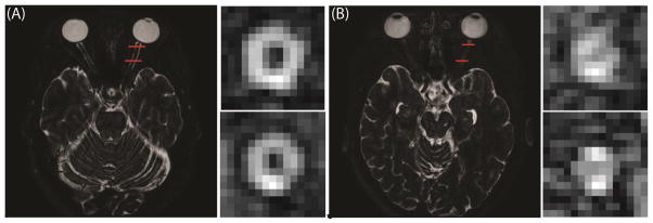
Optic neuritis is a sudden inflammation of the optic nerve (ON) and is marked by pain on eye movement, and visual symptoms such as a decrease in visual acuity, color vision, contrast and visual field defects. The ON is closely linked with multiple sclerosis (MS) and patients have a 50% chance of developing MS within 15 years. Recent advances in multi-atlas segmentation methods have omitted volumetric assessment. In the past, measuring the size of the ON has been done by hand. We utilize a new method of automatically segmenting the ON to measure the radii of both the ON and surrounding cerebrospinal fluid (CSF) sheath to develop a normative distribution of healthy young adults. We examine this distribution for any trends and find that ON and CSF sheath radii do not vary between 20–35 years of age and between sexes. We evaluate how six patients suffering from optic neuropathy compare to this distribution of controls. We find that of these six patients, five of them qualitatively differ from the normative distribution which suggests this technique could be used in the future to distinguish between optic neuritis patients and healthy controls.