Resources : Data
Standard Amygdalar fMRI Probe Tasks
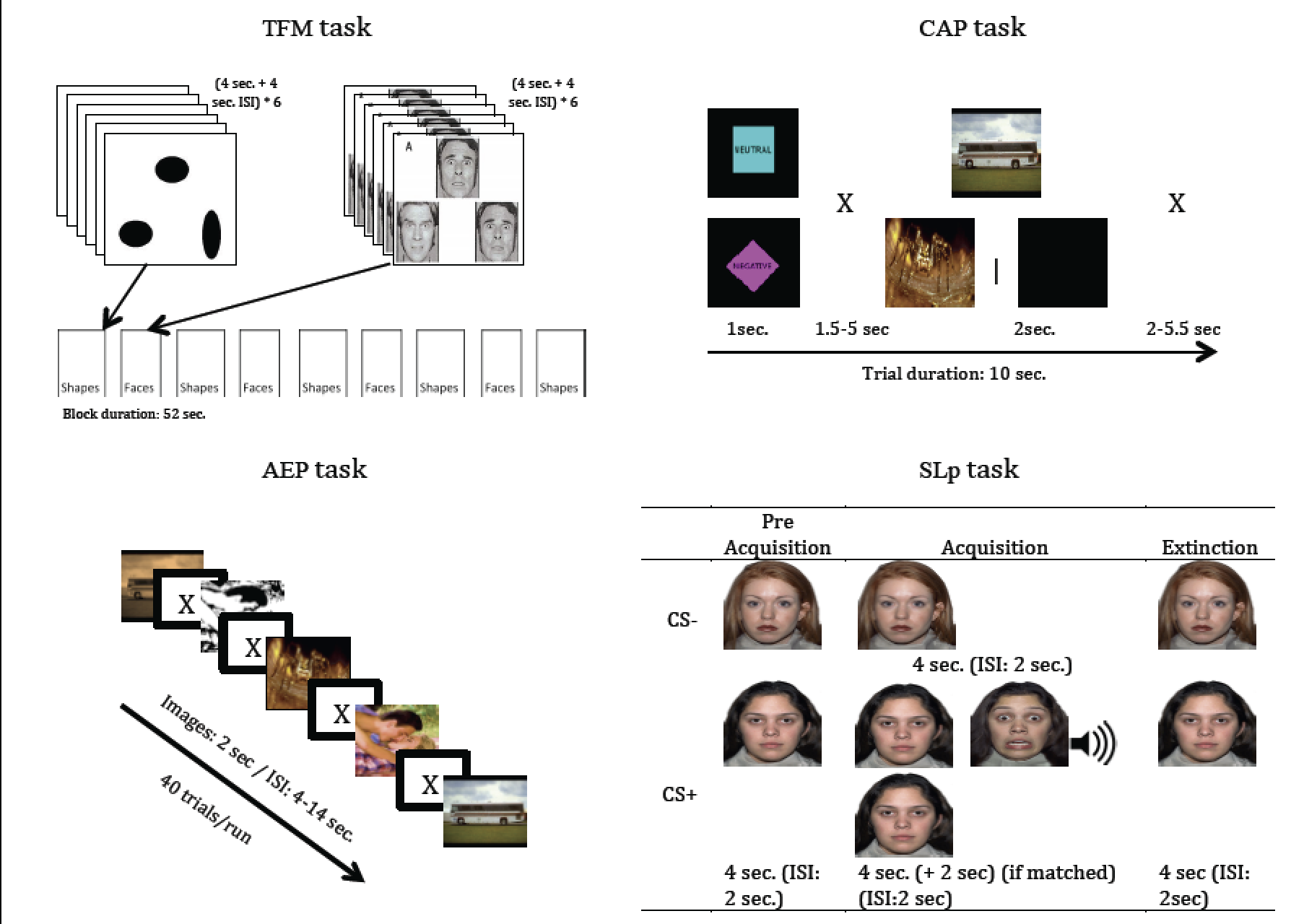
The amygdala (AMG) has been repeatedly implicated in the processing of threatening and negatively valenced stimuli and multiple fMRI paradigms have reported personality, genetic, and psychopathological associations with individual differences in AMG activation in these paradigms. Yet the interchangeability of activations in these probes has not been established, thus it remains unclear if we can interpret AMG responses on specific tasks as general markers of its reactivity. In this study we aimed to assess if different tasks that have been widely used within the Affective Neuroscience literature consistently recruit the AMG.
Method: Thirty-two young healthy subjects completed four fMRI tasks that have all been previously shown to probe the AMG during processing of threatening stimuli: the Threat Face Matching (TFM), the Cued Aversive Picture (CAP), the Aversive and Erotica Pictures (AEP) and the Screaming Lady paradigm (SLp) tasks. Contrasts testing response to aversive stimuli relative to baseline or neutral stimuli were generated and correlations between activations in the AMG were calculated across tasks were performed for ROIs of the AMG.
Total Intracranial Vault (TICV) BC2 Atlases
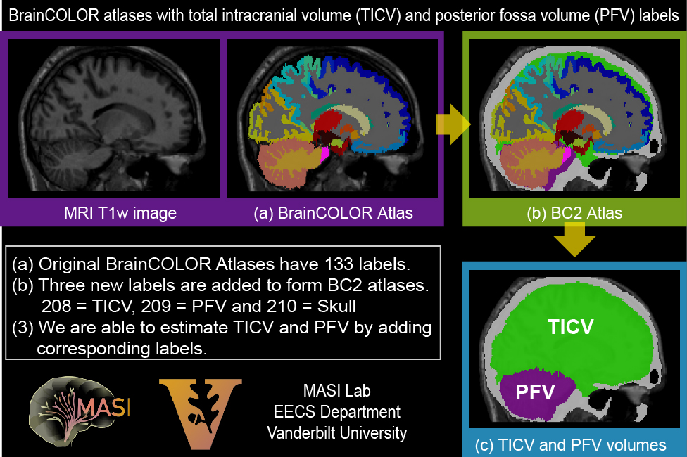 The data is released under the Creative Commons Attribution-NonCommercial (CC BY-NC) with no end date. Users should credit the MRI scans as originating from the OASIS project and the labeled data as “provided by Neuromorphometrics, Inc. (http://Neuromorphometrics.com/) under academic subscription”. These references should be included in all publications. The TICV_BC2 atlases consist of 27 OASIS images released by “MICCAI 2012 Grand Challenge and Workshop on Multi-Atlas Labeling”. The terms of using the TICV_BC2 atlases obey MICCAI 2012 Grand Challenge and Workshop on Multi-Atlas Labeling
Citation: Yuankai Huo, Andrew J. Asman, Andrew J. Plassard, Bennett A Landman “Simultaneous total intracranial volume and posterior fossa volume estimation using multi-atlas label fusion” Human Brain Mapping, 2016.
Multi-Modal MRI Reproducibility Resource
We have acquired scan-rescan imaging sessions on 21 healthy volunteers (no history of neurological disease). Imaging modalities include MPRAGE, FLAIR, DTI, resting state fMRI, B0 and B1 field maps, ASL, VASO, quantitative T1 mapping, quantitative T2 mapping, and magnetization transfer imaging. All data have been converted to NIFTI format.
This is intended to be a resource for statisticians and imaging scientists to be able to quantify the reproducibility of their imaging methods using data available from a generic “1 hour” session at 3T.
Please cite: Bennett. A. Landman, Alan J. Huang, Aliya Gifford, Deepti S. Vikram, Issel Anne L. Lim, Jonathan A.D. Farrell, John A. Bogovic, Jun Hua, Min Chen, Samson Jarso, Seth A. Smith, Suresh Joel, Susumu Mori, James J. Pekar, Peter B. Barker, Jerry L. Prince, and Peter C.M. van Zijl. “Multi-Parametric Neuroimaging Reproducibility: A 3T Resource Study”, NeuroImage. (2010) NIHMS/PMC:252138 doi:10.1016/j.neuroimage.2010.11.047
Rosetta Bit
 The Rosetta Bit project will house public datasets that have been transcoded into multiple formats. This library of valid file format conversions (DICOM->NIFTI, DICOM->PAR/REC, etc.) will provide a reference for tool developers seeking to support multiple sources of data.
Yvernault BC, Theobald CD Jr, Smith JC, Villalta V, Zald DH, Landman BA.
Neuroinformatics. 2014 Oct;12(4):615-7. doi: 10.1007/s12021-014-9230-9.
Validating DICOM Transcoding with an Open Multi-Format Resource.