Aravind R. Krishnan, Kaiwen Xu, Thomas Li, Lucas W. Remedios, Kim L. Sandler, Fabien Maldonado, Bennett A. Landman. “Lung CT harmonization of paired reconstruction kernel images using generative adversarial networks.”Med Phys. 2024;1-14.https://doi.org/10.1002/mp.17028
Abstract
Background
The kernel used in CT image reconstruction is an important factor that determines the texture of the CT image. Consistency of reconstruction kernel choice is important for quantitative CT-based assessment as kernel differences can lead to substantial shifts in measurements unrelated to underlying anatomical structures.
Purpose
In this study, we investigate kernel harmonization in a multi-vendor low-dose CT lung cancer screening cohort and evaluate our approach’s validity in quantitative CT-based assessments.
Methods
Using the National Lung Screening Trial, we identified CT scan pairs of the same sessions with one reconstructed from a soft tissue kernel and one from a hard kernel. In total, 1000 pairs of five different paired kernel types (200 each) were identified. We adopt the pix2pix architecture to train models for kernel conversion. Each model was trained on 100 pairs and evaluated on 100 withheld pairs. A total of 10 models were implemented. We evaluated the efficacy of kernel conversion based on image similarity metrics including root mean squared error (RMSE), peak signal-to-noise ratio (PSNR), and structural similarity index measure (SSIM) as well as the capability of the models to reduce measurement shifts in quantitative emphysema and body composition measurements. Additionally, we study the reproducibility of standard radiomic features for all kernel pairs before and after harmonization.
Results
Our approach effectively converts CT images from one kernel to another in all paired kernel types, as indicated by the reduction in RMSE (p < 0.05) and an increase in the PSNR (p < 0.05) and SSIM (p < 0.05) for both directions of conversion for all pair types. In addition, there is an increase in the agreement for percent emphysema, skeletal muscle area, and subcutaneous adipose tissue (SAT) area for both directions of conversion. Furthermore, radiomic features were reproducible when compared with the ground truth features.
Conclusions
Kernel conversion using deep learning reduces measurement variation in percent emphysema, muscle area, and SAT area.
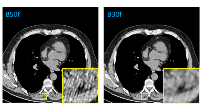
Figure 1. The reconstruction kernel determines the texture appearance of a CT image in a given vendor. In a Siemens vendor, B50f represents a hard kernel and B30f represents a soft kernel image. The difference in texture may lead to substantial differences in measurement during quantitative imaging.
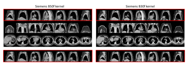
Figure 2. The reconstructions for the hard and soft kernel are obtained from the same projection data for a given subject. However, the scans may not be spatially aligned. For this purpose, the sampled pairs were visually inspected through a manual quality assurance process. The figure above highlights a case that was rejected during the manual inspection. The sagittal view indicates differences in the spatial extent of the reconstructions.
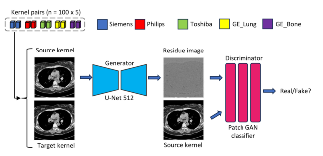
Figure 3. We perform kernel conversion on paired hard and soft CT kernel images using a pix2pix GAN. The generator, a U-Net, takes as input the source CT kernel image and predicts a residue image. The output kernel is obtained by adding the residue image to the input source kernel image. The discriminator, a PatchGAN, takes as input the generated image and the input kernel, and outputs the probability of the generated image being a translated version of the input.
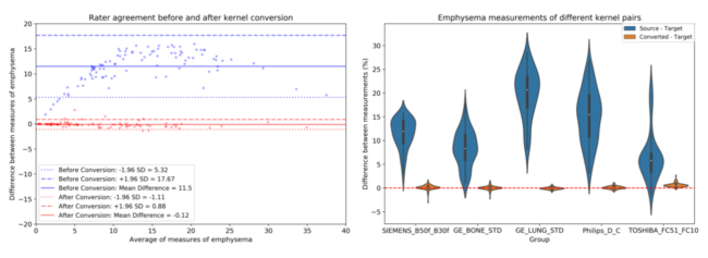
Figure 4. Left: Bland Altman plots are constructed to show agreement between kernels for emphysema measurements before and after conversion for the Siemens vendor. The dashed lines represent the confidence intervals while the solid lines represent the mean bias between the two kernels for a given measurement. Right: Distribution of percent differences before conversion showcases a lack of agreement across the different kernel pairs from different manufacturers. Prior to conversion, there is a disparity between the measurements. Kernel conversion reduces the difference in measurements, bringing the mean difference close to zero.
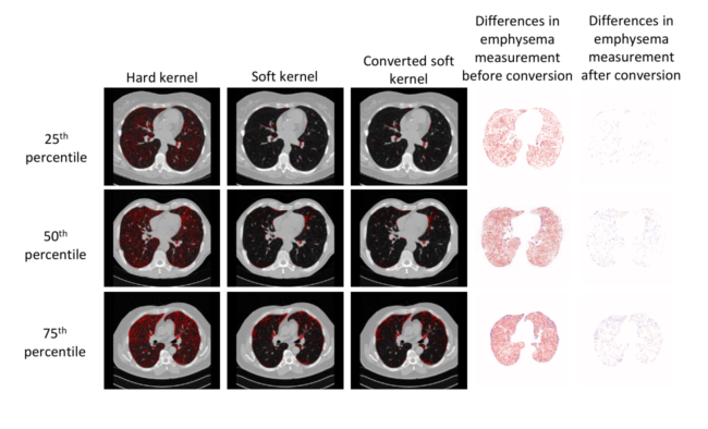
Figure 5. Emphysema measurements in CT images vary across subjects. Furthermore, the reconstruction kernel affects the emphysema measurement, creating a difference in measurements obtained. Soft kernels are favorable for emphysema measurement. The conversion from a hard to soft kernel minimizes the measurement errors, that can be observed with the help of emphysema masks obtained before and after conversion.
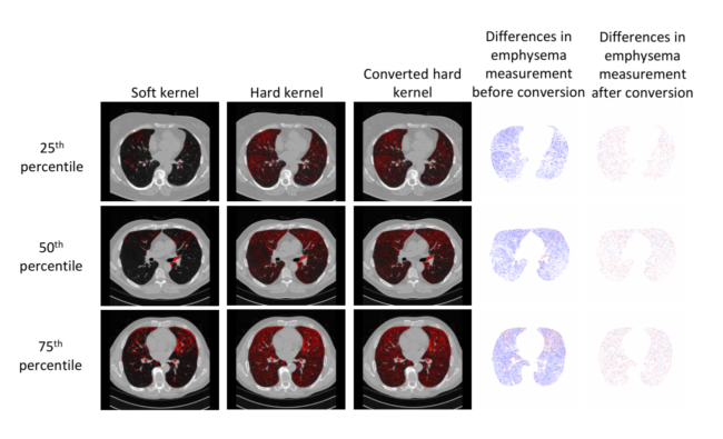
Figure 6. When soft kernels are converted to hard kernels, inconsistencies in the form of noise are introduced in the image. The converted hard kernel overestimates emphysema as expected. Differences in emphysema measurements before and after conversion are observed by taking the difference map between the emphysema measurements.
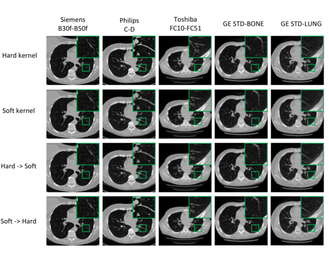
Figure 7: Noise introduced during computed tomography reconstruction leads to differences in textures of underlying anatomical structure. Harmonization between kernels within a given manufacturer enforces consistency in the texture of underlying anatomical structures that benefits quantitative image assessment.