“Resolution of Crossing Fibers with Constrained Compressed Sensing using Traditional Diffusion Tensor MRI”
A. Landman, H. Wan, J. Bogovic, P.-L. Bazin, and J. L. Prince. “Resolution of Crossing Fibers with Constrained Compressed Sensing using Traditional Diffusion Tensor MRI”, In Proceedings of the SPIE Medical Imaging Conference. San Diego, CA, February 2010 (Oral Presentation) PMC2854412
Abstract
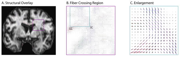
Diffusion tensor imaging (DTI) is widely used to characterize tissue micro-architecture and brain connectivity. Yet, DTI suffers serious limitations in regions of crossing fibers because traditional tensor techniques cannot represent multiple, independent intra-voxel orientations. Compressed sensing has been proposed to resolve crossing fibers using a tensor mixture model (e.g., Crossing Fiber Angular Resolution of Intra-voxel structure, CFARI). Although similar in spirit to deconvolution approaches, CFARI uses sparsity to stabilize estimation with limited data rather than spatial consistency or limited model order. Here, we extend the CFARI approach to resolve crossing fibers through a strictly positive, parsimonious mixture model. Together with an optimized preconditioned conjugate gradient solver, estimation error and computational burden are greatly reduced over the initial presentation. Reliable estimates of intra-voxel orientations are demonstrated in simulation and in vivo using data representative of typical, low b-value (30 directions, 700 s/mm(2)) clinical DTI protocols. These sequences are achievable in 5 minutes at 3 T, and the whole brain CFARI analysis is tractable for routine analysis. With these improvements, CFARI provides a robust framework for identifying intra-voxel structure with traditional DTI and shows great promise in helping to resolve the crossing fiber problem in current clinical imaging studies.