Resolution of Crossing Fibers with Constrained Compressed Sensing using Diffusion Tensor MRI
Bennett A. Landman, John A Bogovic; Hanlin Wan; Fatma El Zahraa ElShahaby; Pierre-Louis Bazin, and Jerry L Prince. “Resolution of Crossing Fibers with Constrained Compressed Sensing using Diffusion Tensor MRI”, NeuroImage. 2012 Feb 1;59(3):2175-86. PMC22019877
Full text: https://www.ncbi.nlm.nih.gov/pubmed/22019877
Abstract
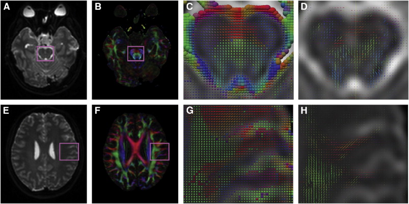
Diffusion tensor imaging (DTI) is widely used to characterize tissue micro-architecture and brain connectivity. In regions of crossing fibers, however, the tensor model fails because it cannot represent multiple, independent intra-voxel orientations. Most of the methods that have been proposed to resolve this problem require diffusion magnetic resonance imaging (MRI) data that comprise large numbers of angles and high b-values, making them problematic for routine clinical imaging and many scientific studies. We present a technique based on compressed sensing that can resolve crossing fibers using diffusion MRI data that can be rapidly and routinely acquired in the clinic (30 directions, b-value equal to 700 s/mm2). The method assumes that the observed data can be well fit using a sparse linear combination of tensors taken from a fixed collection of possible tensors each having a different orientation. A fast algorithm for computing the best orientations based on a hierarchical compressed sensing algorithm and a novel metric for comparing estimated orientations are also proposed. The performance of this approach is demonstrated using both simulations and in vivo images. The method is observed to resolve crossing fibers using conventional data as well as a standard q-ball approach using much richer data that requires considerably more image acquisition time.
