A brain MRI atlas of the common squirrel monkey, Saimiri sciureus
Yurui Gao, Shweta P. Khare, Swetasudha Panda, Ann S Choe, Iwona Stepniewska, Xia Li, Zhoahua Ding, Adam Anderson, Bennett A. Landman, “A brain MRI atlas of the common squirrel monkey, Saimiri sciureus.” In Proceedings of the SPIE Medical Imaging Conference. San Diego, California, February 2014. Oral Presentation. †
Full Text: https://www.ncbi.nlm.nih.gov/pubmed/24817811
Abstract
The common squirrel monkey, Saimiri sciureus, is a New World monkey with functional and microstructural organization of central nervous system similar to that of humans. It is one of the most commonly used South American primates in biomedical research. Unlike its Old World macaque cousins, no digital atlases have described the organization of the squirrel monkey brain. Here, we present a multi-modal magnetic resonance imaging (MRI) atlas constructed from the brain of an adult female squirrel monkey. In vivo MRI acquisitions include T2 structural imaging and diffusion tensor imaging. Ex vivo MRI acquisitions include T2 structural imaging and diffusion tensor imaging. Cortical regions were manually annotated on the co-registered volumes based on published histological sections.
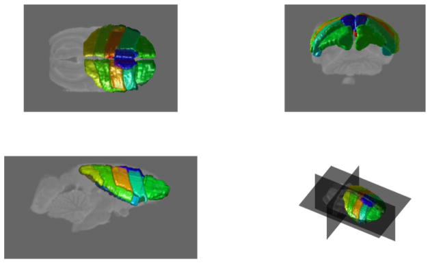
Rendering of the manually labeled cortical regions ().