Disambiguating the Optic Nerve from the Surrounding Cerebrospinal Fluid: Application to MS-related Atrophy
Robert L. Harrigan, Andrew J. Plassard, Frederick W. Bryan, Gabriela Caires, Louise A. Mawn, Lindsey M. Dethrage, Siddharama Pawate, Robert L. Galloway, Seth A. Smith, Bennett A. Landman. “Disambiguating the Optic Nerve from the Surrounding Cerebrospinal Fluid: Application to MS-related Atrophy.” Magnetic Resonance in Medicine. In press 2014.”
Full Text: https://www.ncbi.nlm.nih.gov/pubmed/25754412
Abstract
PURPOSE:
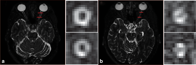
Our goal is to develop an accurate, automated tool to characterize the optic nerve (ON) and cerebrospinal fluid (CSF) to better understand ON changes in disease.
METHODS:
Multi-atlas segmentation is used to localize the ON and sheath on T2-weighted MRI (0.6 mm(3) resolution). A sum of Gaussian distributions is fit to coronal slice-wise intensities to extract six descriptive parameters, and a regression forest is used to map the model space to radii. The model is validated for consistency using tenfold cross-validation and for accuracy using a high resolution (0.4 mm(2) reconstructed to 0.15 mm(2)) in vivo sequence. We evaluated this model on 6 controls and 6 patients with multiple sclerosis (MS) and a history of optic neuritis.
RESULTS:
In simulation, the model was found to have an explanatory R-squared for both ON and sheath radii greater than 0.95. The accuracy of the method was within the measurement error on the highest possible in vivo resolution. Comparing healthy controls and patients with MS, significant structural differences were found near the ON head and the chiasm, and structural trends agreed with the literature.
CONCLUSION:
This is a first demonstration that the ON can be exclusively, quantitatively measured and separated from the surrounding CSF using MRI.