Improving Cerebellar Segmentation with Statistical Fusion
Andrew J. Plassard, Zhen Yang, Swati D. Rane, Jerry L. Prince, Daniel O. Claassen, Bennett A. Landman. “Improving Cerebellar Segmentation with Statistical Fusion. In Proceedings of the SPIE Medical Imaging Conference. San Diego, California, February 2016.
Full Text: https://www.ncbi.nlm.nih.gov/pubmed/?term=Improving+Cerebellar+Segmentation+with+Statistical+Fusion
Abstract
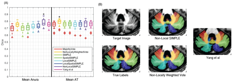
The cerebellum is a somatotopically organized central component of the central nervous system well known to be involved with motor coordination and increasingly recognized roles in cognition and planning. Recent work in multi-atlas labeling has created methods that offer the potential for fully automated 3-D parcellation of the cerebellar lobules and vermis (which are organizationally equivalent to cortical gray matter areas). This work explores the trade offs of using different statistical fusion techniques and post hoc optimizations in two datasets with distinct imaging protocols. We offer a novel fusion technique by extending the ideas of the Selective and Iterative Method for Performance Level Estimation (SIMPLE) to a patch-based performance model. We demonstrate the effectiveness of our algorithm, Non-Local SIMPLE, for segmentation of a mixed population of healthy subjects and patients with severe cerebellar anatomy. Under the first imaging protocol, we show that Non-Local SIMPLE outperforms previous gold-standard segmentation techniques. In the second imaging protocol, we show that Non-Local SIMPLE outperforms previous gold standard techniques but is outperformed by a non-locally weighted vote with the deeper population of atlases available. This work advances the state of the art in open source cerebellar segmentation algorithms and offers the opportunity for routinely including cerebellar segmentation in magnetic resonance imaging studies that acquire whole brain T1-weighted volumes with approximately 1 mm isotropic resolution.