
Peripheral sphingolipids are associated with variation in white matter microstructure in older adults.

Posted by on Sunday, July 31, 2016 in Diffusion Weighted MRI, Neuroimaging, News.

Christopher E. Gonzalez, Vijay K. Venkatraman, Yang An, Bennett A. Landman, Christos Davatzikos, Veera Venkata Ratnam Bandaru, Norman J. Haughey, Luigi Ferruci, Michelle M. Mielke, Susan M. Resnick. “Peripheral sphingolipids are associated with variation in white matter microstructure in older adults.” Neurobiology of Aging. July 2016. Volume 43, Pages 156–163

Full Text: https://www.ncbi.nlm.nih.gov/pubmed/27255825

Abstract

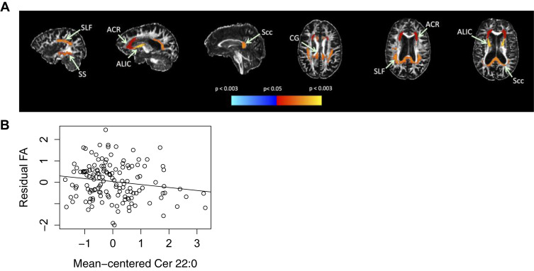
Sphingolipids serve important structural and functional roles in cellular membranes and myelin sheaths. Plasma sphingolipids have been shown to predict cognitive decline and Alzheimer’s disease. However, the association between plasma sphingolipid levels and brain white matter (WM) microstructure has not been examined. We investigated whether plasma sphingolipids (ceramides and sphingomyelins) were associated with magnetic resonance imaging-based diffusion measures, fractional anisotropy (FA), and mean diffusivity, 10.5 years later in 17 WM regions of 150 cognitively normal adults (mean age 67.2). Elevated ceramide species (C20:0, C22:0, C22:1, and C24:1) were associated with lower FA in multiple WM regions, including total cerebral WM, anterior corona radiata, and the cingulum of the cingulate gyrus. Higher sphingomyelins (C18:1 and C20:1) were associated with lower FA in regions such as the anterior corona radiata and body of the corpus callosum. Furthermore, lower sphingomyelin to ceramide ratios (C22:0, C24:0, and C24:1) were associated with lower FA or higher mean diffusivity in regions including the superior and posterior corona radiata. However, although these associations were significant at the a priori p < 0.05, only associations with some regional diffusion measures for ceramide C22:0 and sphingomyelin C18:1 survived correction for multiple comparisons. These findings suggest plasma sphingolipids are associated with variation in WM microstructure in cognitively normal aging.

(A) Significant associations with plasma C22:0 and subsequent FA. (A) Red–yellow indicates higher plasma C22:0 associated with lower FA in white matter regions, while cyan–blue indicates higher plasma C22:0 associated with higher FA. Regional statistics are overlaid onto an FA image from a single subject; results are not corrected for multiple comparisons. (B) ACR FA by mean centered Cer 22:0. (B) Association between FA in ACR and Cer 22:0. The residual FA of ACR was calculated by regressing standardized FA on baseline age, sex, interval between blood draw and DTI, diabetes, BMI, scanner, and ratio of white matter lesions to intracranial volume. This is shown plotted against mean centered Cer 22:0 with a linear fit overlaid. Abbreviations: ACR, anterior corona radiata; ALIC, anterior limb of internal capsule; BMI, body mass index; CG, cingulum of cingulate gyrus; DTI, diffusion tensor imaging; FA, fractional anisotropy; Scc, splenium of corpus callosum; SLF, superior longitudinal fasciculus; SS, sagittal stratum. (For interpretation of the references to color in this figure legend, the reader is referred to the Web version of this article.)

Tags: , , ,