Robust Multi-contrast MRI Spleen Segmentation for Splenomegaly using Multi-atlas Segmentation
Yuankai Huo, Jiaqi Liu, Zhoubing Xu, Robert L. Harrigan, Albert Assad, Richard G. Abramson, and Bennett A. Landman. “Robust Multi-contrast MRI Spleen Segmentation for Splenomegaly using Multi-atlas Segmentation.” IEEE Transactions on Biomedical Engineering (2017).
Full text: onlinelibrary.wiley.com/doi/10.1002/hbm.23432/full
Abstract
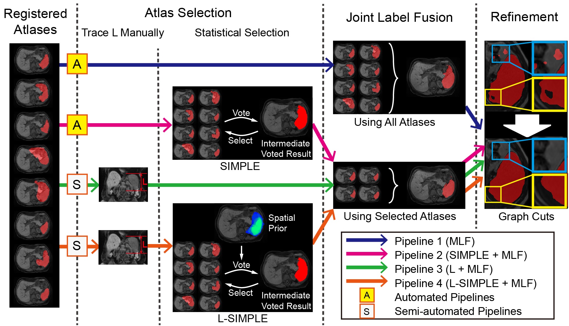
Objective: Magnetic resonance imaging (MRI) is an essential imaging modality in non-invasive splenomegaly diagnosis. However, it is challenging to achieve spleen volume measurement from 3D MRI given the diverse structural variations of human abdomens as well as the wide variety of clinical MRI acquisition schemes. Multi-atlas segmentation (MAS) approaches have been widely used and validated to handle heterogeneous anatomical scenarios. In this paper, we propose to use MAS for clinical MRI spleen segmentation for splenomegaly. Methods: First, an automated segmentation method using the selective and iterative method for performance level estimation (SIMPLE) atlas selection is used to address the concerns of inhomogeneity for clinical splenomegaly MRI. Then, to further control outliers, semi-automated craniocaudal spleen length-based SIMPLE atlas selection (L-SIMPLE) is proposed to integrate a spatial prior in a Bayesian fashion and guide iterative atlas selection. Last, a graph cuts refinement is employed to achieve the final segmentation from the probability maps from MAS. Results: A clinical cohort of 55 MRI volumes (28 T1 weighted and 27 T2 weighted) were used to evaluate both automated and semi-automated methods. Conclusion: The results demonstrated that (1) both methods achieved median Dice > 0.9, (2) outliers were alleviated by the L-SIMPLE (≈1 min manual efforts per scan), which achieved 0.97 Pearson correlation of volume measurements with the manual segmentation. Significance: This work performed spleen segmentation on MRI splenomegaly using MAS.