Electronic Medical Record Context Signatures Improve Diagnostic Classification using Medical Image Computing
Shikha Chaganti, Louise A. Mawn, Hakmook Kang, Josephine Egan, Susan M. Resnick, Lori L. Beason-Held, Bennett A. Landman, Thomas A. Lasko. “Electronic Medical Record Context Signatures Improve Diagnostic Classification using Medical Image Computing”. IEEE Journal of Biomedical and Health Informatics. (In Press)
Abstract
Composite models that combine medical imaging with electronic medical records (EMR) improve predictive power when compared to traditional models that use imaging alone. The digitization of EMR provides potential access to a wealth of medical information, but presents new challenges in algorithm design and inference. Previous studies, such as PheWAS (Phenome Wide Association Study), have shown that EMR data can be used to investigate the relationship between genotypes and clinical conditions. Here, we introduce PheDAS (Phenome-Disease Association Study) to extend the statistical capabilities of the PheWAS software through a custom Python package which creates diagnostic EMR signatures to capture system-wide co-morbidities for a disease population within a given time interval. We investigate the effect of integrating these EMR signatures with radiological data to improve diagnostic classification in disease domains known to have confounding factors because of variable and complex clinical presentation. Specifically, we focus on two studies: (1) a study of four major optic nerve related conditions and (2) a study of diabetes. Addition of EMR signature vectors to radiologically-derived structural metrics improves the area under the curve (AUC) for diagnostic classification using elastic net regression, for diseases of the optic nerve. For glaucoma, the AUC improves from 0.71 to 0.83, for intrinsic optic nerve disease it increases from 0.72 to 0.91, for optic nerve edema it increases from 0.95 to 0.96, and for thyroid eye disease from 0.79 to 0.89. The EMR signatures recapitulate known comorbidities with diabetes, such as abnormal glucose but do not significantly modulate image-derived features. In summary, EMR signatures present a scalable and readily applicable.
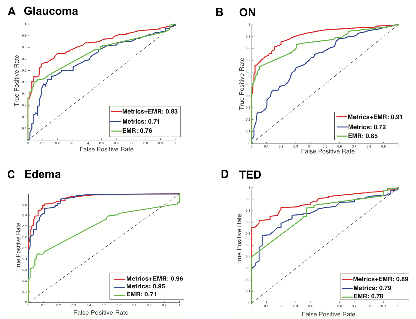
Figure. Disease vs. healthy control results of elastic net classifier for study. Green line indicates the curve for EMR data, blue line indicates the curve for imaging data, and the red line indicates the curve for EMR + imaging data.1. 5A. shows the result for glaucoma, 5B shows the result for intrinsic optic nerve disease, 5C shows the result for optic nerve edema, and 5D shows the result for thyroid eye disease.