Validation and Optimization of Multi-Organ Segmentation on Clinical Imaging Archives
Olivia Tang, Yuchen Xu, Yucheng Tang, Ho Hin Lee, Yunqiang Chen, Dashan Gao, Shizhong Han, Riqiang Gao, Michael R. Savona, Richard G. Abramson, Yuankai Huo, Bennett A. Landman, “Validation and Optimization of Multi-Organ Segmentation on Clinical Imaging Archives”, SPIE IP:MI 2020. Houston, TX.
https://arxiv.org/abs/2002.04102
Abstract
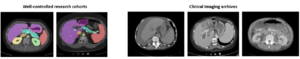
Segmentation of abdominal computed tomography (CT) provides spatial context, morphological properties, and a framework for tissue-specific radiomics to guide quantitative Radiological assessment. A 2015 MICCAI challenge spurred substantial innovation in multi-organ abdominal CT segmentation with both traditional and deep learning methods. Recent innovations in deep methods have driven performance toward levels for which clinical translation is appealing. However, continued cross-validation on open datasets presents the risk of indirect knowledge contamination and could result in circular reasoning. Moreover, “real world” segmentations can be challenging due to the wide variability of abdomen physiology within patients. Herein, we perform two data retrievals to capture clinically acquired deidentified abdominal CT cohorts with respect to a recently published variation on 3D U-Net (baseline algorithm). First, we retrieved 2004 deidentified studies on 476 patients with diagnosis codes involving spleen abnormalities (cohort A). Second, we retrieved 4313 deidentified studies on 1754 patients without diagnosis codes involving spleen abnormalities (cohort B). We perform prospective evaluation of the existing algorithm on both cohorts, yielding 13% and 8% failure rate, respectively. Then, we identified 51 subjects in cohort A with segmentation failures and manually corrected the liver and gallbladder labels. We re-trained the model adding the manual labels, resulting in performance improvement of 9% and 6% failure rate for the A and B cohorts, respectively. In summary, the performance of the baseline on the prospective cohorts was similar to that on previously published datasets. Moreover, adding data from the first cohort substantively improved performance when evaluated on the second withheld validation cohort.