Joint analysis of structural connectivity and cortical surface features: correlates with mild traumatic brain injury
Cailey I. Kerley, Leon Y. Cai, Chang Yu, Logan M. Crawford, Jason M. Elenberger, Eden S. Singh, Kurt G. Schilling, Katherine S. Aboud, Bennett A. Landman, Tonia S. Rex, “Joint analysis of connectivity and cortical surface features: correlates with mild traumatic brain injury.” (2021, Feb) SPIE Medical Imaging. San Diego, CA. (Accepted)
Full text: NIHMSID, arXiv
Abstract
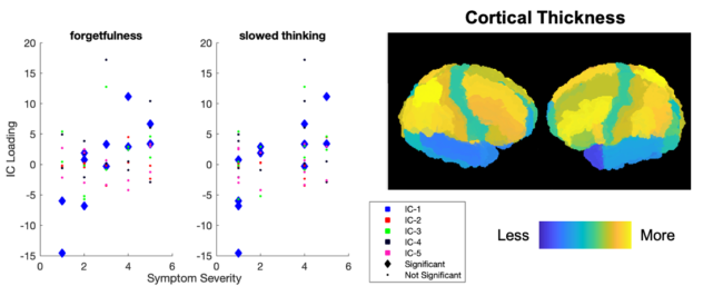
Mild traumatic brain injury (mTBI) is a complex syndrome that affects up to 600 per 100,000 individuals, with a particular concentration among military personnel. About half of all mTBI patients experience a diverse array of chronic symptoms which persist long after the acute injury. Hence, there is an urgent need for better understanding of the white matter and gray matter pathologies associated with mTBI to map which specific brain systems are impacted and identify courses of intervention. Previous works have linked mTBI to disruptions in white matter pathways and cortical surface abnormalities. Herein, we examine these hypothesized links in an exploratory study of joint structural connectivity and cortical surface changes associated with mTBI and its chronic symptoms. Briefly, we consider a cohort of 12 mTBI and 26 control subjects. A set of 588 cortical surface metrics and 4,753 structural connectivity metrics were extracted from cortical surface regions and diffusion weighted magnetic resonance imaging in each subject. Principal component analysis (PCA) was used to reduce the dimensionality of each metric set. We then applied independent component analysis (ICA) both to each PCA space individually and together in a joint ICA approach. We identified a stable independent component across the connectivity-only and joint ICAs which presented significant group differences in subject loadings (p<0.05, corrected). Additionally, we found that two mTBI symptoms, slowed thinking and forgetfulness, were significantly correlated (p<0.05, corrected) with mTBI subject loadings in a surface-only ICA. These surface-only loadings captured an increase in bilateral cortical thickness.
Keywords: mild traumatic brain injury, multi-modal MRI, independent component analysis