Construction of a Multi-Phase Contrast Computed Tomography Kidney Atlas
Ho Hin Lee, Yucheng Tang, Kaiwen Xu, Shunxing Bao, Agnes B. Fogo, Raymond Harris, Mark P. de Caestecker, Mattias Heinrich, Jeffrey Spraggins, Yuankai Huo, Bennett A, Landman, Construction of a Multi-Phase Contrast Computed Tomography Kidney Atlas, SPIE 2021 Medical Imaging
Full Text
Abstract
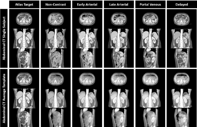
The Human BioMolecular Atlas Program (HuBMAP) seeks to create a molecular atlas at the cellular level of the human body to spur interdisciplinary innovations across spatial and temporal scales. While the preponderance of effort is allocated towards cellular and molecular scale mapping, differentiating and contextualizing findings within tissues, organs and systems are essential for the HuBMAP efforts. The kidney is an initial organ target of HuBMAP, and constructing a framework (or atlas) for integrating information across scales is needed for visualizing and integrating information. However, there is no abdominal atlas currently available in the public domain. Substantial variation in healthy kidneys exists with sex, body size, and imaging protocols. With the integration of clinical archives for secondary research use, we are able to build atlases based on a diverse population and clinically relevant protocols. In this study, we created a computed tomography (CT) phase-specific atlas for the abdomen, which is optimized for the kidney organ. A two-stage registration pipeline was used by registering extracted abdominal volume of interest from body part regression, to a high-resolution CT. Affine and non-rigid registration were performed to all scans hierarchically. To generate and evaluate the atlas, multi-phase CT scans of 500 control subjects (age: 15 – 50, 250 males, 250 females) are registered to the atlas target through the complete pipeline. The abdominal body and kidney registration are shown to be stable with the variance map computed from the result average template. Both left and right kidneys are substantially localized in the high-resolution target space, which successfully demonstrated the sharp details of its anatomical characteristics across each phase. We illustrated the applicability of the atlas template for integrating across normal kidney variation from 64 cc to 302 cc.