Establishing Surface Correspondence for Post-surgical Cortical Thickness Changes in Temporal Lobe Epilepsy
Yue Liu, Dario J. Englot, Victoria L. Morgan, Warren D. Taylor, Ying Wei, Ipek Oguz, Bennett A. Landman, Ilwoo Lyu. Establishing Surface Correspondence for Post-surgical Cortical Thickness Changes in Temporal Lobe Epilepsy. SPIE Medical Imaging, 2021.
Full Text
Abstract
In pre- and post-surgical surface shape analysis, establishing shape correspondence is necessary to investigate the post-operative surface changes. However, structural absence after the operation accompanies focal non-rigid changes, which leads to challenges in existing surface registration methods. In this paper, we present a fully automatic particle-based method to establish surface correspondence that can handle partial structural abnormality in the temporal lobe resection. Our method optimizes the coordinates of points which are modeled as particles on surfaces in a hierarchical way to reduce a chance of being trapped in a local minimum during the optimization. In the experiments, we evaluate the effectiveness of our method in comparison with conventional spherical registration (FreeSurfer) on two scenarios: cortical thickness changes in healthy controls within a short scan-rescan time window and patients with temporal lobe resection. The post-surgical scan is acquired at least 1 year after the presurgical scan. In region of interest-wise (ROI-wise) analysis, no changes on cortical thickness are found in both methods on the healthy control group. In patients, since there is no ground truth available, we instead investigated the disagreement between our method and FreeSurfer. We see poorly matched ROIs and large cortical thickness changes using FreeSurfer. On the contrary, our method shows well-matched ROIs and subtle cortical thickness changes. This suggests that the proposed method can establish a stable shape correspondence, which is not fully captured in a conventional spherical registration.
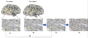
Figure 2. A schematic overview of the proposed method. (a) shows the fixed particles on pre-surgical surface. (b) shows the initial particles on post-surgical surfaces. (c) shows the updated particles on post-surgical surfaces after optimization. (d) shows the established correspondence between two particles. The particles with the same index have same locations on pre- and post-surgical surface. (e) shows the icosahedral subdivision to get a better initial guess of higher levels of optimization