Quantifying emphysema in lung screening computed tomography with robust automated lobe segmentation
Thomas Z. Li, Ho Hin Lee, Kaiwen Xu, Riqiang Gao, Benoit M. Dawant, Fabien Maldonado, Kim L. Sandler, Bennett A. Landman. Journal of Medical Imaging 10(4), 044002 (2023), doi: 10.1117/1.JMI.10.4.044002.
Abstract
Introduction: Anatomy-based quantification of emphysema in a lung screening cohort has the potential to improve lung cancer risk stratification and risk communication. Segmenting lung lobes is an essential step in this analysis, but leading lobe segmentation algorithms have not been validated for lung screening computed tomography (CT).
Methods: In this work, we develop an automated approach to lobar emphysema quantification and study their association with lung cancer incidence. We combine self-supervised training with level-set regularization and finetuning with radiologist annotations on three datasets to develop a lobe segmentation algorithm that is robust for lung screening CT. Using this algorithm, we extract quantitative CT measures for a cohort (n=1189) from the National Lung Screening Trial and analyze the multivariate association with lung cancer incidence.
Results: Our lobe segmentation approach achieved an external validation dice of 0.93, significantly outperforming a leading algorithm at 0.90 (p<0.01). Percentage of low attenuation volume in the right upper lobe was associated with increased lung cancer incidence (Odds ratio: 1.97; 95% CI: [1.06, 3.66]) independent of PLCOm2012 risk factors and diagnosis of whole lung emphysema. Quantitative lobar emphysema improved the goodness-of-fit to lung cancer incidence (x2=7.48, p=0.02).
Conclusion: We are the first to develop and validate automated lobe segmentation algorithm that is robust to smoking related pathology. We discover a novel quantitative risk factor, lending further evidence that regional emphysema is independently associated increased lung cancer incidence. The algorithm is provided at https://github.com/MASILab/EmphysemaSeg.
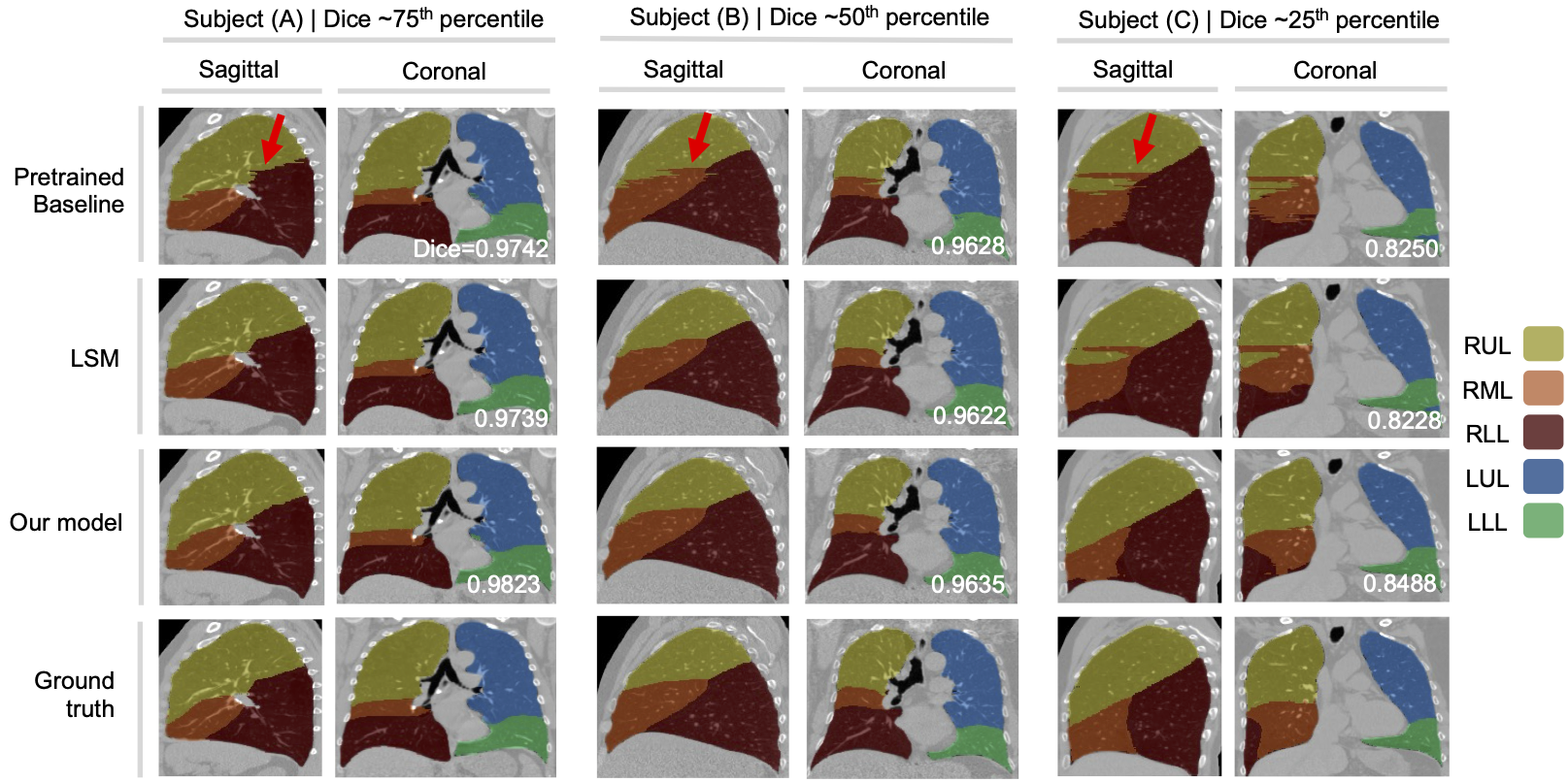
Lobe segmentations and ground truth annotations for three subjects using three methods: a pretrained baseline model, the baseline model post-processed with the LSM, and our proposed model. Segmentations of subject A, B, and C come from the 75’th, 50’th, and 25’th percentiles of our proposed model’s Dice performance. The LSM resolves small-scale border artifacts and achieves smoothly contoured fissures, but large-scale defects such as those seen in subject C’s RML remain unresolved. Our proposed method effectively resolves large-scale defects and segments the RML more accurately than the baseline method.