Reproducibility and Variation of Diffusion Measures in the Squirrel Monkey Brain, In Vivo and Ex Vivo
Kurt Schilling, Yurui Gao, Iwona Stepniewska, Ann S. Choe, Bennett A. Landman, and Adam W Anderson. “Reproducibility and Variation of Diffusion Measures in the Squirrel Monkey Brain, In Vivo and Ex Vivo”. Magnetic Resonance Imaging. In press August 2015
Full text: https://www.ncbi.nlm.nih.gov/pubmed/27587226
Abstract
Purpose: Animal models are needed to better understand the relationship between diffusion MRI (dMRI) and the underlying tissue microstructure. One promising model for validation studies is the common squirrel monkey, Saimiri sciureus. This study aims to determine (1) the reproducibility of in vivo diffusion measures both within and between subjects; (2) the agreement between in vivo and ex vivo data acquired from the same specimen and (3) normal diffusion values and their variation across brain regions.
Methods: Data were acquired from three healthy squirrel monkeys, each imaged twice in vivo and once ex vivo. Reproducibility of fractional anisotropy (FA), mean diffusivity (MD), and principal eigenvector (PEV) was assessed, and normal values were determined both in vivo and ex vivo.
Results: The calculated coefficients of variation (CVs) for both intra-subject and inter-subject MD were below 10% (low variability) while FA had a wider range of CVs, 2-14% intra-subject (moderate variability), and 3-31% inter-subject (high variability). MD in ex vivo tissue was lower than in vivo (30%-50% decrease), while FA values increased in all regions (30-39% increase). The mode of angular differences between in vivo and ex vivo PEVs was 12 degrees.
Conclusion: This study characterizes the diffusion properties of the squirrel monkey brain and serves as the groundwork for using the squirrel monkey, both in vivo and ex vivo, as a model for diffusion MRI studies.
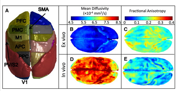
Fig. 6.Diffusion measures across the in vivo and ex vivo squirrel monkey cortex, averaged across animals. 3D rendering of gray matter ROIs (A), and voxel-wise renderings of MD (B,D) and FA (C,E) across the cortex.