Author
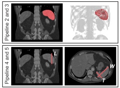
Improving Spleen Volume Estimation via Computer Assisted Segmentation on Clinically Acquired CT Scans
Oct. 31, 2016—Zhoubing Xu, Adam L. Gertz, Ryan P. Burke, Neil K. Bansal, Hakmook Kang, Bennett A. Landman, Richard G. Abramson. Improving Spleen Volume Estimation via Computer Assisted Segmentation on Clinically Acquired CT Scans. Acad Radiol. 2016 Oct;23(10):1214-20. PMC5026951 Full text: https://www.ncbi.nlm.nih.gov/pubmed/27519156 Abstract Objectives: Multi-atlas fusion is a promising approach for computer-assisted segmentation of anatomic structures. The...
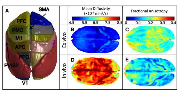
Reproducibility and Variation of Diffusion Measures in the Squirrel Monkey Brain, In Vivo and Ex Vivo
Aug. 31, 2016—Kurt Schilling, Yurui Gao, Iwona Stepniewska, Ann S. Choe, Bennett A. Landman, and Adam W Anderson. “Reproducibility and Variation of Diffusion Measures in the Squirrel Monkey Brain, In Vivo and Ex Vivo”. Magnetic Resonance Imaging. In press August 2015 Full text: https://www.ncbi.nlm.nih.gov/pubmed/27587226 Abstract Purpose: Animal models are needed to better understand the relationship between diffusion...
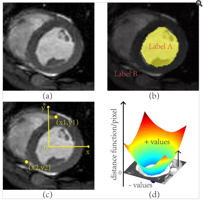
Investigation of Bias in Continuous Medical Image Label Fusion
Jun. 30, 2016—Fangxu Xing; Jerry Prince; Bennett Landman. Investigation of Bias in Continuous Medical Image Label Fusion. PLoS One. 2016 Jun 3;11(6):e0155862. PMC4892597 Full text: https://www.ncbi.nlm.nih.gov/pubmed/27258158 Abstract Image labeling is essential for analyzing morphometric features in medical imaging data. Labels can be obtained by either human interaction or automated segmentation algorithms, both of which suffer from errors....
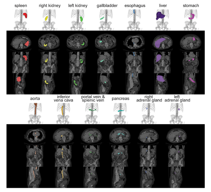
Evaluation of Five Registration Methods for the Human Abdomen on Clinically Acquired CT
Jun. 30, 2016—Zhoubing Xu, Christopher P. Lee, Marc Modat, Daniel Rueckert, Sebastien Ourselin, Richard G. Abramson, Bennett A. Landman. “Evaluation of Five Registration Methods for the Human Abdomen on Clinically Acquired CT”. IEEE Transactions of Biomedical Engineering. 2016 Aug;63(8):1563-72. PMC4972188 Full text: https://www.ncbi.nlm.nih.gov/pubmed/27254856 Abstract Objective: This work evaluates current 3-D image registration tools on clinically acquired abdominal...
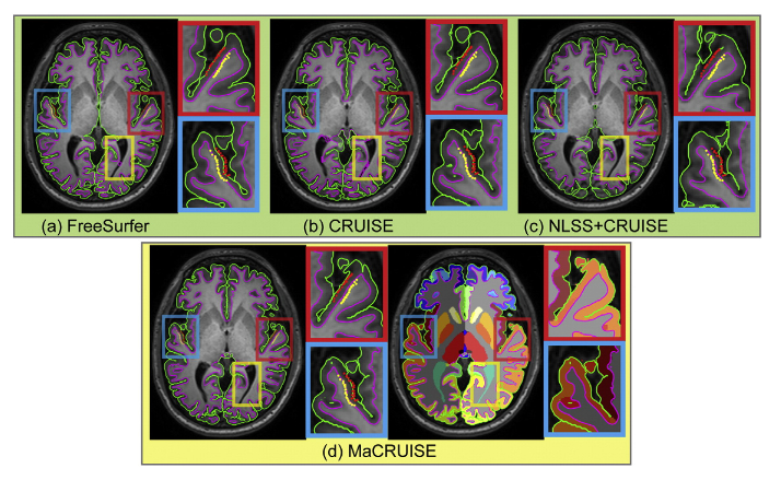
Consistent Cortical Reconstruction and Multi-atlas Brain Segmentation
Apr. 30, 2016—Yuankai Huo, Aaron Carass, Susan M. Resnick, Dzung L. Pham, Jerry L. Prince, Bennett A. Landman. “Consistent Cortical Reconstruction and Multi-atlas Brain Segmentation”. NeuroImage. Volume 138, September 2016, Pages 197–210 PMC4927397 Full text: https://www.ncbi.nlm.nih.gov/pubmed/27184203 Abstract Whole brain segmentation and cortical surface reconstruction are two essential techniques for investigating the human brain. Spatial inconsistences, which can...
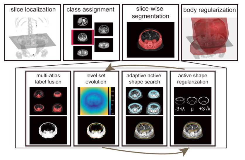
Abdomen and spinal cord segmentation with augmented active shape models
Feb. 27, 2016—Xu, Zhoubing; Benjamin Conrad; Rebeccah Baucom; Seth Smith; Benjamin Poulose; Landman, Bennett. “Abdomen and spinal cord segmentation with augmented active shape models.” Journal of Medical Imaging. 3(3) 036002 Full text: https://spie.org/Publications/Journal/10.1117/1.JMI.3.3.036002 Abstract The abdominal wall is an important structure differentiating subcutaneous and visceral compartments and intimately involved with maintaining abdominal structure. Segmentation of the whole...
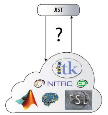
Integration of the Java Image Science Toolkit with E-Science Platform
Jan. 31, 2016—S. Damon, S. Panjwani, S. Bao, P. Kochunov, B. Landman, Integration of the Java Image Science Toolkit with E-Science Platform. 2016. InSight Journal. #963 Full text: http://insight-journal.org/browse/publication/963 Abstract Medical image analyses rely on diverse software packages assembled into a “pipeline”. The Java Image Science Toolkit (JIST) has served as a standalone plugin into the Medical...