Diffusion Weighted MRI Category
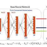
Harmonizing 1.5 T/3T diffusion weighted MRI through development of deep learning stabilized microarchitecture estimators
Jan. 17, 2020—Nath V, Remedios S, Parvathaneni P, Hansen CB, Bayrak RG, Bermudez C, Blaber JA, Schilling KG, Janve VA, Gao Y, Huo Y. Harmonizing 1.5 T/3T diffusion weighted MRI through development of deep learning stabilized microarchitecture estimators. In Medical Imaging 2019: Image Processing 2019 Mar 15 (Vol. 10949, p. 109490O). International Society for Optics and Photonics....

Tractography reproducibility challenge with empirical data (TRAceD): The 2017 ISMRM diffusion study group challenge
Jan. 17, 2020—Nath V, Schilling KG, Parvathaneni P, Huo Y, Blaber JA, Hainline AE, Barakovic M, Romascano D, Rafael‐Patino J, Frigo M, Girard G. Tractography reproducibility challenge with empirical data (traced): The 2017 ISMRM diffusion study group challenge. Journal of Magnetic Resonance Imaging. 2020 Jan;51(1):234-49. Full text: https://www.ncbi.nlm.nih.gov/pubmed/31179595 Abstract BACKGROUND: Fiber tracking with diffusion-weighted MRI has become an...
Deep learning reveals untapped information for local white-matter fiber reconstruction in diffusion-weighted MRI
Jan. 17, 2020—Nath V, Schilling KG, Parvathaneni P, Hansen CB, Hainline AE, Huo Y, Blaber JA, Lyu I, Janve V, Gao Y, Stepniewska I, Anderson AW, Landman BA. Deep learning reveals untapped information for local white-matter fiber reconstruction in diffusion-weighted MRI. Magnetic resonance imaging. 2019 Oct 1;62:220-7. Abstract PURPOSE: Diffusion-weighted magnetic resonance imaging (DW-MRI) is of critical importance...
Improved gray matter surface based spatial statistics in neuroimaging studies
May. 21, 2019—Prasanna Parvathaneni; Ilwoo Lyu; Yuankai Huo; Baxter P. Rogers; Kurt G. Schilling; Vishwesh Nath; Justin A Blaber; Allison E Hainline; Adam W Anderson; Neil D. Woodward; Bennett A Landman. “Improved gray matter surface based spatial statistics in neuroimaging studies.” Magnetic Resonance Imaging, 61, 285-295, 2019. Full text Abstract Neuroimaging often involves acquiring high-resolution anatomical images along with...
A fiber coherence index for quality control of B-table orientation in diffusion MRI scans.
May. 16, 2019—Kurt G Schilling, Fang-Cheng Yeh, Vishwesh Nath, Colin Hansen, Owen Williams, Susan Resnick, Adam W. Anderson, Bennett A. Landman. “A fiber coherence index for quality control of B-table orientation in diffusion MRI scans”. Magnetic Resonance Imaging. 2019. doi: 10.1016/j.mri.2019.01.018 Full text: https://www.ncbi.nlm.nih.gov/pubmed/?term=fiber+coherence+b-table Abstract Purpose The diffusion MRI “b-vector” table describing the diffusion sensitization direction can be flipped and...
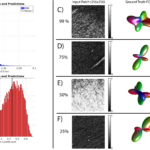
Learning 3D White Matter Microstructure from 2D Histology
Apr. 1, 2019—Histological analysis is typically the gold standard for validating measures of tissue microstructure derived from magnetic resonance imaging (MRI) contrasts. However, most histological investigations are inherently 2-dimensional (2D), due to increased field-of-view, higher in-plane resolutions, ease of acquisition, decreased costs, and a large number of available contrasts compared to 3-dimensional (3D) analysis. Because of this,...
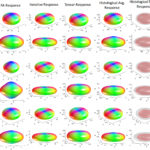
Histologically derived fiber response functions for diffusion MRI vary across white matter fibers-An ex vivo validation study in the squirrel monkey brain.
Mar. 16, 2019—Kurt G. Schilling, Yurui Gao, Iwona Stepniewska, Vaibhav Janve, Bennett A. Landman, Adam W. Anderson. “Histologically derived fiber response functions for diffusion MRI vary across white matter fibers – an ex vivo validation study in the squirrel monkey brain”. NMR in Biomedicine. 2019 Jun;32(6):e4090. doi: 10.1002/nbm.4090. Epub 2019 Mar 25. Full text: https://www.ncbi.nlm.nih.gov/pubmed/30908803 Abstract Understanding the...
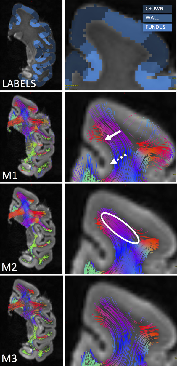
Confirmation of a Gyral Bias in Diffusion MRI Fiber Tractography
Dec. 15, 2018—Kurt G Schilling, Yurui Gao, Iwona Stepniewska, Bennett A. Landman, and Adam W Anderson. “Confirmation of a Gyral Bias in Diffusion MRI Fiber Tractography”. Human Brain Mapping. 2018 Mar;39(3):1449-1466. doi: 10.1002/hbm.23936. Full text: https://www.ncbi.nlm.nih.gov/pubmed/?term=Confirmation+of+a+Gyral+Bias+in+Diffusion+MRI+Fiber+Tractography Abstract Diffusion MRI fiber tractography has been increasingly used to map the structural connectivity of the human brain. However, this technique...
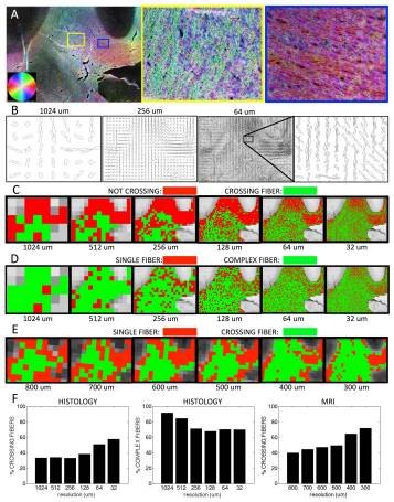
Can increased spatial resolution solve the crossing fiber problem for diffusion MRI?
Dec. 15, 2018—Kurt G Schilling, Yurui Gao, Vaibhav Janve, Iwona Stepniewska, Bennett A Landman, Adam W Anderson. “Can increased spatial resolution solve the crossing fiber problem for diffusion MRI?”. NMR in Biomedicine. (2017) 30(12),e3787. https://doi.org/10.1002/nbm.3787. Full text: https://www.ncbi.nlm.nih.gov/pubmed/?term=Can+increased+spatial+resolution+solve+the+crossing+fiber+problem+for+diffusion+MRI%3F Abstract It is now widely recognized that voxels with crossing fibers or complex geometrical configurations present a challenge for...
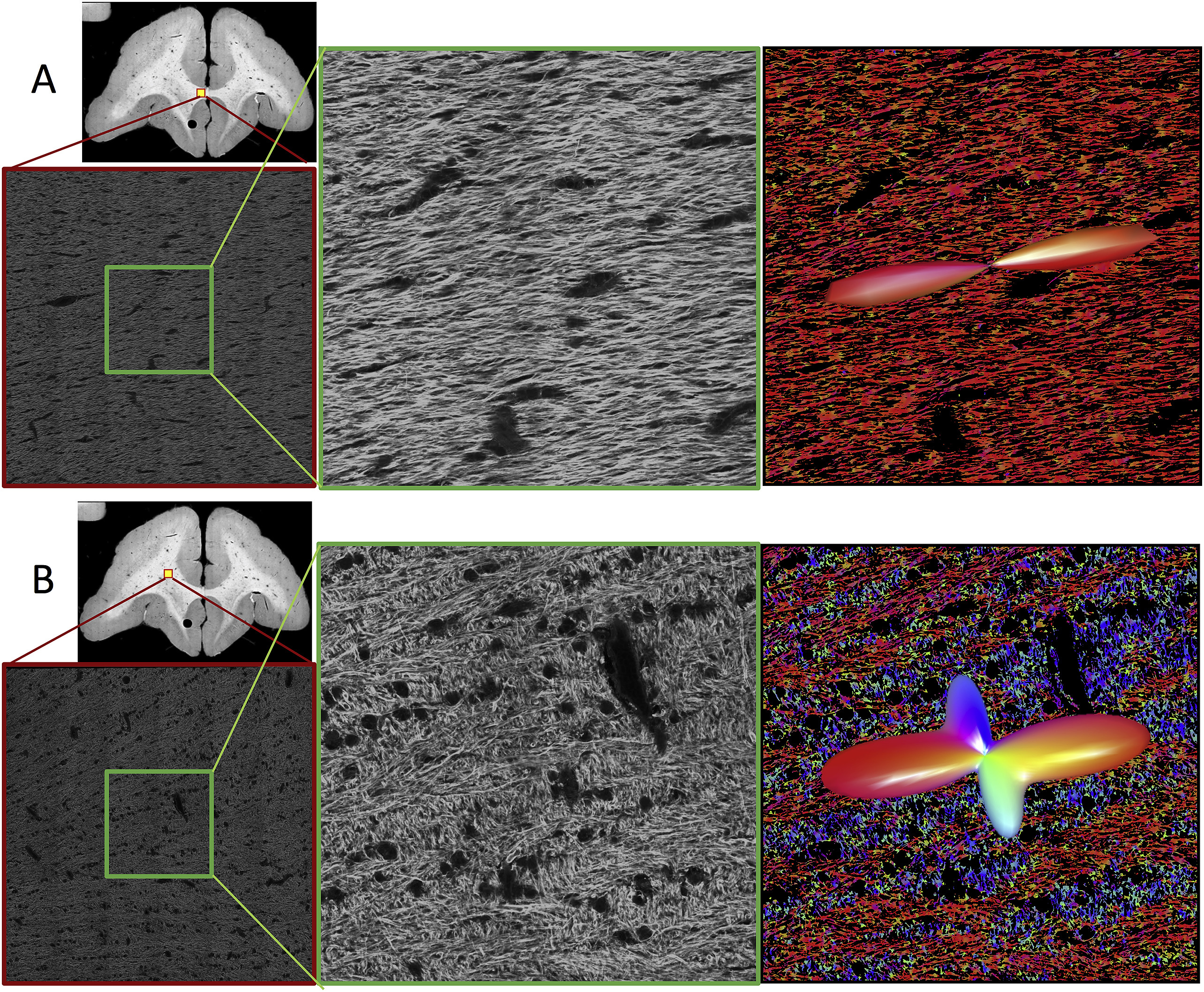
Histological Validation of Diffusion MRI Fiber Orientation Distributions and Dispersion
Dec. 15, 2018—Kurt G Schilling, Vaibhav Janve; Yurui Gao; Iwona Stepniewska; Bennett A Landman; Adam W Anderson. “Histological Validation of Diffusion MRI Fiber Orientation Distributions and Dispersion”. NeuroImage. 2018 Jan 15;165:200-221. doi: 10.1016/j.neuroimage.2017.10.046. Full text: https://www.ncbi.nlm.nih.gov/pubmed/?term=Histological+Validation+of+Diffusion+MRI+Fiber+Orientation+Distributions+and+Dispersion Abstract Diffusion magnetic resonance imaging (dMRI) is widely used to probe tissue microstructure, and is currently the only non-invasive way to...