Computed Tomography Category

Distributed Deep Learning Across Multisite Datasets for Generalized CT Hemorrhage Segmentation
Jan. 2, 2020—Remedios, S. W., Roy, S., Bermudez, C., Patel, M. B., Butman, J. A., Landman, B. A., & Pham, D. L. (2019). Distributed Deep Learning Across Multi‐site Datasets for Generalized CT Hemorrhage Segmentation. Medical physics. Full Text: Pubmed Link Abstract Purpose: As deep neural networks achieve more success in the wide field of computer vision, greater emphasis is...
Distributed deep learning for robust multi-site segmentation of CT imaging after traumatic brain injury
Jan. 2, 2020—Remedios, Samuel, et al. “Distributed deep learning for robust multi-site segmentation of CT imaging after traumatic brain injury.” Medical Imaging 2019: Image Processing. Vol. 10949. International Society for Optics and Photonics, 2019. Full text: PubMed Link Abstract Machine learning models are becoming commonplace in the domain of medical imaging, and with these methods comes an ever-increasing need...
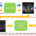
Semi-Supervised Multi-Organ Segmentation through Quality Assurance Supervision
Dec. 19, 2019—Ho Hin Lee, Yucheng Tang, Olivia Tang, Yuchen Xu, Yunqiang Chen, Dashan Gao, Shizhong Han, Riqiang Gao, Michael R. Savona, Richard G. Abramson, Yuankai Huo, Bennett A. Landman, “Semi-Supervised Multi-Organ Segmentation through Quality Assurance Supervision”, SPIE MI:IP 2020. Houston, TX. Link: https://arxiv.org/abs/1911.05113 Abstract Human in-the-loop quality assurance (QA) is typically performed after medical image segmentation to ensure that...
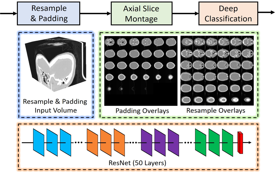
Montage based 3D Medical Image Retrieval from Traumatic Brain Injury Cohort using Deep Convolutional Neural Network
Dec. 10, 2018—Cailey I. Kerley, Yuankai Huo, Shikha Chaganti, Shunxing Bao, Mayur B. Patel, Bennett A. Landman. “Montage based 3D Medical Image Retrieval from Traumatic Brain Injury Cohort using Deep Convolutional Neural Network.” In SPIE Medical Imaging, International Society for Optics and Photonics, 2019. Full text: NIHMSID Abstract Brain imaging analysis on clinically acquired computed tomography (CT) is essential...
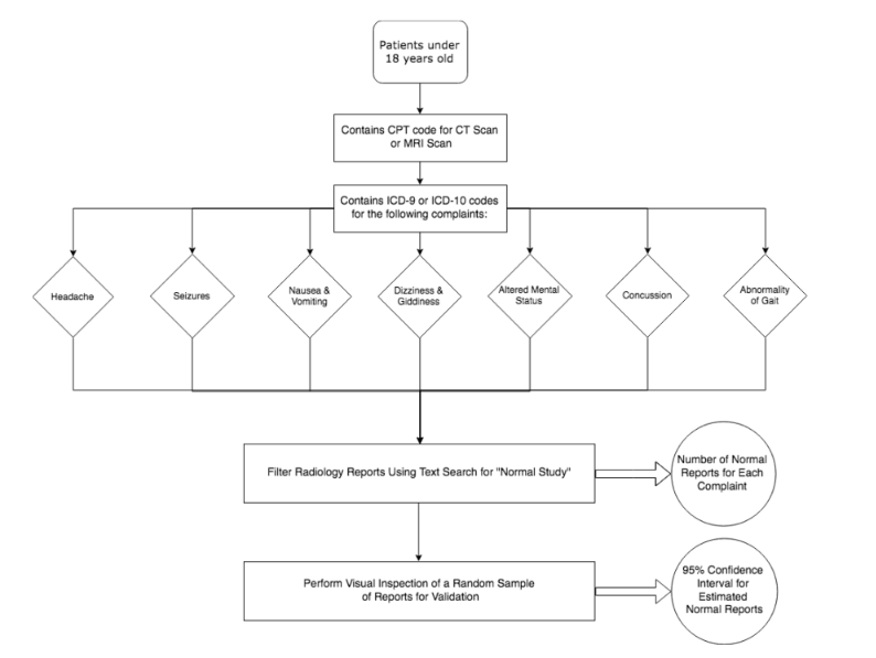
Opportunities for Mining Radiology Archives for Pediatric Control Images
Dec. 17, 2017—Bermudez, C., Probst, V. N., Davis, L. T., Lasko, T., & Landman, B. A. (2017). Opportunities for Mining Radiology Archives for Pediatric Control Images. arXiv preprint arXiv:1712.02728. Full Text: https://arxiv.org/ftp/arxiv/papers/1712/1712.02728.pdf Abstract A large database of brain imaging data from healthy, normal controls is useful to describe physiologic and pathologic structural changes at a population scale....
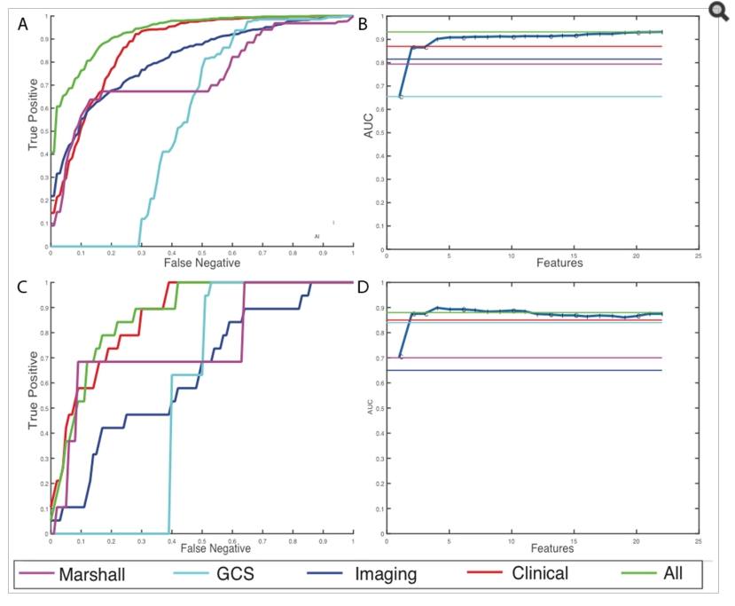
A Bayesian Framework for Early Risk Prediction in Traumatic Brain Injury
Feb. 15, 2016—Shikha Chaganti, Andrew J. Plassard, Laura Wilson, Miya A. Smith, Mayur B. Patel, Bennett A. Landman. A Bayesian Framework for Early Risk Prediction in Traumatic Brain Injury. In Proceedings of the SPIE Medical Imaging Conference. San Diego, California, February 2016. Full Text: https://www.ncbi.nlm.nih.gov/pmc/articles/PMC4845965/ Abstract Early detection of risk is critical in determining the course...
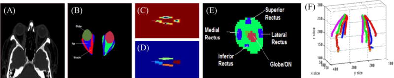
Structural Functional Associations of the Orbit in Thyroid Eye Disease: Kalman Filters to Track Extraocular Muscles
Feb. 10, 2016—Shikha Chaganti, Katrina Nelson, Kevin Mundy, Yifu Luo, Robert L. Harrigan, Steve Damon, Daniel Fabbri, Louise Mawn, Bennett Landman. “Structural Functional Associations of the Orbit in Thyroid Eye Disease: Kalman Filters to Track Extraocular Muscles”. In Proceedings of the SPIE Medical Imaging Conference. San Diego, California, February 2016. Oral presentation. Full text: https://www.ncbi.nlm.nih.gov/pmc/articles/PMC4845964/ Abstract Pathologies of...
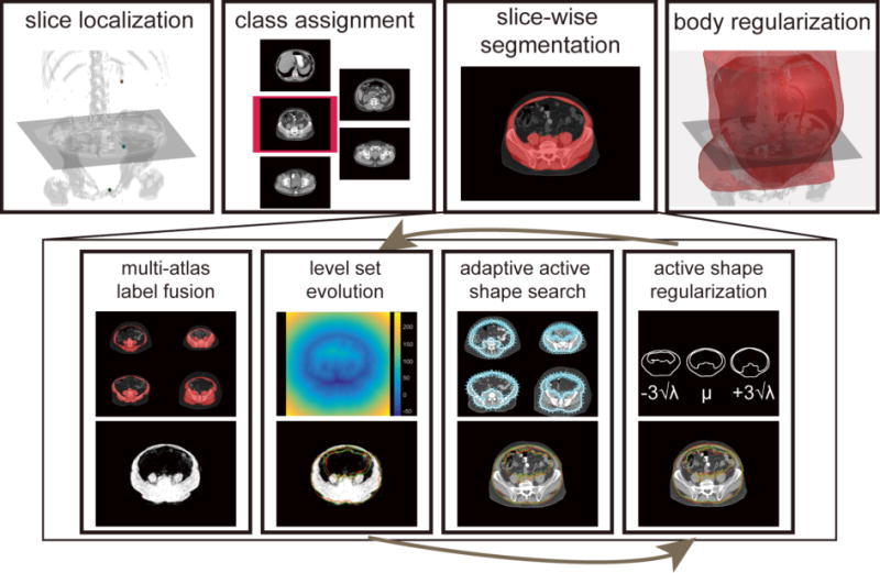
Whole Abdominal Wall Segmentation using Augmented Active Shape Models (AASM) with Multi-Atlas Label Fusion and Level Set
Feb. 1, 2016—Zhoubing Xu, Rebeccah B. Baucom, Richard G. Abramson, Benjamin K. Poulose, Bennett A. Landman, “Whole Abdominal Wall Segmentation using Augmented Active Shape Models (AASM) with Multi-Atlas Label Fusion and Level Set”, In Proceedings of the SPIE Medical Imaging Conference. San Diego, California, February 2016. Oral presentation. Full text: https://www.ncbi.nlm.nih.gov/pmc/articles/PMC4845968/ Abstract The abdominal wall is an...
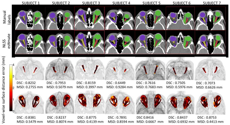
Robust Optic Nerve Segmentation on Clinically Acquired CT
Feb. 1, 2014—Swetasudha Panda, Andrew J. Asman, Michael P. DeLis, Louise A. Mawn, Robert L. Galloway, Bennett A. Landman. “Robust Optic Nerve Segmentation on Clinically Acquired CT.” In Proceedings of the SPIE Medical Imaging Conference. San Diego, California, February 2014. Oral Presentation. † Full Text: https://www.ncbi.nlm.nih.gov/pubmed/24817810 Abstract The optic nerve is a sensitive central nervous system structure,...
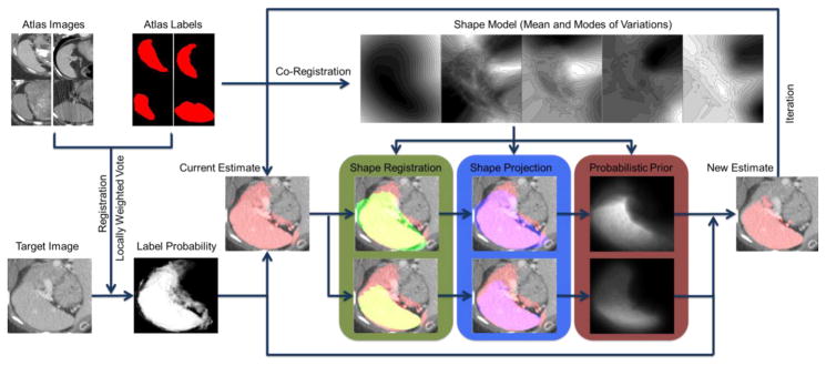
Shape-Constrained Multi-Atlas Segmentation of Spleen in CT
Feb. 1, 2014—Z. Xu, B. Li, S. Panda, A. Asman, Kristen. L. Merkle, Peter L. Shanahan, Richard G. Abramson, B. Landman. “Shape-Constrained Multi-Atlas Segmentation of Spleen in CT.” In Proceedings of the SPIE Medical Imaging Conference. San Diego, California, February 2014† Full Text: https://www.ncbi.nlm.nih.gov/pubmed/?term=Shape-Constrained+Multi-Atlas+Segmentation+of+Spleen+in+CT Abstract Spleen segmentation on clinically acquired CT data is a challenging problem given...